Lecture series Gastrointestinal tract Professor Shraddha Singh Department


")
 are secreted by pancreas in response to protein")
")


 • Medical Physiology, Updated second edition (walter")
- Slides: 28
Lecture series Gastrointestinal tract Professor Shraddha Singh, Department of Physiology, KGMU, Lucknow
Learning Objectives: • Understand the composition of protein • Understand the enzymes responsible for digestion of proteins • What are sites for absorption Molecular basis of protein transportation • Learn about diseases related to protein digestion
What are Protiens ? �Proteins are a sequence of amino acids �One amino acids is joined to the next by a PEPTIDE bond �Provide energy substrate for metabolism (4 kcals/g). � Protein load received by the gut is derived from two primary sources: 70 -100 g dietary protein, and 35 -200 g endogenous protein,
Amino acids • Of the 20 amino acids that exist, 9 are essential amino acids, and 11 are nonessential
AMINO ACID: Sequence Dipeptide – 2 amino acids Tripeptide – 3 amino acids Oligopeptides – 4 -10 amino acids Polypeptide – more than 10 amino acids Proteins in the body and diet are long polypeptides (100 s of amino acids)
AMINO ACID: Sequence Dipeptide – 2 amino acids Tripeptide – 3 amino acids Oligopeptides – 4 -10 amino acids Polypeptide – more than 10 amino acids Proteins in the body and diet are long polypeptides (100 s of amino acids)
AMINO ACIDS: Structure Consist of a central carbon atom bonded to: a hydrogen, a carboxylic acid, an amino group, and an additional side group that is unique to each amino acid
Digestion of proteins
Initial digestion of protein in stomach �No digestion of protein takes place in the mouth, �Hydrochloric acid denatures protein and also converts pepsinogen to pepsin �Pepsin breaks the protein down into peptides of various lengths and some amino acids �Pepsin act only at p. H 1. 6 -3. 2 �Pepsin completes ~ 10 -20% of digestion
Denaturing of Proteins • Acid, alkaline, heat and alcohol, can disrupt the chemical forces that stabilize proteins and can cause them to lose their shape (denature) • Denaturing of proteins happens during food preparation (cooking, whipping, adding acids) or digestion (in the stomach with hydrochloric acid)
Action of Proteolytic enzymes �Pepsin hydrolyses the bonds between aromatic amino acids(phenylanine or tyrosine) and a second amino acid �So the product of pepsin hydrolysis is polypeptides of diverse sizes
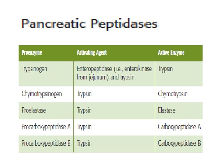
Polypeptidases • Trypsinogen and chymotrypsinogen (proenzymes) are secreted by pancreas in response to protein in the small intestine • They will be activated to trypsin and chymotrypsin (now called proteases)
Peptidases hydrolyse proteins �These enzymes can either cleave internal peptide bonds (i. e. endopeptidases) �exopeptidases cleave off one amino acid at a time from either the –COOH or –NH 2 terminal of the polypeptide (i. e. they are carboxypeptidases , and aminopeptidases, respectively)
• The endopeptidases cleave the large polypeptides to smaller oligopeptides, which can be acted upon by the exopeptidases to produce the final products of protein digestion, amino acids, di- and tripeptides, which are then absorbed by the enterocytes
Further hydrolysis by Peptidases • By the action of endo and exopeptidases some free amino acids are liberated in the intestinal lumen, • But others are liberated at the cell surface by the aminopeptidases, carboxypeptidases, endopeptidases, and dipeptidases in the brush border of the mucosal cells.
Absorption of proteins
Transport of amino acids and polypeptides in the enterocytes The di- and tripeptides are actively transported into enterocytes by a system known as peptide transporter 1) that requires H + instead of Na +
At basolateral membrane • The movement of any one amino acid can occur through one or more amino acid transporters. • At least five amino acid transporters are present in the basolateral membrane. • Three amino acid transport processes on the basolateral membrane mediate amino acid exit from the cell into the blood • Two other amino acid transporters mediate uptake from the blood for the purposes of cell nutrition.
Amino acid transport at basolateral • Individual amino acids are transported across the basolateral membrane without the need for cotransport. • Many different amino acid transporters are located on the basolateral membrane and provide specificity
Diseases associated with absorption of proteins • Hartnup disease and cystinuria are hereditary disorders of amino acid transport across the apical membrane. • These autosomal recessive disorders are associated with both small intestine and renal tubule abnormalities • the absorption of neutral amino acids in the case of Hartnup disease and of cationic (i. e. , basic) amino acids and cystine in the case of cystinuria.
References • Lippincott’s Illustrated Reviews: Physiology (2013) • Medical Physiology, Updated second edition (walter F. Boron, MD, phd) • Berne & levy, physiology, sixth edition, updated edition • Ganong’s Review of Medical Physiology, 26 t h e d i t i o n