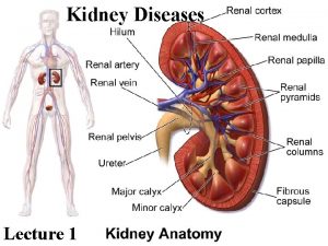
Kidney diseases Dr Manoj Radhakrishnan Addl Professor Dept


- Slides: 25

Kidney diseases Dr. Manoj Radhakrishnan Addl Professor Dept. Of Pathology

The main clinical renal syndromes �Acute Nephritic syndrome �Nephrotic syndrome �Asymptomatic hematuria, proteinuria, or both �Acute renal failure �Chronic renal failure �Renal tubular defects �Urinary infections �Obstructive uropathy �Urinary stones �tumors

Principal laboratory findings in Uremia(endstage kidney disease) �Azotemia(incrsd. BUN blood urea nitrogen, & serum creatinine) �Electrolyte abnormalities(retention of sodium, potassium and phosphate with secondary changes in calcium) �Acidosis �Anemia �Prolonged bleeding time

Acute nephritic syndrome �Typically presents 1 -2 weeks after an upper respiratory tract infection caused by streptococci Ø Oliguria : due to reduced GFR Ø Hematuria : due to damaged GBM, brownish- red urine, RBCs in urine sediment Ø Protienuria : increased permeability of GBM, maybe mild or severe Ø Generalized edema: “ puffy eyes” due to hypo albuminemia Ø Hypertension : reduced GFR leads to secretion of renin Ø Low complement levels in blood: immune complexes bind the complement Ø Azotemia : high bld urea & serum creatinine

Nephrotic syndrome �Protienuria : massive > 3. 5 g/day �There is hypoalbuminemia �Generalized edema : due to reduced oncotic pressure (hypoalbuminemia) �Hyperlipidemia(increased LDL, lipid casts in urine �Affected patients are prone to infection & thrombotic events because of increased urinary loss of serum protiens

Derangements of urine volume �Anuria : reduced urine output (< 100 ml urine per day ) reflecting renal injury �Oliguria : reduced urine output below 400 ml / day sign of renal failure �Polyuria: increased volume of urine ( > 3 L of urine per day) may result from excessive fluid intake, osmotic diuresis( eg Diabetes insipidus) or impaired tubular concentration (eg tubular necrosis)

Features of Acute renal failure �Is characterised most often b reversible detrioration of renal function Ø Oliguria Ø Azotemia ( increased Blood Urea, Serum Creatinine) Ø Electrolyte disturbances �Most patients recover without dialysis

Chronic renal failure �Develops insidiously in stages Ø Diminished renal reserve : develop high urea, creatinine levels during inter-current illness Ø Renal insufficiency : GFR ( 20% - 50% of normal)azotemia anemia, polyuria Ø Renal failure : (<20% of normal GFR) & edema metabolic acidosis hypocalcemia, and multi-sstem signs of uremia Ø End-stage renal failure : <5% GFR , clinical signs of uremia

Features of urinary tract Infection �Charcterised by bacteriuria & pyuria �>100, 000 bacteria/ml of cultured urine �> 10 wbcs /HPF in urine sample �Leucocytes are counted in urinary sediment �Signs of urinary irritation- urgency, pain

Antibody associated glomerular Injury �Antibodies to endogenous antigens of the GBM : This mechanism accounts for the renal inury in Goodpasture syndrome a disease caused by antibodies to collagen type IV �Antibodies to non-glomerular antigens : Antigen-antibody complexes found in glomeruli may result from two pathogenetic mechanisms. Ø In situ immune complex formation results from the binding of circulating antibodies– seen in PSGN ( post streptococcal GN) where streptococcal antigens are implantedin the GBM during the infection Ø Circulating immune complexes formed from soluble antigens and corresponding Ab – eg seen in SLE

PSGN �Group A beta- hemolytic streptococci ( streptococcus pyogenes) account for 90% of all GN cases �Typically occurs 1 -4 weeks after a strep throat infection or skin Infection(Impetigo) �Occasionally same clinical-pathological findings may follow staphlococcal infection or even some viral diseases –HBV, HCV or HIV Infection �.

PSGN �The increase in cellularity is due to increasi in epithelial endothelial and mesangial cells �As well as neutrophils in and around capillary loops.

PSGN �Showing increased number of neutrophils , in post-infectious GN

sub-epithelial humps in acute PSGN �Represent epimembranous deposits of immune complexes on the GBM, seen under Electron microscope. �Immunofluorescence shows many other immune complexes not dense enough to be seen by EM �Smaller immune complexes in mesangium

Clinical features of acute PSGN(nephritic syndrome) �Childhood disease �Presents with fever, nausea, oliguria, hematuria, RBC casts in urine, mild protienuria(usually less than < 1 g/ day) peri-orbital edema and mild to moderate HT �As immune complex deposition is assoc. with complement activation C 3 is the complement consumed in both classical and alternate complement pathway. Hence there will be hypo-complementemia

Outcome & long term consequences of acute PSGN �CHILDREN Ø 90% recover within 2 -3 months with conservative therapy aimed at maintaining sodium & water balance. Ø 5 -8% have persistent GN, in much milder form with abnormal urinary findings for 6 -8 months. Ø <1% cases develop Rapidly progressive GN (RPGN) �ADULTS Ø 60% recover promptly Ø 3 -5% develop RPGN

Rapidly progressive glomerulonephritis is reflective of advaned renal pathology �Mostly caused by immunologic mechanisms �Is assoc. with 25% mortality & 40 % progression to chronic end- stage kidney disease � 3 groups Ø A. N. C. A + Cases(Anti-neutrophil cytoplasmic Ab) , most Adult cases , Wegener’s granulomatosis, P. A. N Ø Immune complex mediated – most common form seen in children & young adults < 20 years – seen in post-infectious GN(PSGN) , SLE Ø Anti-GBM Ab – Good Pasteur's syndrome

RPGN or crescentic GN �Crescents are found in more than 50 % of glomeruli represent a sin of severe glomerular injury �Usually starts as focal and segmental necrosis – resulting inrupture of the GBM which allows the entry of inlammatory cells and fibrinogen into theurinary space

Crescentic GN �Early crescents are composed of fibrin , inflammatory cells exudated into the urinary spaces and proliferated epithelial cells of the bowman’s capsule � later these early crescents may get fully fibrotic

Nephrotic syndrome �Protienuria : massive > 3. 5 g/day �There is hypoalbuminemia �Generalized edema : due to reduced oncotic pressure (hypoalbuminemia) �Hyperlipidemia(increased LDL, lipid casts in urine �Affected patients are prone to infection & thrombotic events because of increased urinary loss of serum protiens

Differences between Nephrotic syndrome & Nephritic syndrome �Protienuria ++ to +++ �Hypoalbuminemia + �Edema + to ++ �Hematuria ++ and RBC casts �Oliguria ++++ �No Hyperlipidemia �No Lipiduria �Hypertension + �Nephrotic syndrome �Protienuria in nephrotic range(> 3. 5 g/dl) �Hypoalbuminemia +++ �Edema + to ++ �No hematuria �No Oliguria �Hyperlipidemia �Lipiduria �No Hypertension

Membranous Nephropathy �Is the most common primary cause of Nephrotic syndrome in Adults �Etiol. : Immune complex deposition in glomeruli �Primary – Idiopathic , unknown �Secondary– prolonged antigenemia- SLE, chronic viral hepatitis B & C, cancer pts �Pathology : Diffuse thickening of BM, but no proliferation �No response to steroids �Progresses to end stage renal failure over 10 -15 years

Minimal change disease(Lipoid nephrosis) �Most common cause of nephrotic syndrome in children ( 90 % cases < 5, 50 % of < 10 yrs) �Unknown etiology related to Type IV hypersensitivity �EM scopy shows loss of foot processes of epithelial cells �Responds to steroid therapy

How does SLE affect the kidneys? �Signs of renal disease are found in 50 % of patients at the time of diagnosis � 80% will develop some signs of renal disease at some time during the life span. �SLE is an immune complex mediated disease, immune complexes are deposited in Ø Glomeruli : most frequently affected part, Immune complexes maybe trapped on the sub-endothelial side, inside the GBM, or on the sub epithelial side & mesangium basement membranes lined with sub-endothelial deposits appear thickened , called wireloops Ø Blood vessels Ø Tubules & interstitial spaces

Classification/ stages of lupus nephritis ( SLE) �Class I �Class-II-mesangial GN �Class III- Focal proliferative GN �Class IV- diffuse proliferative GN �Class V- membranous GN �Class VI- Chronic GN �No deposits �Mesangial deposists , mild hematuria �Mesangial & sub-endothelial moderate GN �widespread deposits, severe GN �Subepithelial- nephrotic syndrome �Chronic GN
 Manoj radhakrishnan
 Sarvepalli radhakrishnan
 Jai radhakrishnan
 Veena radhakrishnan
 Promotion from associate professor to professor
 Measurement and scaling
 Manoj abichandani
 Given signs signify something and on that basis
 Manoj toshniwal dubai
 Panama real estate law
 Manoj magician
 Dr manoj das products
 Manoj mehta ficci
 Department of finance and administration
 Nys dept of homeland security
 Florida dept of agriculture and consumer services
 Vaginal dept
 Worcester ma building department
 Florida dept of agriculture and consumer services
 Nebraska dept of agriculture
 Albany county dept of social services
 Dept of education
 Gome dept
 Oviposition
 Dept. name of organization (of affiliation)
 La dept of revenue
