Introduction to Anatomy Chapter 3 ANATOMICAL POSITION 3

 2. Sagittal (Vertical) 3.")

 3. Superficial")


 5. Protraction and Retraction (Jaw and shoulder blades)")
 7. Circumduction (shoulder or hip)")


 B. Between bones")
: Great mobility Hinge or multiaxial joints (ball & socket) Synovial")
 Saddle Joint-the joint")


- Slides: 16
Introduction to Anatomy Chapter 3
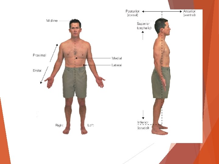
ANATOMICAL POSITION 3 Planes of the body: 1. Frontal (Coronal) 2. Sagittal (Vertical) 3. Transverse (Horizontal)
Reference Terms 1. Anterior and posterior 2. Medial and lateral 3. Proximal and distal
Reference Terms 1. Superior and inferior 2. Dorsal and ventral (hand foot) 3. Superficial and deep
MOVEMENTS 1. Flexion and Extension 2. Abduction and Adduction
Pronation and Supination
4. Inversion and Eversion (foot) 5. Protraction and Retraction (Jaw and shoulder blades)
6. Rotation (head) 7. Circumduction (shoulder or hip)
BONES Purpose? How many? Categories? 1. Axial skeleton 2. Appendicular skeleton
Bones Long-femur Short-metacarpal Flat-scapula Irregular-vertebra Epiphysis—Growth Plate
Cartilage 1. Location: A. Covers the ends of bones. (Articular Cartilage) B. Between bones Hyaline (Cartilage) 2. Functions A. Joins structures B. Absorb shock C. Permit smooth bone movement
JOINTS A. Diarthrodial (synovial): Great mobility Hinge or multiaxial joints (ball & socket) Synovial membrane Hyaline cartilage B. Amphiarthodial: Cartilage attaching two bones together (cartilaginous joints) C. Synarthrodial: Fibrous joints – held together by tough connective tissue.
6 types of joints Hinge-permit motion in 1 plane (knee, elbow) Saddle Joint-the joint is shaped like a saddle and rider (carpometacarpal joint-thumb) Ball and Socket-made up of a spherical shape and the other of a cup-like socket (hip, shoulder) Gliding joint-allows joint to glide in any direction (carpals of hands) Pivot joint-central bony cylinder with a ring like structure (proximal joint of radius and ulna) Condyloid-Oval articular surface of one bone fits into a complementary depression (
Tendons and Ligaments TENDON: Attach muscle to bone. LIGAMENT: Connects bone to bone (forms joints)
SKIN First line of defense. Layers: 1. Epidermis – most superficial 2. Dermis – next layer in 3. Hypodermis – holds the skin to the bone and muscle.