Infrared spectroscopy IR regions The infrared portion of


 for C- H bond, knowing")
, bond strength and mass The vibrational frequency of")


- Slides: 18
Infra-red spectroscopy
IR regions The infrared portion of the electromagnetic spectrum is usually divided into three regions: Ø 1. The near-infrared; The higher-energy near-IR, approximately 14000– 4000 cm− 1 (0. 8– 2. 5 μm wavelength) can excite overtone or harmonic vibrations. Ø 2. The mid-infrared; approximately 4000– 400 cm− 1(2. 5– 25 μm) may be used to study the fundamental vibrations and associated rotational-vibrational structure. Ø Ø 3. The far-infrared, approximately 400– 10 cm− 1 (25– 1000 μm), lying adjacent to the microwave region, has low energy and may be used for rotational spectroscopy.
Hooke’s law § Bonds can be thought like a spring, and wavenumbers can be approximated by Hooke’s law § This study of vibrations of bonds between different atoms and varied multiplicities which depending on: a. The electronegativity (force constant of the bond) b. The relative masses of the atoms c. Their geometry vibrate at different c = speed of light (3 x 1010 cm/s) k = force constant m = reduced mass of the atoms Mx = mass of atom x in kg My = mass of atom y in kg
Hooke’s law-Example Calculate the predicted vibrational frequency (in cm-1) for C- H bond, knowing that: The force constant for single bond is 5 x 10⁵ dyne/ cm, the velocity of light is 3 X 108 cm/s, the mass of carbon atom is 20 x 10 -24 g, the mass of hydrogen is 1. 6 x 10 -24 g, and what are the relationship between wave number (v), bond strength and mass?
The relationship between wave number (v), bond strength and mass The vibrational frequency of a bond would increase with the increase in bond. 1 strength. Consequently, we can expect that C=C and C=O > C-C and C-O, respectively The vibrational frequency of a bond would increase with the decrease in reduced mass of the system. C-H and O-H Similarly, > C-C and C-O, respectively O-H > O-D
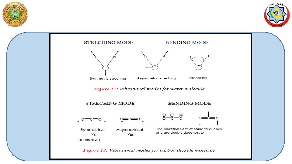
Types of molecular vibrations The two atoms joined together by a chemical bond (may be single, double or triple bond), macroscopically can be composed as two balls joined by a spring. (i) Stretching of one or both the atoms is a rhythmical movement along the band axis such that interatomic distance is increasing or decreasing its either Symmetric Antisymmetric
Number of vibrational modes Ø A molecule can vibrate in many ways, and each way is called a vibrational mode. Ø In order for a vibrational mode in a sample to be "IR active", it must be associated with changes in the dipole moment. A permanent dipole is not necessary, as the rule requires only a change in dipole moment. Ø For molecules with N number of atoms, q Linear molecules have 3 N – 5 degrees of vibrational modes, q Nonlinear molecules have 3 N – 6 degrees of vibrational modes (also called vibrational degrees of freedom). Symmetric Bending Asymmetric Example H 2 O, will have (3 × 3 – 6 = 3) degrees of vibrational freedom, or modes.
ØBending of one of the atoms either vertically or horizontally and then release of the force results in the vibrations on the two balls (atoms). Ø Changing of in bond angles between bonds with common atom of the movement of group of atom with respect to the reminder of the molecule without movement of the atoms in the group with respect to another These vibrations depend on the strength of the spring and also the mode (stretching or bending) in which the force is being applied.
Symmetric stretching Rocking Asymmetric stretching Wagging Scissoring Twisting
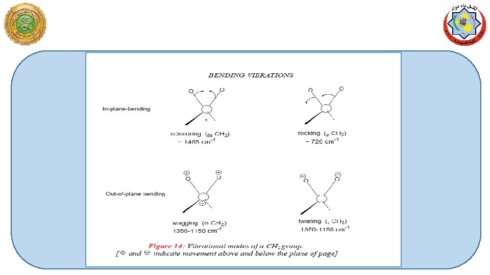
Bending Vibrations of a CH 2 Group In plane
Bending Vibrations of a CH 2 Group Out of plane
Stretching CH 2 Ø The stretching and bending modes for an AX 2 group appearing as a portion of molecule, for example, the CH 2 group in a hydrocarbon molecule. Ø Any atom joined to two other atoms will undergo comparable vibrations for example NH 2 or NO 2. Each of different vibration modes may give rise to a different absorption band so that CH 2 groups give rise to two C-H stretching bands i. e. υsym and υasym. Ø Some of the vibrations may have the same frequency i. e. they are degenerate and their absorption bands will appear at same position.
The atoms in a CH 2 X 2 group, commonly found in organic compounds and where X can represent any other atom, can vibrate in nine different ways. Six of these vibrations involve only the CH 2 portion: symmetric and asymmetric stretching, scissoring, rocking, wagging and twisting. Structures that do not have the two additional X groups attached have fewer modes because some modes are defined by specific relationships to those other attached groups. For example, in water, the rocking, wagging, and twisting modes do not exist because these types of motions of the H represent simple rotation of the whole molecule rather than vibrations within it.
Effects of hydrogen bonding IR spectroscopy v Intermolecular hydrogen bonding involves association of two or more molecules of the same different compounds. v Intermolecular hydrogen bonding may result in dimer molecules (as observed for carboxylic acid) or in polymeric molecular chains. v Intramolecular hydrogen bonds are formed when the proton donor and acceptor are present in a single molecule under spatial conditions that allow the of a five or six membered ring the extent of both inter and intra molecular bonding is temperature dependent. v Hydrogen bonding to a C=O group withdraws electrons from oxygen and lowers the C=O double bond character. This results in lowering of C=O absorption frequency. More effective is the hydrogen bonding, higher will be the lowering in C=O absorption frequencies v The monomeric carboxylic acids (in very dilute solutions) absorb at ~1760 cm -1. The dimerization of carboxylic acids in their concentrated solutions or in solid state lowers the carboxyl carbonyl frequency to 1710 cm-1.
Instrumentation IR spectroscopy Ø source of energy Ø Sampling area Ø Photometer Ø Grating (monochromator ) Ø Detector
Sample Preparation § For recording an IR spectrum, the sample may be gas, a liquid, a solid or a solution of any of these. The samples should be perfectly free of moisture, since cell materials (Na. Cl, KBr, Cs. Br etc. ) are usually spoiled by the moisture. § Liquids are studied neat or in solution. In case of neat liquid, a thin film of < 0. 01 mm thickness is obtained by pressing the liquid between two sodium chloride plates and plates are subjected to IR beam. Spectra of solutions are obtained by taking 110 % solution of the sample in an appropriate solvent in cells of 0. 1 -1 mm thickness. § A compensating cell, containing pure solvent is placed in the reference beam of the instrument. The choice of solvent depends on the solubility of the sample and its own minimal absorption in IR region. Carbon tetrachloride, chloroform and carbon disulfide are preferred solvents.