Human Semen Analysis Dr Surojit Das Ph D


 are very small mucus secreting")


 for Semen Characterestics")


 reflect")


 Eosin")


 • Simple, quick staining method. • Moderate")


 üLifestyle: Factors like stress, heat, obesity &")
 Genetics: There a number of genetic disorders")
 Infection: Viral and bacterial infections – both")
- Slides: 39
Human Semen Analysis Dr Surojit Das, Ph. D Assistant Professor Vidyasagar University
Introduction • A semen analysis measures the amount of semen a man produces and determines the number and quality of sperm in the semen sample. • A semen analysis is usually one of the first tests done to help determine whether a man has a problem fathering a child (infertility). • A problem with the semen or sperm affects more than one-third of the couples who are unable to have children (infertile).
Purpose of seminal fluid analysis There are basically four indications for the examination of seminal fluid: 1. The investigation of fertility: male infertility is primarily responsible in 30 -50% of infertile marriages. 2. To determine the effectiveness of vasectomy (a surgical intervention for male sterilization or permanent contraception). 3. To determine the suitability of semen for artificial insemination (the medical procedure of injecting semen into vagina or uterus). 4. Medico-legal testes to detect semen are frequently requested in alleged rape or in association with other sexual crimes of violence.
Fluid Fractions 1. Bulbourethral & Urethral glands (2 -5%) are very small mucus secreting glands, add alkaline mucus to neutralize prostatic acid and vaginal acidity. 2. Prostate: (produce about 13 -33% of the fluid volume of semen) Prostate glands secretion is a milky, alkaline fluid that plays a role in activating sperm, the secretion contains acid phosphatase and proteolytic enzymes that act on the fluid from the seminal vesicles, resulting in the coagulation and liquefaction of the semen. 3. Seminal vesicles (produce about 46 -80% of the fluid volume of semen) Viscous, yellowish secretion is rich in fructose, vitamin C, prostaglandin, and other substances, which nourish and activate the sperm passing through the tract. This component has high flavin content, which is largely responsible for the fluorescence of semen. 4. Testis & Epididymis: (5%) Spermatozoa are produced in the testis under the influence of testosterone, and then the epididymis (is the first part of the duct system) provides a temporary storage site for the immature sperm that enter it from testis. This fraction still in the inactive form until ejaculation due to the high content of carnitine, glyceryle-phosphorylcholine and diminished oxygen supply.
Factors Effecting the Semen Analysis Results
Time-line of Semen Analysis
Nomenclature Related to Semen Quality
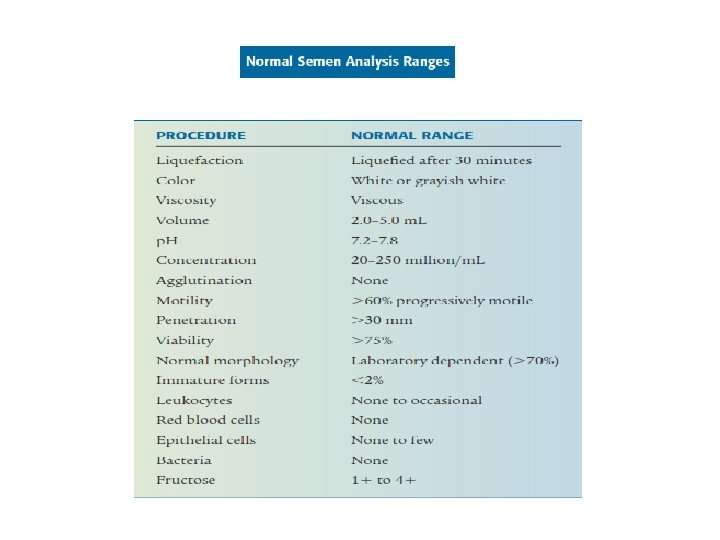
Lower Reference Limits (5 th percentiles & 95% confidence intervals) for Semen Characterestics
Sample Collection
Semen Collection Good and reliable SA results starts from semen collection, preferably by masturbation • Abstinence days 2 -6 • Pass urine • Wash hands with soap, dry • Collect the entire sample into the wide mouth sterile container, 70% of sperms is in the first part of the ejaculate • Keep the sample at body temperature, no sunlight • Deliver the sample within one hour of ejaculation
Ways to Collect Semen • Masturbation - directing the semen into a clean sample cup. Do not use a lubricant which can kill sperms. • Coitus interruptus - withdrawing the penis from the partner just before ejaculating follow by ejaculating into a clean sample cup. • Coitus - by using a condom. A special (silicon) condom that does not contain any substance that kills sperm (spermicide). After ejaculation, carefully remove the condom from the penis. Tie a knot in the open end of the condom and place it in a container that can be sealed in case the condom leaks or breaks. Ordinary condoms should not be used since they usually contain spermicides. • Assisted ejaculation – electro-ejaculation used in paralegics (paralysis that affects all or part of the torso, legs & pelvic organs).
Macroscopic Examination WHO criteria 2010 Description Appearance Normal: Whitish to grey opalescent Yellow (urine, jaundice); Pink/Reddish/Brown (RBCs) Liquefaction Normal: 15– 30 minutes after collection Lumpy >60 min – sperms may be trapped in unliquefied jelly; may be sign of prostatic infection, lack of prostatic protease Viscosity Normal Smooth and watery Abnormal – thick with long threads. High viscosity impede sperm movements Normal semen viscosity test MIX SEMEN THOROUGHLY BEFORE ANALYSIS –USE A PIPETTE
Macroscopic Examination Volume Normal: 1. 5 ml per ejaculation Low volume (<1 ml) reflect a problem with the seminal vesicles and prostate – a block, retrograde ejaculation, infection or lack of androgen. Low semen volume cannot neutralize vaginal acidity High semen volume dilute sperms/ active infection p. H Normal: =/>7. 2 (alkaline) Acidic p. H (<7. 0) in a low volume & density sample indicates – congenital bilateral absence of vas deferens (in which seminal vesicles are also poorly developed) and ejaculatory duct obstruction. p. H increases with time as natural buffering capacity of semen decreases – therefore high p. H is not clinically useful
Microscopic Examination
Sperm Agglutination
Sperm Motility
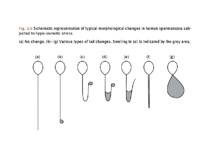
Sperm Vitality 1. 2. 3. 4. • • Eosin-nigrosin (dead sperm stain pink/red) Eosin (1%) (dead sperm stain pink/red) Trypan (0. 4%) blue (dead sperm stain blue) Hypo-osmotic swelling test (HOS) (live sperm shows tail curling) Test 1, 2 and 3 for diagnostic uses. Usually 1: 1 ratio of semen to dye mixture, mix well and smear onto a slide. Read immediately at x 40 objective, count 200 sperms Test 4 is use to choose live (immotile) sperm for ICSI. Dead sperms will not react in HOS while live sperm will take up fluids causing their tails to curl within 5 min and stabilize at 30 min. Therefore viable sperms may be selected for ICSI.
Sperm Counts Neubauer Haemocytometer
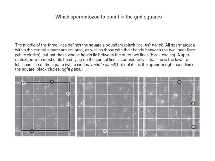
Counting Cells with a Hemacytometer The example at right shows red lines where cells on the line would be counted. If red dots represent cells, one would count 3 cells in the top middle large square. All 25 large squares can be counted, or a counting pattern using fewer squares can be used like the ones below. It is important to distribute the counting areas in a non-biased manner since cells can be more concentrated on one side of the chamber. If you count over only 5 of the 25 large squares, then multiply that value by 5 to obtain the number of cells per central counting area. At least two chambers should be counted, including at least 100 cells within each central counting area of each chamber. For higher precision, additional sperm can be counted and the average used to calculate cell concentration. Each of the nine squares on the grid, including the central counting area of 25 large squares, has an area of 1 mm 2, and the coverglass rests 0. 1 mm above the floor of the chamber. Thus, the volume over the central counting area is 0. 1 mm 3 (1/10 cm x 1/100 cm=10 -4 cc) [1 cc=1 ml]. You can thus multiply the average number of sperm over each central counting area by 10, 000 to obtain the number of sperm per ml of diluted sample. Sperm/ml= ave. no. of sperms for each chamber x 104 x dil. factor Example: Assume that you dilute the original semen sample by adding 0. 1 ml of semen to 9. 9 ml of diluent (1: 100 dilution factor). You then count the number of sperm in 5 of the 25 large squares within the central counting area of two chambers, obtaining counts of 132 and 128 cells. 1. The mean number of sperm per chamber is thus 130 (132+128/2) x 5 or 650 cells per counting area (650 cells per 0. 1 microliter). 2. Multiply the 650 cells per counting area by 10, 000 to obtain the number of cells per ml of diluted sample (answer = 6, 500, 000) 3. Multiply 6, 500, 000 cells per ml of diluted sample by 100 (the dilution factor) to obtain 650, 000 per ml of original semen sample.
• Several terms are used to describe both sperm concentration and sperm count: • Azoospermia describe a total absence of spermatozoa in semen. (After centrifuge sperm count is zero/HPF). • Oligozoospermia refers to a reduced number of spermatozoa in semen and is usually used to describe a sperm concentration of less than 20 million/ml. Sperm count 5 -10 sperm/HPF. • Severe oligospermia, sperm count 1 -2 sperm/HPF. • Polyzoospermia denotes an increased number of spermatozoa in semen and is usually refers to a sperm concentration in excess of 350 million/ml.
Sperm Morphology ü This describes the shape of the sperm. ü 70% of the sperm should be normal by these criteria. ü Generally accepted that a high incidence of morphologically abnormal spermatozoa in a semen sample is associated with reduced fertility. ü Human sperm can be visualized using bright field microscopy on fixed stained specimens. üExamples of fixed stained preparations (Papanicolaou stain, Vital staining with eosin/nigrosin, giemsa stain). üNormal spermatozoa should have an oval shaped head (4 -5. 5µm long and 2. 5 -3. 5µm wide). üThe midpiece should be cylindrical (3 -5µm long and 1. 0µm wide). üThe tail should also be cylindrical (45 -50µm long and 0. 5µm wide) with a narrower terminal segment (46µm long). üThere should be no head, midpiece or tail defects, and no cytoplasmic droplet more than one-third the size of a normal sperm head.
• Normal spermatozoa structure
Defects to be scored • Head shape/size defects - such as large, small, tapering, pinhead form, amorphous, vacuolated, multiple heads or any combination of these. • Neck and midpiece defects - such as non-inserted or bent tail, distended, irregular / bent midpiece, thin midpiece (no mitochondrial sheath), absent tail (free or loose heads) or any combination of these. • Tail defects - such as short, multiple, hairpin, broken, irregular width, coiled tails, tails with terminal droplets or any combination of these. • Cytoplasmic droplets - greater than one-third the size of a normal sperm head.
• Each spermatozoa is scored as either normal or abnormal with each of the defects being tallied separately. If a majority of the cells have a particular morphological defect this should also be noted. • In stained preparations 100 -200 sperm should be scored using a x 100 oil-immersion bright field objective
Hematoxylin-Eosin Staining o o o • Fairly good Hematoxylin-Eosin differentiation Fix slide in Et. OH/Me. OH 95% 20 min • The acrosomal area Wash in running tap water 5 min and cytoplasmic fragments is stained Dry on absorbent paper pink and the post. Hematoxylin (Sigma, HHS 20 min acrosomal area is 128) stained dark purple. Wash in running tap water 5 min • Abnormally stained Acid alcohol (99 ml 70% Dip (2) sperms (nuclear/ Et. OH + 1 ml H 2 SO 4) chromatin material) Eosin (Sigma, HT 1102128) 5 min may be differentiated. Et. OH 70% 2 min Et. OH 90% 2 min (2) • Takes longer and need experience to Absolute Et. OH (99. 9%) 2 min (2) produce good Xylene 2 min (2) staining
Diff-Quik Staining • Diff-Quik Staining (Baxter, Australia) • Simple, quick staining method. • Moderate differentiation of structures --the acrosome is stained red and the post acrosomal area dark red. Very thin smears are necessary as the background is influenced by the presence of protein in the seminal fluid and tends to be dark. • The slides are fixed with fixative (Cytospray, Kinetik, Australia). • Diff Quik 1 for 5 -6 seconds. • Clean water x 2. Removed excess moisture. • Diff Quik 2 for 10 seconds • 2 -3 dips in clean water. • It is left to dry by standing the slide on one end on absorbent paper towel. • Sperms stained by Diff-Quik are larger by comparison to H&E staining as the cells did not undergo dehydration by alcohol.
Abnormal Sperms
Abnormal Sperms 1. Triple head sperm 2. Acrosome reacted sperm 3. Sperm with no acrosome 4. Sperm with a tapering head and swollen midpiece
Morphology Assessment § Assessment is subjective due to the wide variation in sperm sizes and shapes. § % NF correlated well with the fertilization rate in-vitro and pregnancy rate [Kruger et al, 1996; Morgenthaler et al, 1995] § Sperms with defective heads are more likely to be immotile than sperms without defects, and defective motile sperms tend to show sluggish motility as compared to normal sperms [Aitken et al, 1995].
Chemical Analysis
p. H üShould be measured within an hour of collection (acidic: lactic acid production with high sperm count; alkaline: loss of CO 2 over time). üNitrozine paper is used to measure p. H. üp. H of fresh semen: 7. 2 -7. 8. üCongenital aplasia of vas deferentia & seminal vesicles >>> Acidic p. H üMale reproductive infection (Ureaplasma urealyticum, Mycoplasma hominis, Chlamydia trachomatis, Herpes simplex) >>> Alkaline p. H
Acid Phosphatase üIt is used to evaluate the secondary function of the prostate. üNormal level: ≥ 200 units/ejaculate üIt is used to establish the validity of alleged sexual assault in vaginal fluid, skin washings, or clothing.
Fructose üFructose provides energy for spermatozoa. üSemen fructose is produced by the seminal vesicles, with normal levels ≥ 13 µmol/ejaculate & comprises 99% of reducing sugar found in semen. üLow level >>> indicates presence of ejaculatory duct obstruction or abnormalities in the vas deferens & is accompanied by azoospermia (absence of sperm). üLow level >>> correlate with androgen deficiency & decreased testosterone levels.
Common Causes of Low Sperm Count (Oligospermia) üLifestyle: Factors like stress, heat, obesity & toxins (SHOT) can reduce sperm counts drastically. üVaricocele: Varicose veins (enlarged veins) in the scrotum, known as varicoceles, have been known to impact testosterone and sperm production as well as sperm health (motility, morphology). Varicoceles are relatively common, occurring in about 15% of all men, and impact men differently. Some men can have large varicoceles with little to no impact on fertility, while other men are more sensitive and even small varicoceles can dramatically impact semen parameters. Varicoceles are repairable through surgery; it often takes 6 -12 months post-surgery to fully improve fertility.
Common Causes of Low Sperm Count (Oligospermia) Genetics: There a number of genetic disorders that contribute to male infertility. Because the molecular science is still relatively young, new genetic markers for male fertility are still being discovered. The good news is that most genetic causes of infertility are relatively rare. The most common disorders are: Klinefelter’s Syndrome: Genetic condition in which a man has an extra X chromosome causing him to be XXY rather than the normal XY. Y chromosome microdeletions: The deletions of genes from the Y chromosome that are responsible for sperm production.
Common Causes of Low Sperm Count (Oligospermia) Infection: Viral and bacterial infections – both in childhood and as an adult – can cause scarring on tiny microtubes of the male reproductive tract leading to a partial or complete blockage. Leading offenders include: mumps, tuberculosis, chlamydia, gonorrhea, mycoplasma, urinary tract infections and prostate infections. Hormone imbalances: Hormones play a critical role in the production and maturation of sperm cells. For reproductive health, the most critical hormones include – Testosterone, Follicle Stimulating Hormone (FSH), and Luteinizing Hormone (LH). Hormonal imbalances can occur for a number of reasons including: impaired testicular function, thyroid disorders, diabetes, pituitary disorders or growths and abuse of androgens for athletic performance.