General Embryology By Dr Ahmed Abo Ahmed B

General Embryology By Dr. Ahmed Abo. Ahmed

B- Spermatogenesis

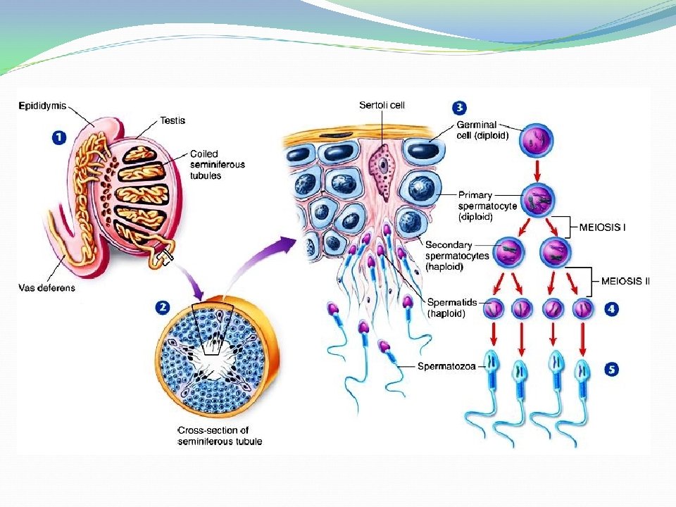
B- Spermatogenesis - It is the process of formation and maturation of the spermatozoa in the testis. - The production of spermatozoa in seminiferous tubules is continuous from puberty to death.
 Mt Spermatogonium A (2 N)")
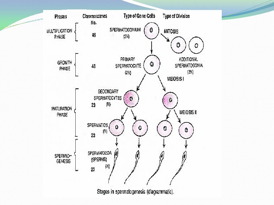
Developmental stages of spermatogenesis: Primordial germ cell (2 N) Mt Spermatogonium A (2 N) Secondary spermatocyte Spermatid (1 N) Secondary spermatocyte Me. II (1 N) Mt Spermatogonium B (2 N) Mt Primary spermatocyte Me. I (2 N) Spermatozoon (1 N) (Mt = Mitosis; Me. I = Meiosis I; Me. II = Meiosis II)
 Spermatocytogenesis: - It starts")
- Spermatogenesis can be subdivided into 2 successive stages: I) Spermatocytogenesis: - It starts from primordial germ cell to spermatid. - It consists of three phases: 1 - Period of proliferation (from PGC to spermatogonia type B). 2 -Period of growth (from spermatogonia type B to primary spermatocyte). 3 -Period of maturation ( from primary spermatocyte to spermatid). II) Spermiogenesis: - Starts from spermatid to spermatozoon.
 . . Spermatogonia type B (2 N)")
. Dormant Spermatogonia type A (2 N) . . Spermatogonia type B (2 N) . Maturation Primary spermatocyte (2 N) Secondary spermatocyte (1 N) Spermatid (1 N) Spermiogenesis Sperm (1 N) Meiosis I Growth Meiosis II Spermatocytogenesis Proliferation . Primordial germ cells (2 N) Mitosis Spermatogonia type A (2 N)
 Spermatocytogenesis: A) Proliferation stage: - The PGCs (2 N) in the germinal epithelium")
I) Spermatocytogenesis: A) Proliferation stage: - The PGCs (2 N) in the germinal epithelium of the convoluted seminiferous tubules form spermatogonia type A (2 N) which remain inactive in the basal layer of seminiferous tubules. - At puberty, spermatogonia type A proliferate by mitosis forming 2 types of cells: 1) Dormant spermatogonia type A: which remains in a stage of rest to act as a stem cell to renew the stock of type A cells. This can explain why the male can produce sperms along his life. 2) Spermatogonia type B: which enters the stage of growth which is very short then divides by mitosis giving primary spermatocyte (2 N) which characterized by large size of nucleus.
 Maturation stage: - In which the primary spermatocyte (2 N) divides by")
B) Maturation stage: - In which the primary spermatocyte (2 N) divides by 1 st meiotic division giving 2 equal size secondary spermatocytes (haploid, 1 N). - Each secondary spermatocyte (1 N) divides by 2 nd meiotic division producing 2 spermatids (1 N) with haploid number of chromosomes. (This means each primary spermatocyte gives 4 spermatids with haploid set of chromosomes). - The spermatids enter stage of spermiogenesis to become movable sperms.
 Spermiogenesis: - It is the process of transformation of spermatids into movable sperms.")
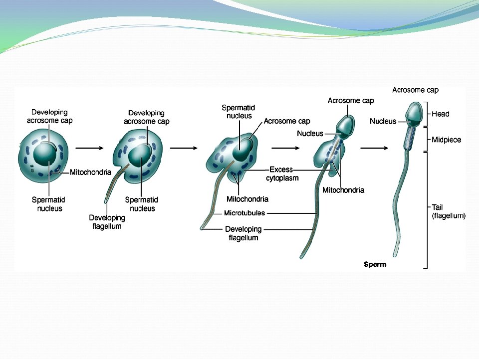
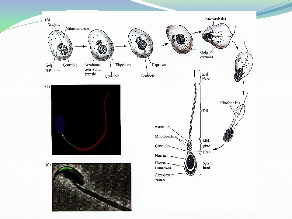
II) Spermiogenesis: - It is the process of transformation of spermatids into movable sperms. - It includes the following changes: 1. Nuclear condensation: the nucleus begins to lose fluid, becomes smaller with condensation of its chromatin and moves to an eccentric position and the cytoplasm appears as tail-like stream. 2. Flagellum formation: the anterior centriole forms flagellum (axial filament) while the posterior centriole takes a shape of a ring encircling this flagellum. 3. The mitochondria begin to concentrate around the proximal part of the axial filament (flagellum) which will become the middle piece of the sperm.

4. Acrosome formation: Part of the cytoplasm containing the Golgi apparatus concentrates at the apical end of the elongated nucleus to form the acrosome or the anterior cap. 5. Cytoplasmic reduction: elimination of unnecessary cytoplasm (called residual cytoplasm) by phagocytosis. 6. So, the sperm becomes mature without all non-essential materials, it free itself from the sertoli cells and go to lumen of seminiferous tubule. It consists only of a head containing the concentrated nuclear material bearing genes and a tail, which gives it mobility.

mitochondria Stages of Spermiogenesis Tail
 Head: contains – Nucleus (1 N) which forms the")
Structure of mature sperm: 1) Head: contains – Nucleus (1 N) which forms the bulk of head. - Acrosome: a cap like organelle covering the anterior 2/3 of nucleus containing enzymes which help sperm to penetrate the ovum during fertilization. - Post acrosomal plasma membrane: covers the posterior 1/3 of nucleus. 2) Neck: the weakest part contains 2 centrioles (proximal & distal, the distal one forms the flagellum). 3) Middle piece: It is tubular structure in which ring-shaped mitochondria are spirally arranged. It is called power house of sperm because it provides energy for flagellar motility. 4) Tail: It provides motility to help its transport to site of fertilization. It contains axial filament and consists of principle piece and end piece.

Structure of mature sperm

Abnormalities of sperms 1. Morphological Abnormalities: The head & tail may be abnormal, they may be: a) giants. b) Dwarfs. c) Joined head and tail. Lack motility and don’t fertilize the egg. 2. Numerical Abnormalities: Oligospermia: few number of sperms in semen. Aspermia: no sperms at all in semen. Necrospermia: sperms found dead.

Comparison between sperm and ovum Sperm Ovum Size Small Large Shape Elongated Round Quantity Large no. (million) One or more (species var. ) Sex determination X or Y chromosome Only X chromosome Motility Vigorous (by tail) Lack (no tail) Nucleus Eccentric, condensed chromatin Not condensed Yolk No Little to much Golgi complex Form acrosomal cap Diffused Mitochondria Aggregate in middle piece Diffused Centrioles Retained forming flagellum Disappeared

Comparison between oogenesis and spermatogenesis Oogenesis Spermatogenesis 1 - Occurrence In the ovarian cortex In seminiferous tubules of testis 2 - Start time Before birth and ovulation ends at menopause Before birth and is continuous until death 3 - Stage of growth Very prolonged Very short 4 - Motility Stored food material makes ovum inactive and depends on surroundings for its transportation High degree of independent motility due to development of flagellum 5 - Species variation Oogenesis differs according to amount of stored food material between species Nearly similar in all vertebrates

Oogenesis Spermatogenesis 6 - Direction of Mature cells move towards cell movement the surface of ovary Mature cells move towards the lumen or center of seminiferous tubules 7 - Proliferation Starts and ends before birth or mitosis so number of oocyte is limited - Gives only one type of oogonia Starts late (at puberty) and continues until death so unlimited cell no. - 2 types of spermatogonia A and B 8 - Meiosis I Starts before birth and Occurs at any time from completed few hours before puberty to death ovulation - Gives 2 equal size - Gives one large secondary spermatocytes oocyte and a small polar body

Oogenesis 9 - Meiosis II Begins at ovulation and completed after fertilization in uterine tube Spermatogenesis Occurs at any time from puberty to death 10 -Transformation No Occurs (spermiogenesis) 11 - End result Each spermatogonium gives 4 sperms (1 N, either X or Y chromosome) Each oogonium gives one large ovum (1 N, X chromosome) and 3 polar bodies

Results of gametogenesis: 1 - The reduction of the number of chromosomes to half of their number. 2 - The redistribution of the hereditary materials. 3 - The reproductive cells obtain special form and function to be ready for fertilization.

II- Fertilization

II- Fertilization - It is the union of haploid male and female gametes, followed by the joining of their nuclei to produce a diploid zygote. Types: 1. External fertilization: occurs outside the body as in frogs and bony fish. 2. Internal fertilization: occurs inside the body as in mammals, birds, cartilaginous fish and reptiles. They can be classified into: oviparous, in which zygote developed outside the body in eggs as in birds and viviparous, zygote developed inside the body in the uterus as in mammals. Site of fertilization: Fertilization occurs at the proximal third (ampulla) of uterine tube (uterine tube in animals or fallopian tube in human or oviduct in birds and reptiles).
 of the ovum - Following the ovulation, the mature ovum, (secondary oocyte)")
Journey (Transport) of the ovum - Following the ovulation, the mature ovum, (secondary oocyte) which is surrounded by its corona radiate is set free into the body cavity close to the abdominal opening of the uterine tube. It will be captured or taken-up and transported in the uterine tube by: Ø Fimbriae of infundibulum of uterine tube engulf the ovulated ovum and the currents produced by their cilia move the ovum into infundibulum. Ø Peristaltic or muscular contraction of the wall of the tube move the secondary oocyte into the ampulla of the uterine tube. - The ovum is capable for fertilization for a maximal time of 24 hrs.

Ampulla Fimbriae of uterine tube Peritoneal cavity
 of the sperm - The newly formed sperms in testis are not")
Journey (Transport) of the sperm - The newly formed sperms in testis are not able to fertilize the ovum because they should undergo several maturation and activation stages in different locations to be able to penetrate the ovum. These include: 1) Storage in the epididymis for maturation 2) During ejaculation Activation 3) Ascending till the ampulla Capacitation 4) Near the oocyte Acrosomal reaction

1. Sperm maturation in the epididymis: - After the sperms move from lumen of seminiferous tubules, they pass to rete testis then epididymal duct then to tail of epididymis in which: a) Head becomes smaller, compact due to DNA condensation. b) Addition of inhibitory substances to plasma membrane to prevent hypermotility. 2. Sperm activation during ejaculation: -During ejaculation, sperms become more motile due to: a) Mechanical stimulation. b) The seminal plasma which nourishes sperms and forms alkaline medium in the vagina.

3. Sperm capacitation: -permits hypermotility due to removal of inhibitory substances from the cell membrane of sperm head which previously added during storage in tail of epididymis. - Capacitation starts in the uterus and ends in isthmus of uterine tube. Sperm transport in the female genital tract helped by: - Muscular contraction of the wall of female genital tract. - Prostaglandins in the semen stimulates uterine motility. Ø Sperms reach site of fertilization 1 -3 hrs after coitus and they don’t survive for more than 48 hrs in female genital tract. However in birds, sperms can survive for 10 -14 days in the female vagina.

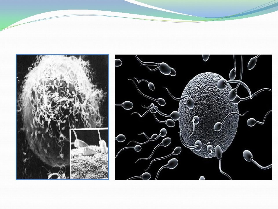
4. Acrosomal reaction: -It means release of acrosomal contents when sperms contact with corona radiata and zona pellucida including hyaluronidase and acrosin enzymes to penetrate corona radiata and zona pellucida. Ø Getting together of the two gametes is purely accidental in mammals and there are chemical substances secreted by the ovum and sperm, which play an important role in completing the fertilization process. ØThe fertilized ovum reaches the uterus with in 3 days in cattle, sheep, pig, cat, rat and rabbit and with in 8 -10 in case of dog and mare.

Substances secreted by the ovum: As the nucleus of the ovum begins its first maturation division, the ovum produces the gynagamon I & II. - Gynagmon I plays an important role in activation of the sperms to reach the ovum. - Gynagamon II is responsible for the effect of fertilizin which is a species specific glycoprotein and causes agglutination of the corresponding sperms (adhesion of the sperms to the surface of the ovum of the same species). Fertilizin is very important for external fertilization (fishes).

Substances secreted by the sperm: - Androgamon I The acrosomal cap of the head of the sperm is responsible for the effects of anti-fertilizin, in addition, it controls the movement of the sperms, so that the sperms stay vital for fertilization for along time. - Androgamon II is responsible for the effect of hyaluronidase enzyme which dissolves the cement substance between the cells of the corona radiate and opens a canal in the zona pellucida. q For this reason, the fertilization needs numerous numbers of healthy sperms.
 of fertilization A. Penetration of the ovum: - Penetration of corona radiata")
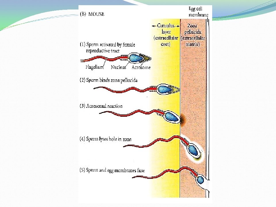
Mechanism (Sequences) of fertilization A. Penetration of the ovum: - Penetration of corona radiata - Binding to zona pellucida - Penetration of zona pellucida - Fusion with plasma membrane B. Reaction of ovum to prevent polyspermy: - Plasma membrane reaction - Cortical reaction - Zona pellucida reaction C. Sperm after penetration D. Formation of zygote
 of fertilization: A. Penetration of the ovum: 1) Penetration of corona radiata:")
Mechanism (Sequences) of fertilization: A. Penetration of the ovum: 1) Penetration of corona radiata: by - Hyaluronidase enzyme produced by the acrosomal cap dissolves the muco-polysaccharide hyaluronic acid (the cement substance between the corona radiate cells). -Motility of sperms tails: pushes sperms through corona radiata. 2) Binding to zona pellucida: -After the sperm penetrates between the corona radiate cells, it makes a narrow canal through zona pellucida to reach the surface of the ovum.
 Penetration of zona pellucida: by help of - Acrosin enzyme produced by the")
3) Penetration of zona pellucida: by help of - Acrosin enzyme produced by the acrosome to digest zona pellucida. -Motility of sperms tails. Ø After penetration of zona pellucida, the sperm enters perivitelline space and the central part of the acrosome elongates and becomes transformed into a long thin filament which protrudes forward from the sperm and is called acrosomal filament. 4) Fusion with plasma membrane: -Acrosomal cell membrane fuses with cell membrane of oocyte then the cytoplasm of the ovum bulge at the point of contact producing a process known as fertilization cone.

- This fertilization cone begins to retract and engulfs the sperm contents carrying the sperm inward. - In some cases, the head and neck of sperm enter the ovum; the tail is dropped off. In other cases, the whole of the sperm penetrates into the cytoplasm. B. Reaction of the ovum to prevent polyspermy: -The entry of the sperm in the ovum results in a marked reaction in its peripheral cytoplasm and vitelline membrane as follows: � Plasma membrane reaction: In which the receptors on plasma membrane of oocyte changed and so no other sperms can fuse with it.
 Cortical reaction: -Swelling of the cortical granules in the cytoplasm of the egg;")
2) Cortical reaction: -Swelling of the cortical granules in the cytoplasm of the egg; then ejecting their contents onto the surface of the vitelline membrane, transforming it to a very much thick fertilization membrane. -The fertilization membrane becomes lifted from the surface of the egg and a fluid filled space appears between them (the perivitelline space). -The fertilization membrane prevents the entrance of additional sperms. 3) Zona pellucida reaction: - Blocking the specific binding receptors for other sperms. - Hardening of its structure.

Acrosomal, Cortical Reactions

. Leishmania spp Leishmaniasis Kinetoplastids

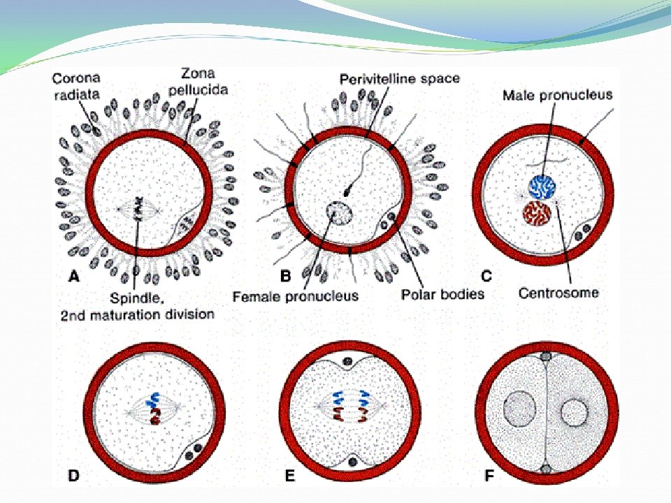
C. Sperm after penetration: -After penetration of the ovum, the sperm moves with acrosome at its front. The nucleus and centrosome arranged behind the acrosome. - The centrosome rotates in front of nucleus and the nucleus turns 180° and the sperm loses its remaining parts. - The ovum resumes Meiosis II forming second polocyte or polar body and mature oocyte. Female nucleus now is called female pronucleus. - The sperm head begins to move toward the female pronucleus, on its way it absorbs fluids from the ovum cytoplasm and the nucleus becomes enlarged, vesicular shape and known as male pronucleus.

D. Formation of zygote: - The female pronucleus moves centrally to meet the male pronucleus with help of 2 centrioles. When the 2 pronuclei approach to each other, their nucleic membranes dissolve and their chromosomes align themselves and the cell now is called zygote (diploid, 2 N) and its sex type either male or female is determined.

Results of fertilization: 1 - Re-association of the male and female sets of chromosomes, thus restoring the full diploid number. 2 -Determination of the sex of the new individual. 3 - Activation of the ovum into cell division, or cleavage.

Fertilization

ØMonospermy: Means fertilization of ovum by only one sperm. ØPolyspermy: Means fertilization of ovum by more than one sperm. It may be: Pathological polyspermy -Due to high concentration of sperm around egg or due to retarded reaction of the egg after contact with 1 st sperm. -Development is abnormal causing death of zygote Physiological polyspermy - Several spermatozoa enter the egg but only one participates in formation of viable zygote while the others degenerate. -Occurs normally in animals with yolky eggs as birds, reptiles, insects.

ØParthenogenesis: ﺍﻟﺘﻜﺎﺛﺮ ﺍﻟﻌﺬﺭﻱ Development of embryo from an ovum that has been activated by means other than sperm. It may be: Natural (Virginal reproduction) -Eggs develop without fertilization -e. g in frog, bees, wasps in which fertilized egg produces female individual while unfertilized egg produces male. Artificial - Certain treatment of ripe eggs may induce them to develop without fertilization such as X-rays, UV irradiation, needle pinching, heat shock, some salts and acids.

- Slides: 50