Genomic equivalence Various models were used to explain
- Slides: 26
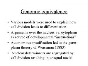
Genomic equivalence • Various models were used to explain how cell division leads to differentiation • Arguments over the nucleus vs. cytoplasm as source of developmental “instructions” • Autonomous specification led to the germplasm therory of Weissman (1883) • Nuclear determinants are segregated by cell division resulting in unequal nuclei
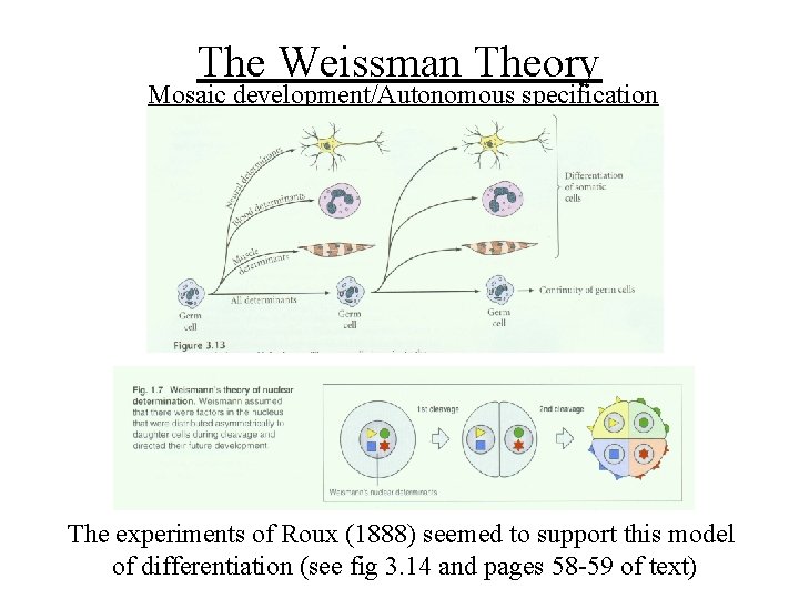
The Weissman Theory Mosaic development/Autonomous specification The experiments of Roux (1888) seemed to support this model of differentiation (see fig 3. 14 and pages 58 -59 of text)
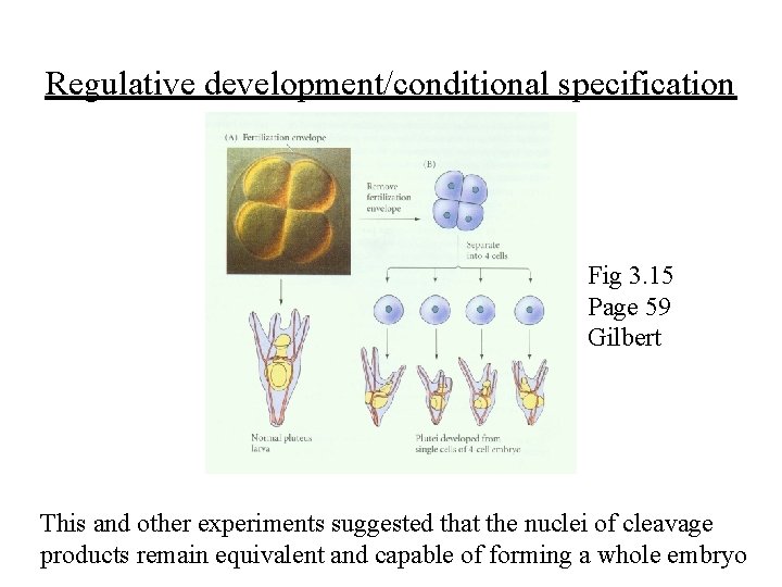
Regulative development/conditional specification Fig 3. 15 Page 59 Gilbert This and other experiments suggested that the nuclei of cleavage products remain equivalent and capable of forming a whole embryo
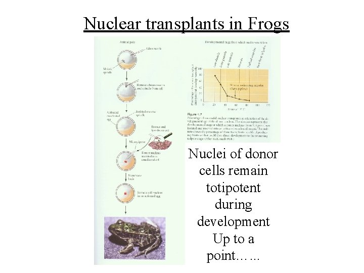
Nuclear transplants in Frogs Nuclei of donor cells remain totipotent during development Up to a point…. . .
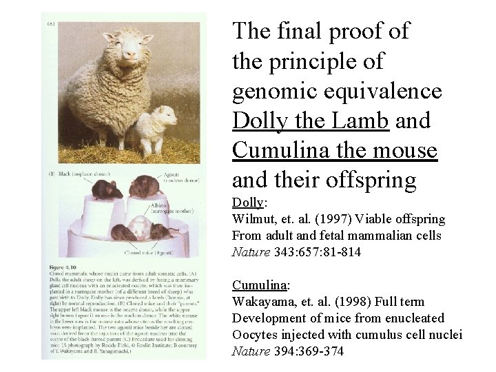
The final proof of the principle of genomic equivalence Dolly the Lamb and Cumulina the mouse and their offspring Dolly: Wilmut, et. al. (1997) Viable offspring From adult and fetal mammalian cells Nature 343: 657: 81 -814 Cumulina: Wakayama, et. al. (1998) Full term Development of mice from enucleated Oocytes injected with cumulus cell nuclei Nature 394: 369 -374
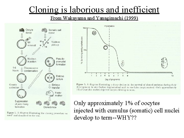
Cloning is laborious and inefficient From Wakayama and Yanagimachi (1999) Only approximately 1% of oocytes injected with cumulus (somatic) cell nuclei develop to term--WHY? ?
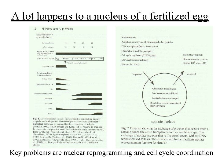
A lot happens to a nucleus of a fertilized egg Key problems are nuclear reprogramming and cell cycle coordination
Cell cycle coordination • Donor nucleus must adopt the cell cycle parameters of a zygote • Donor nucleus needs to be in G 0 or G 1 phases of the cell cycle (quiescent-starved) • (S or G 2 phases: BAD) (G 2/M phase: BAD) • Therefore initial cell cycles can be controlled by the recipient (oocyte) cytoplasm (as normally occurs)
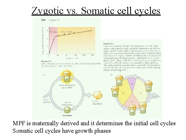
Zygotic vs. Somatic cell cycles MPF is maternally derived and it determines the initial cell cycles Somatic cell cycles have growth phases
Nuclear reprogramming • Somatic cells are transcriptionally active whereas a fertilized egg is NOT • All of the epigenetic changes that created the somatic cells need to be “erased” so that the state of the chromatin of the transplanted nucleus is more “neutral” • Epigenetic changes which normally occur during embryogenesis start anew
Changes in chromatin structure • 75% of pre-existing protein is lost from the somatic nucleus following transplantation • Involves changes in the following: – Histone components and modifications – Chromatin remodeling complexes – DNA methylation status – Transcription factors – Repressive chromatin (heterochromatin) proteins
Cloning and development • Events following nuclear transplantation mimic normal events which initiate embryonic development • Genomes are equivalent • Differences in “use” of the genome occurs through epigenetic regulation
Exceptions to genomic equivalence • B-lymphocytes and T-lymphocytes • Both cell types utilize somatic DNA rearrangement to create antigen receptors • Results in PERMANENT LOSS of DNA from the genome • Lymphocytes utilize multiple strategies to generate antigen receptor diversity. Many of these make permanent changes to DNA
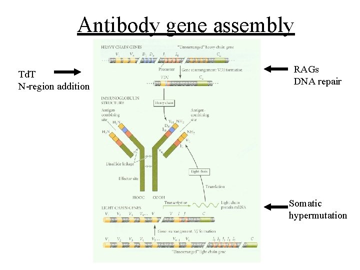
Antibody gene assembly Td. T N-region addition RAGs DNA repair Somatic hypermutation
DNA altering processes in antigen receptor generation • Somatic recombination (V-D-J) – Involves specific recombination enzymes (RAG 1 and RAG 2) – Involves DNA repair machinery (DNA-PK, Ligase) • N-region addition (Td. T enzyme) • Somatic hypermutation (linked to transcription) • Class switch recombination (unknown)
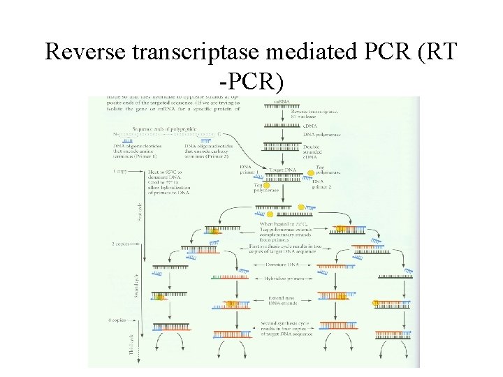
Techniques to detect gene expression (m. RNA) • • • Northern blot RNase protection assay Reverse transcriptase-mediated PCR In situ hybridization Subtractive hybridization – Allows cloning of unknown genes that are differentially expressed
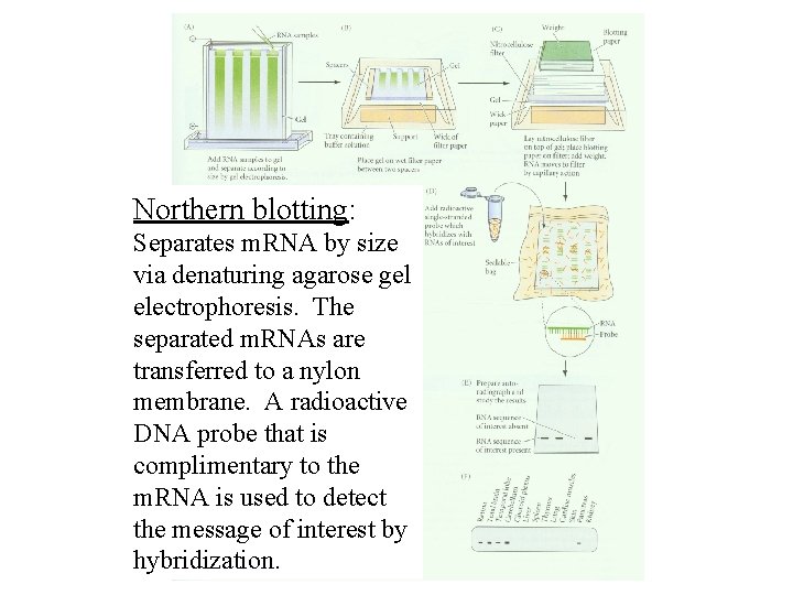
Northern blotting: Separates m. RNA by size via denaturing agarose gel electrophoresis. The separated m. RNAs are transferred to a nylon membrane. A radioactive DNA probe that is complimentary to the m. RNA is used to detect the message of interest by hybridization.
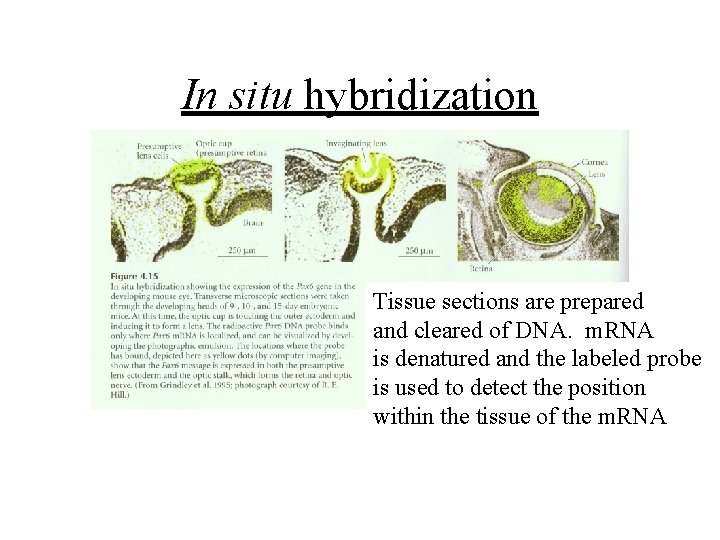
In situ hybridization Tissue sections are prepared and cleared of DNA. m. RNA is denatured and the labeled probe is used to detect the position within the tissue of the m. RNA
Reverse transcriptase mediated PCR (RT -PCR)
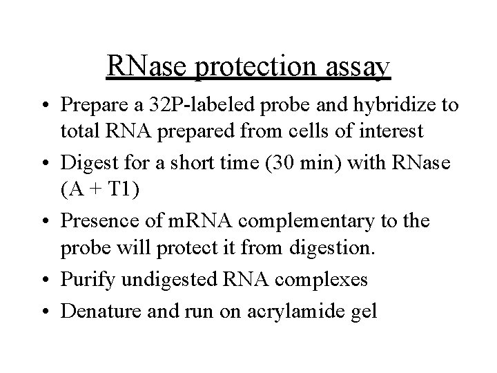
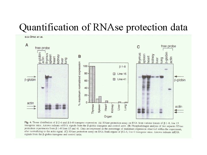
RNase protection assay • Prepare a 32 P-labeled probe and hybridize to total RNA prepared from cells of interest • Digest for a short time (30 min) with RNase (A + T 1) • Presence of m. RNA complementary to the probe will protect it from digestion. • Purify undigested RNA complexes • Denature and run on acrylamide gel
Quantification of RNAse protection data
Subtractive m. RNA screen strategies • Subtractive hybridization • Differential display • Representational difference analysis (RDA) • Gene chip analyses
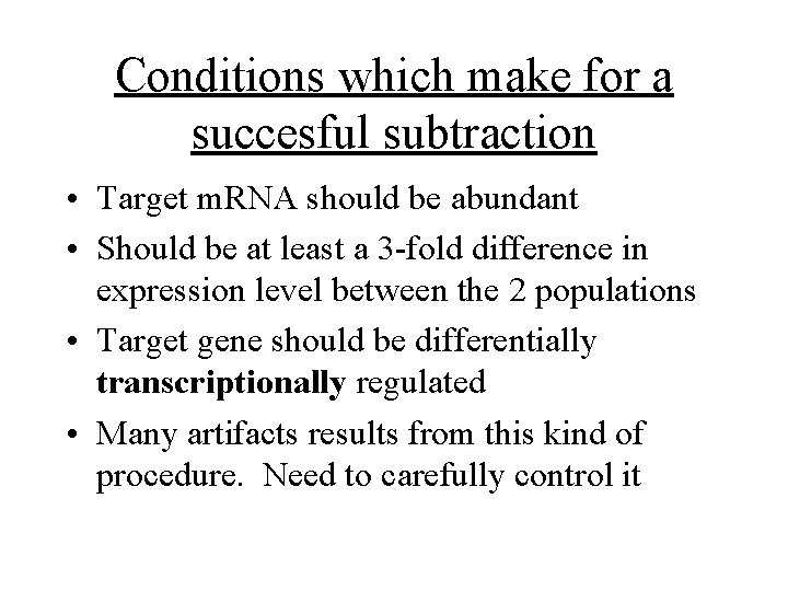
Conditions which make for a succesful subtraction • Target m. RNA should be abundant • Should be at least a 3 -fold difference in expression level between the 2 populations • Target gene should be differentially transcriptionally regulated • Many artifacts results from this kind of procedure. Need to carefully control it
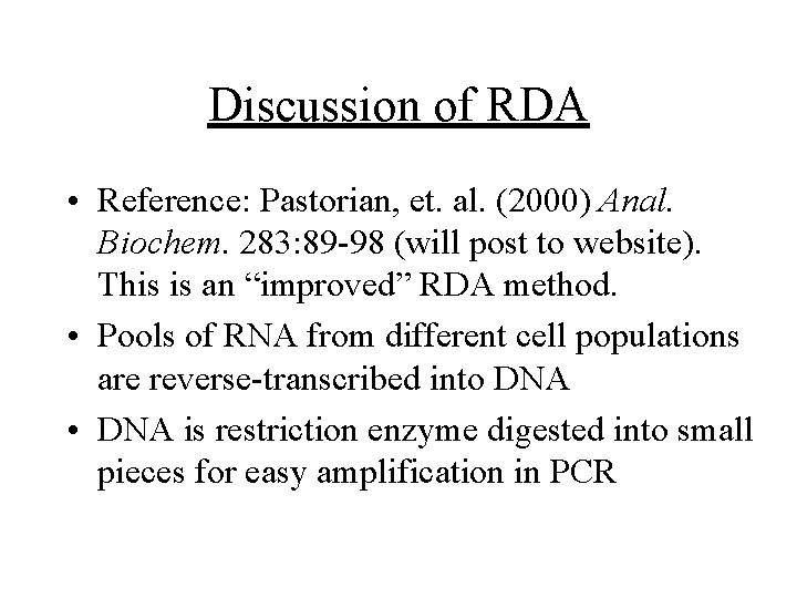
Discussion of RDA • Reference: Pastorian, et. al. (2000) Anal. Biochem. 283: 89 -98 (will post to website). This is an “improved” RDA method. • Pools of RNA from different cell populations are reverse-transcribed into DNA • DNA is restriction enzyme digested into small pieces for easy amplification in PCR
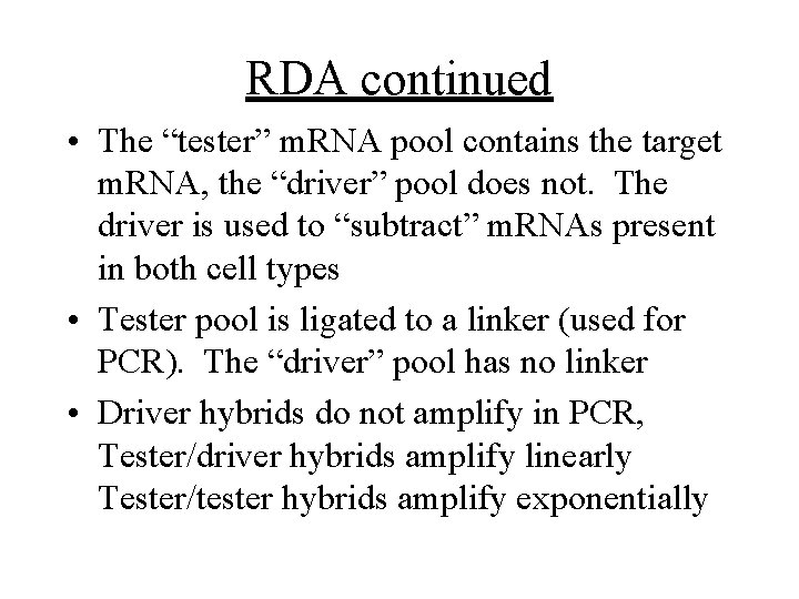
RDA continued • The “tester” m. RNA pool contains the target m. RNA, the “driver” pool does not. The driver is used to “subtract” m. RNAs present in both cell types • Tester pool is ligated to a linker (used for PCR). The “driver” pool has no linker • Driver hybrids do not amplify in PCR, Tester/driver hybrids amplify linearly Tester/tester hybrids amplify exponentially
Next lecture: techniques used to study the role of genes in develpoment • • • Random genetics followed by screening Targeted mutagenesis (gene knockout) Transgenic animal models Dominant negative mutant molecules Antisense RNA interference (c. elegans-website 4. 8)