GAS EXCHANGE Dr JAWAD NAWAZ Diffusion Random movement


- Slides: 19
GAS EXCHANGE Dr. JAWAD NAWAZ
Diffusion �Random movement of molecules of gas by their own kinetic energy �Net diffusion from higher conc. to lower conc �Molecules try to equilibrate in all empty places
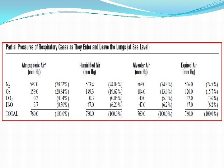
Partial pressure �The pressure exerted by the gas molecules on a surface In atmospheric air �PO 2 160 mm. Hg �PCO 2 0. 3 mm. Hg �PN 2 600 mm. Hg
Pressure of gases dissolved in water and tissues �Partial pressure in fluid develop same way as in air �Partial pressure= conc. of dissolved gas/solubility coefficient HENRY’S LAW � Solubility coefficients of different gases O 2=0. 024 CO 2=O. 57 CO=0. 018 N 2=0. 012 H=0. 008 �Water solubility of CO 2 20 times more than that of O 2 �Partial pressure of carbon dioxide is less than one twentieth that exerted by oxygen.
Water vapor pressure �In airway passage air gets humidified, water vapors mixed up with inspired air �At body temp. 370 C p. H 2 O =47 mm Hg �p. H 2 O directly proportional to temperature �In fever p. H 2 O is more
Rate of diffusion D=Δ P×A×S/d×√MW Δ P=Partial pressure difference A=cross-sectional area S=solubility of gas d= distance √MW=molecular weight �Diffusion coefficient=S/ √MW �Two gases at same partial pressure, rate of diffusion proportional to diffusion coefficient
Expired Air
Respiratory Unit �Respiratory Lobule 1. Respiratory bronchiole 2. Alveolar ducts 3. Atria 4. Alveoli
� 300 millions alveoli �Diameter 0. 2 milliliter �Sheet of flowing blood
Respiratory Membrane or Pulmonary Membranes of all the terminal portions of the lungs
Factors That Affect the Rate of Gas Diffusion Through the Respiratory Membrane 1. Thickness of membrane 1. Edema & Fibrosis 2. Surface area of membrane 2. Emphysema 3. Diffusion coefficient 3. Solubility of gas/ √ Mol. Weight 4. partial pressure of gas in the 4. Partial pressure difference of alveoli and partial pressure of the gas in the pulmonary capillary blood
Diffusion Capacity Volume of a gas that will diffuse through the membrane each minute for a partial pressure difference of 1 mm. Hg �Diffusing capacity for oxygen 21 ml/min/mm Hg at rest 65 ml/min/mm Hg during exercise �Diffusing capacity for carbon dioxide 20 times more than O 2 400 to 450 ml/min/mm Hg at rest 1200 to 1300 ml/min/mm Hg during exercise
Measurement of Diffusing Capacity 1. Alveolar Po 2 2. Po 2 in the pulmonary capillary blood 3. Rate of oxygen uptake by the blood Diffusing capacity(DC) of CO=Volume of CO absorbed p. CO DC of O 2 = DC of CO × 1. 23 = 17× 1. 23= 21 ml/min/mm. Hg
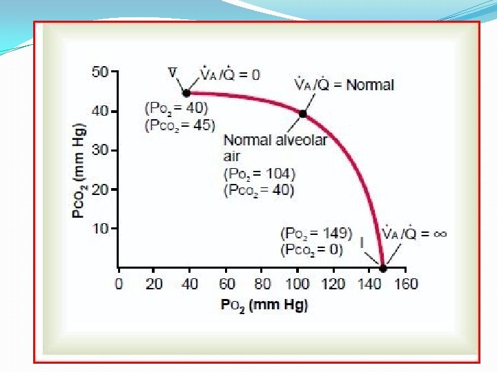
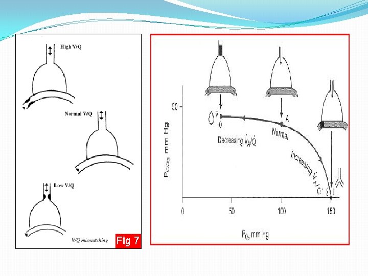
Ventilation – Perfusion Ratio The imbalance between alveolar ventilation and alveolar blood flow �Va Alveolar ventilation �Q Blood flow �Va/Q �When the ventilation(Va) is zero, yet there is still perfusion (Q) of the alveolus, Va/Q is zero �When there is adequate ventilation (Va) but zero perfusion (Q), Va/Q is infinity.
Physiological Shunt �When Va/Q is below normal �Shunted blood �Bronchial vessels �The total quantitative amount of shunted blood per minute is called the physiologic shunt �The greater the physiologic shunt, the greater the amount of blood that fails to be oxygenated as it passes through the lungs. �Lower part of lung Va/Q is 0. 6 times below normal
Physiological Dead Space � When Va/Q is ∞ � Alveolar wasted ventilation or alveolar dead space � Anatomical dead space � The sum of these two types of wasted ventilation is called the physiologic dead space � When the physiologic dead space is great, much of the work of ventilation is wasted effort because so much of the ventilating air never reaches the blood � Upper part of Lung Va/Q 2. 5 times more than normal