FOOT ANKLE 4 Motions of the Foot Plantarflexion


 – talus to fibula;")
 Posterior tibiotalar (PTT) Tibionavicular (TN) Tibiocalcaneal")
 n n internal rotation, plantar flexion (talus has to")

 n n n attaches to sustentaculum tali – “shelf” on calcaneous")


 0 1 2 3 4 5 no mm contraction, no")
- Slides: 27
FOOT & ANKLE
4 Motions of the Foot Plantarflexion Dorsiflexion Inversion Eversion
Joints of the ankle Talocrual n n Plantarflexion Dorsiflexion
Joints of the ankle Subtalar – between talus & calcaneus n n Inversion Eversion Talus Calcaneus
Most ankle sprains happen in PF because of anterior movement of talus because of no bony congruence If limited at one joint – have excessiveness in another
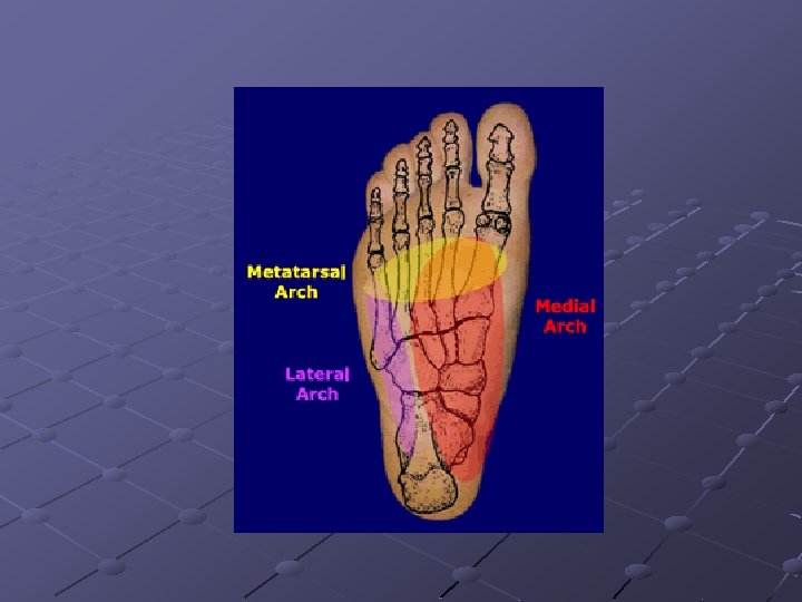
4 Arches of the Foot medial longitudinal – 1 st metatarsal head to the talus; most common n pronated – flattened (pes planus) n supinated - excessive (pes cavus) lateral longitudinal – base of 5 th metatarsal up to calcaneus anterior metatarsal – across metararsal heads; if falls, increases distance between calcaneus & metatarsal heads; which leads to plantar fasciitis transverse arch – over tarsal bones; medial to lateral (navicular to cuboid)
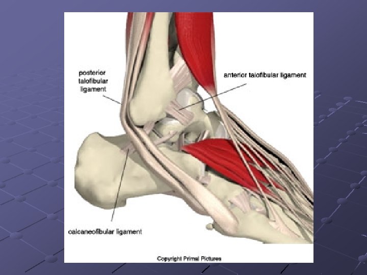
Why more inversion ankle sprains? Lateral malleolus is more distal than medial malleolus The deltoid complex is stronger than the ligaments on the lateral side
Lateral Complex Ligaments n n n Anterior talofibular ligaments (ATF) – talus to fibula; 1 st to be injured Calcaneal fibular ligament (CF) Posterior talofibular ligament (PTF) Ligamentous injuries ATF – plantar flexion; inversion CF – inversion, either dorsiflexion or neutral PTF – inversion, dorsiflexion
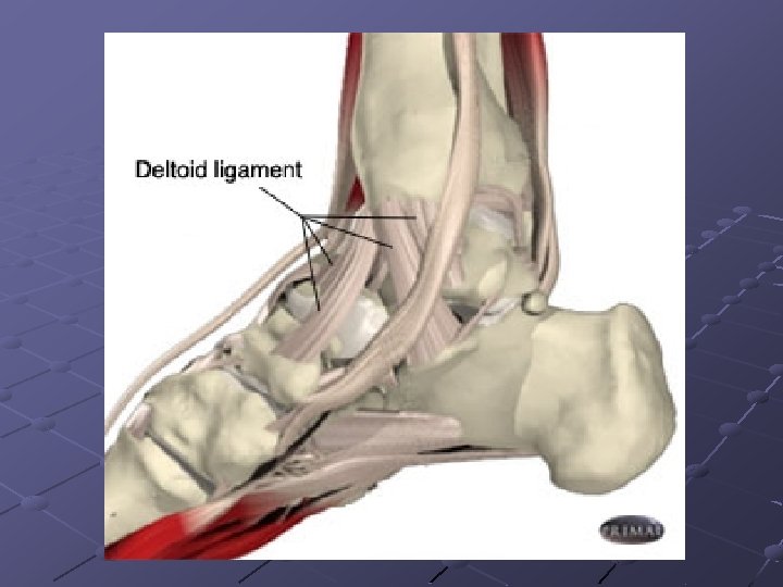
Medial Complex Ligament n Deltoid Anterior tibiotalar (ATT) Posterior tibiotalar (PTT) Tibionavicular (TN) Tibiocalcaneal (TC)
Other ligaments Anterior tibiofibular (ATF) n n internal rotation, plantar flexion (talus has to come out) High ankle sprain – long time to heal b/c can tape for DF, PF, INV, EV; but not rotation
Bifurcate n n attaches calcaneus & cuboid; sits on dorsum of foot Midfoot sprain – extreme forefoot varus in plantar flexed position
Spring (plantar calcanonavicular) n n n attaches to sustentaculum tali – “shelf” on calcaneous Chronic problem normally with gait problems; normally not injured in an acute injury
Muscles & Tendons Medial longitudinal arch n Flexor hallucis longus muscle starts on tibia & fibula runs posterior to malleolus under sustentaculum tali and in between sesmoids attaches to great toe; dynamic stabilizer of medial longitudinal arch n tibialis anterior starts as muscle and attached anterior to navicular n tibialis posterior long muscle; attaches into navicular
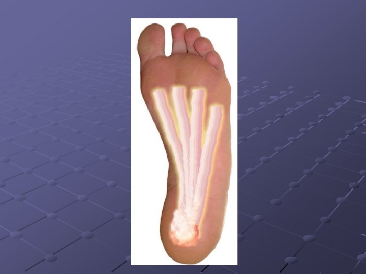
plantar fascia thick band of fascia covering the plantar aspect of the foot starts at medial calcaneal tubercle and attaches above MTP joint as toes go into extension; fascia (arch) increases wind last mechanism – “spring” to maintain normal alignment
Lower Leg Compartments Anterior n Muscles Tibialis anterior – dorsiflexion Extensor hallucis longus – extension of great toe Extensor digitorum longus – extension of toes Peroneus tertius – eversion n Nerve Deep peroneal nerve
Lateral n Muscles Peroneus brevis – eversion Peroneus longus – eversion n Nerve Superficial peroneal nerve Deep posterior n Muscles Flexor digitorum longus – flexion of toes Tibialis posterior – inversion, plantar flexion Flexor hallucis longus – flexion of great toe Popliteus n Nerve Tibial nerve
Superficial posterior n Muscle Gastrocnemius – plantarflexion Soleus – plantarflexion Plantaris – plantarflexion n Nerve Tibial nerve
Range of Motion
Dorsiflexion 0 -20 How you measure n n n SA: fibular head MA: 5 th MT Axis: below joint – calcaneus Muscles involved n n 1 st – Tibialis Anterior 2 nd – EHL & ED Nerve n Deep Peroneal
Plantarflexion 0 -45 How you measure n n n SA: fibular head MA: 5 th MT Axis: below joint – calcaneus Muscles involved n n 1 st – Gastroc/Soleus 2 nd – Tibialis Posterior Nerve n Tibial
Inversion 0 -15 How you measure n n Lie prone SA: bisect calf MA: bisect calcaneus Axis: @ subtalar joint Muscles involved n n 1 st – Tibialis Posterior 2 nd – Tibialis Anterior Nerve n Tibial & Deep Peroneal
Eversion 0 -10 How you measure n n Lie prone SA: bisect calf MA: bisect calcaneus Axis: @ subtalar joint Muscles involved n 1 st – Peroneus longus & peroneus brevis Nerve n Superficial Peroneal
Manual Muscle Test (MMT) 0 1 2 3 4 5 no mm contraction, no joint mov’t take out gravity, mov’t “fair”, no resistance against gravity “good”, some resistance “normal”