Ankle Injuries Ankle injuries fall into the same


 2. strapping 3. shoe padding -")
 -sports orthotics (lighter in")


 (pump bumps) A. Mechanism of Injury 1. direct or microtrauma")


 2. hyperflexion 3. valgus")


 1. Possible Responsible Factors a. heredity b. shoes")


 Calluses (tylomas) 1. Possible Responsible Factors a. Cavus foot - toes")


 1. Possible Responsible Factors a. shoes too tight b. shoes")

 1. Possible Responsible Factors a. hyperhidrosis b. abrasions to plantar surface")


 A. Possible Responsible Factors 1.")


 Previous history of injury to that area?")
 medial: deltoid / anterior, medial, and posterior portions (2)")


 Check tibia and fibula 7. Functional tests a)")


- Slides: 85
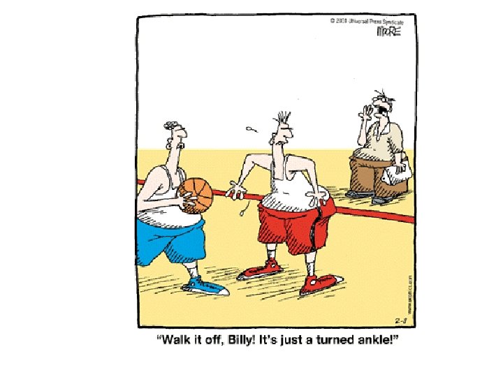
Ankle Injuries Ankle injuries fall into the same basic categories as do all athletic injuries: • Contusions • Sprains • Strains • Fractures
85% of all ankle sprains involve some plantar flexion of the ankle and inversion of the foot. The remaining 15% consist of eversion mechanisms which are often the result of an outside force such as being fallen on from the outside.
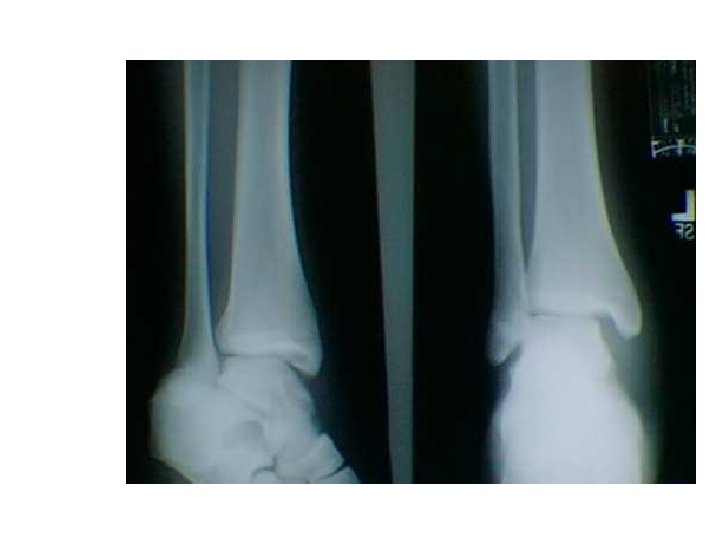
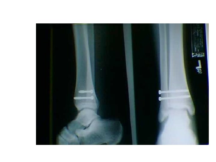
The syndesmosis ligament is often also injured with an eversion force. If the tibia and fibula spread on the talus, the ankle mortise is disrupted and the ankle can become very unstable. It is also not unusual to see an associated fibula fracture with an eversion mechanism. (see x-rays below) Assessment of a syndesmosis sprain will be difficult for the initial 24 to 48 hours. If the ankle is quite swollen and edematous assessment of a syndesmosis sprain may be difficult until the pain and swelling have isolated to individual areas or x-rays show some spreading of the ankle mortise.
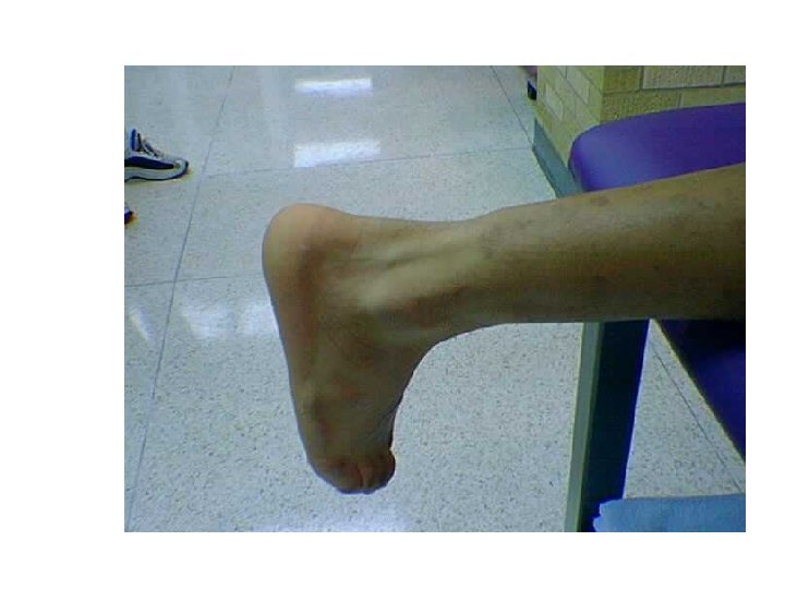
Ankle Ecchymosis
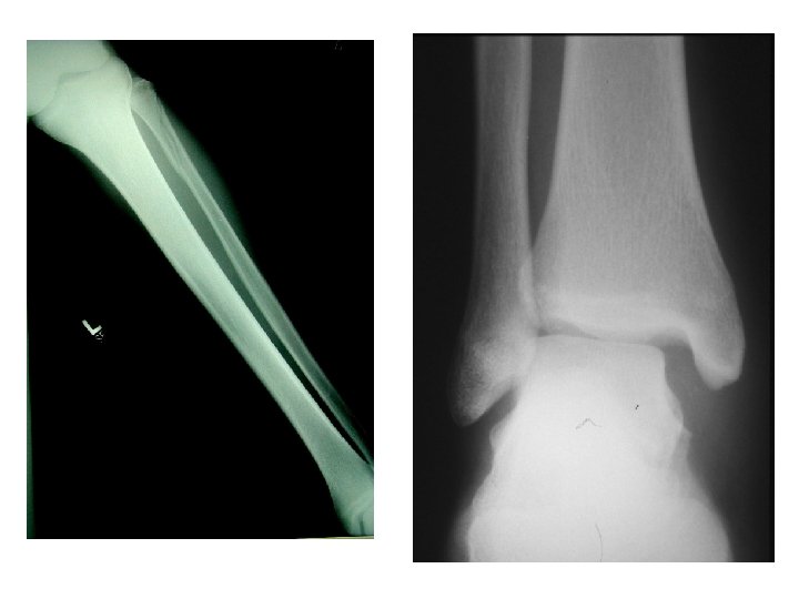
Maison - Neuve type fracture.
Distal Fibula fracture with associated medial deltoid ligament disruption. This injury is frequently the result of the foot being planted with a valgus load applied to the leg.
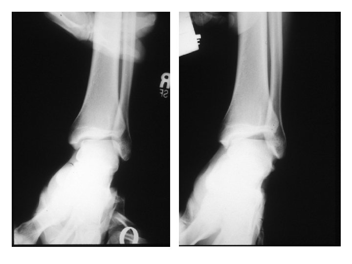
Notice the disruption of the medial deltoid ligament and the widening between the medial malleolus and the talus. This is indicative of a ruptured deltoid ligament.
Os Trigonum
This fracture requires surgical fixation of the fibula using a screw and plate system. The plate should be removed prior to return to competitive athletic activity as it will cause stress areas in the bone at each end of the plate. Recovery time (return to athletic activity) for a generally healthy patient with this type of fracture will be in the 6 month range.
Name the Injury
Talar Dome - AVN
Talar Dome - AVN
Ankle dislocation with no fractures. This takes a high degree of trauma and force. In this case this was generated as the result of a high flip off of a trampoline and impact with the ground. The ankle was in a plantar flexion and inverted position upon impact. This was an open dislocation.
Negative Thompson Test
Positive Thompson Test
Achilles Tendon Tear and Repair
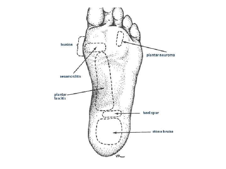
Foot Injuries
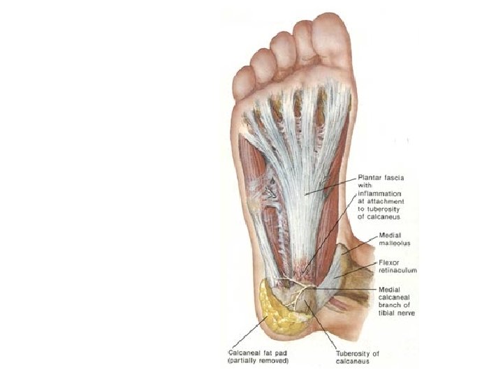
Plantar Fasciitis/Arch Strain A. Mechanism of Injury 1. strain of plantar fascia-usually at medial insertion into calcaneus 2. middle strip of plantar fascia sometimes involved 3. lateral strip almost never involved B. Possible Responsible Factors 1. shoes 2. artificial turf 3. severe pronation 4. excessive weight 5. leg length discrepancy 6. tight bed sheets
Treatment 1. stretching, Achilles, plantar fascia (night splints) 2. strapping 3. shoe padding - medial heel wedge -saddle pads -arch supports -lift type / padded heel pad -heel lift for short leg
4. orthotics-rigid (for heavy lineman, need more support & control) -sports orthotics (lighter in weight with more flexibility for backs and wide receivers) 5. Non Steroidal Anti Inflammatories 6. Steroid Injections - once weekly for 3 weeks 7. Surgical intervention - fasciotomy last resort, after 1 year of conservative treatment
Chronic plantar fasciitis can lead to formation of heel spurs. Plantar Fasciitis is the most common injury seen among long distance runners. It is very painful and can be chronic, extending over several years. The heel spur does not cause the plantar fasciitis, the fasciitis causes the heel spur.
Morton's Neuroma A. Mechanism of Injury 1. direct or microtrauma to an interdigital nerve 2. 90% of neuromas involve the 3 rd common digital nerve approximately 10% involve the 2 nd common interdigital nerve - 3 & 4 metatarsals B. Possible Responsible Factors 1. poorly cushioned and or tight shoes, high heels 2. pronation - nerve gets pinched between the heads of the 3 rd and 4 th metatarsals and the base of the proximal phalanx of the 3 rd & 4 th toes 3. hard surfaces 4. leg length discrepancy
Calcaneal Apophysitis (Severs Disease) (pump bumps) A. Mechanism of Injury 1. direct or microtrauma to the growth center of the posterior calcaneus 2. causes avascularity to the apophysis 3. Usually 8 - 12 year olds
B. Possible Responsible Factors 1. hard playing surfaces 2. shoes - poorly padded - cleats - poor support 3. cavus type foot 4. tight Achilles and or plantar fascia
C. Treatment 1. get out of cleats 2. shock absorbent heel pads 3. strapping - to help support plantar fascia 4. orthotic or heel stabilizers 5. in resistant cases immobilization for 4 - 6 weeks may be needed
Sesamoiditis/Sesamoid Fractures Sesamoid fractures must be differentiated from a normal bipartite sesamoid. A. Mechanism of Injury 1. direct trauma to tibial (medial) sesamoid - most common 2. direct trauma to fibular (lateral) sesamoid - rare 3. overuse - chronic microtrauma
B. Possible Responsible Factors 1. hard playing surfaces 2. hallux valgus - tibial sesamoid directly under mp joint 3. lack of cushioning in shoes C. Structures involved 1. sesamoids 2. joint capsule 3. flexor brevis 4. plantar 1 st metatarsal head
Treatment Sesamoiditis 1. shoe padding - transfer weight away from sesamoid 2. super cushion inner soles 3. ice, elevation, compression 4. possible post-op shoe 5. steroid injection Sesamoid fracture 1. cast for 3 weeks - BK 2. post-op shoe
Surgical excision of affected sesamoid in resistant cases very often will not heal. If hallux valgus present should correct at time of surgery, because weakening of flexor apparatus will increase deformity. This is a last resort in most cases as it changes the bio mechanical forces on the flexor tendons and if a single sesamoid is left in place, the weight bearing mechanics of the foot are greatly altered.
Turf Toe A. Mechanism of Injury 1. hyperextension (most common) 2. hyperflexion 3. valgus injury - usually from sudden acceleration B. Possible Responsible Factors 1. artificial turf - no give, can be like playing on hard asphalt 2. shoes - too much forefoot flexion (no turf toe plate) 3. combination of turf & shoes
Specific Structures Involved 1. capsular & ligamentous structures 2. flexor apparatus 3. possibly sesamoids
D. Treatment 1. rest, ice, elevation, compression 2. possible immobilization and non weight bearing 3. shoe modifications - spring steel splint 4. activity is resumed within the limits of pain Starting with flat foot walking, then normal gait, then jogging, then straight ahead running at full speed, next running from stance, last performing cutting maneuvers.
5. taping the to prevent injury from recurring 6. anti-inflammatories 7. surgery - for capsular repair in non responsive cases
Misc. Aggravations A. Hallux Valgus (bunions) 1. Possible Responsible Factors a. heredity b. shoes - irritate but don't cause c. pronation - accentuates 2. Specific Structures Involved a. 1 st MP - all structures b. sesamoids c. lst metatarsal - medial cuneiform joint
3. Treatment a. accommodate in wider shoes b. shoe stretching c. surgical correction in off season if chronically painful (may cause some limitation of joint movement)
Hallux Limitus 1. Possible Responsible Factors a. heredity b. trauma to joint c. foot type - plantar flexed 1 st digit 2. Specific Structures Involved a. 1 st MP - degeneration of joint cartilage with osteophytic limping of 1 st metatarsal head and base of proximal phalanx b. sesamoids - in advanced cases
3. Treatment a. rigid soled shoes which limit dorsiflexion b. taping c. injection with local and steroid when symptoms acute d. when condition becomes debilitating & conservative measures fail then surgical intervention is necessary - usually with placement of plastic implant (will weaken push off)
Corns (digital clavi) Calluses (tylomas) 1. Possible Responsible Factors a. Cavus foot - toes hammer - plantar flexion of forefoot causes excess pressure on metatarsal heads b. pronated foot - abnormal weight transfer c. poor fitting shoes
Specific Structures Involved a. interphalangeal joints of toes b. extensor & flexor tendons c. metatarsal heads 3. Treatment a. deep & wide toe box b. débride hyperkeratotic tissue regularly c. Vaseline d. padding - Spenco 2 nd skin - moleskin
Ingrown Nails 1. Possible Responsible Factors a. improper cutting of nails b. heredity c. injury d. tight shoes 2. Specific Structures Involved a. tibial & fibular borders, usually hallux nails b. nail groove 3. Treatment a. packing cotton under affected border b. wedge resection of affected border c. partial radical nail procedure with matrix destruction (phenol method)
Black Toe (Subungual Hematoma) 1. Possible Responsible Factors a. shoes too tight b. shoes too loose c. low toe box d. long 2 nd toe e. cleats f. kicking g. direct trauma
Specific Structures Involved a. pedal nails b. nail bed c. distal phalanx - possible formation of subungual exostosis 3. Treatment a. drain hematoma as soon as possible b. if nail partially avulsed - remove nail completely & débride the nail bed - start soaks & topical antibiotics c. if chronic, hypertrophied nail - keep nail débrided back & thinned as much as possible - complete avulsion of nail plate with destruction of matrix
Plantar Verruca (Warts) 1. Possible Responsible Factors a. hyperhidrosis b. abrasions to plantar surface of the foot c. exposure to verruca virus - showers- locker rooms - brothers & sisters d. age - most commonly seen in teen years
Specific Structure Involved a. skin - warts do not penetrate the basement membrane of the skin b. metatarsal heads and/or calcaneus- areas of most pressure in weight bearing 3. Treatment a. mechanical debridement prn. b. topical acids c. cryotherapy d. surgical removal - does not leave scar e. laser
Name the Injury
Rodeo Clown Foot
Foot Fractures
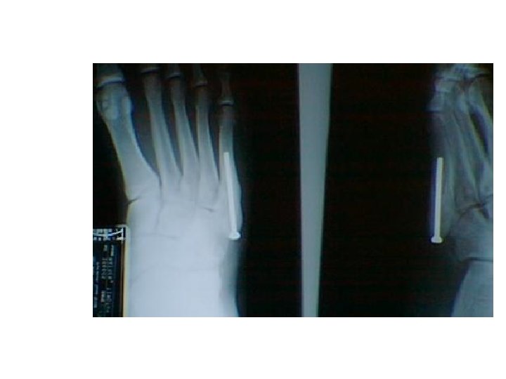
This is a ballerina type fracture of the 5 th metatarsal. The etiology involves and avulsion of the proximal tip of the 5 th metatarsal where the peroneus brevis muscle tendon attaches.
Fracture of Styloid process of 5 th metatarsal A. Mechanism of injury 1. severe inversion ankle sprain causes peroneus brevis tendon to pull away the base of the 5 th metatarsal (Ballerina fracture)
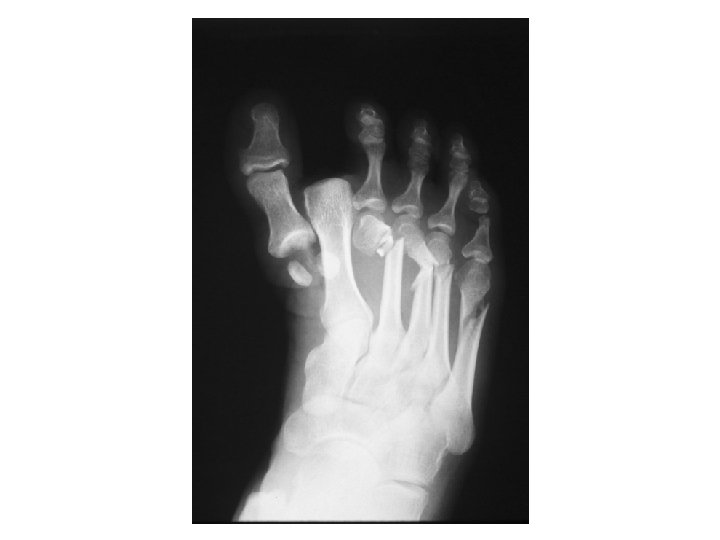
These X-Rays show a fracture of the proximal end of the 5 th Metatarsal. This fracture is commonly called a "Jones Fracture".
Direct trauma to base of 5 th (Jones fracture) A. Possible Responsible Factors 1. Cavus foot type 2. chronic ankle sprains 3. poor shoe and/or tape support B. Specific structures involved 1. peroneus brevis tendon 2. styloid process 5 th metatarsal base
Treatment 1. ice, elevation, compression and lift under 5 th metatarsal base 2. short leg walking brace 3. if severe avulsion of fragment, open reduction screw fixation
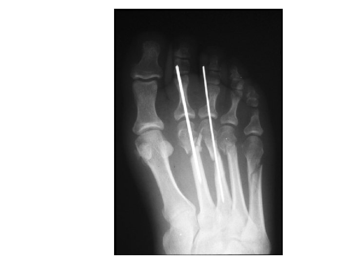
Treatment of Jones Fractures includes several options. Option 1 - immobilization of foot and ankle with non weight bearing for a period of 1 month to 6 plus weeks and more time may be required if the bone healing is delayed. The peroneus brevis tendon attaches at the proximal end of the 5 th Metatarsal and treatment without ankle immobilization is not effective. Every time the muscle contracts and pulls on the tendon, the fracture site is disrupted. This type of fracture is known to form a non union.
Option 2 - insertion of a intramedullary screw into the fracture to compress the fragment and the bones back together. May or may not be used with a bone graft. Many physicians will also opt to use a bone growth stimulator on this fracture to insure that healing occurs. Option 2 is certainly preferred in the authors opinion since the fracture site is stabilized and the ends of the fracture approximated. The screw fixation allows for earlier return to weight bearing and decreased immobilization time. (Dr. Joe Milne, Dr. Steve Brotherton)
Ankle Evaluation How did it happen? (etiology) Previous history of injury to that area? Sight evaluation a) swelling? b) deformities? c) discoloration?
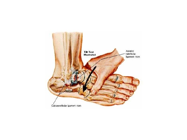
Palpation evaluation examine ligaments (1) medial: deltoid / anterior, medial, and posterior portions (2) lateral: Anterior talo-fib
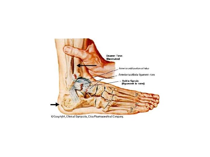
Anterior drawer tests should always be performed with the knee bent to eliminate the Achilles and Gastrocnemius muscles from providing any stability to the ankle. A lateral talar tilt test can be conducted at the same time.
Talar Tilt
Inversion Stress Test
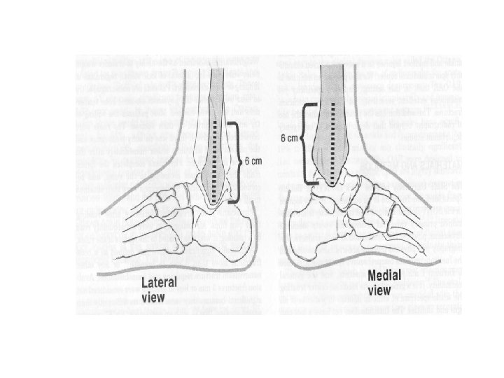
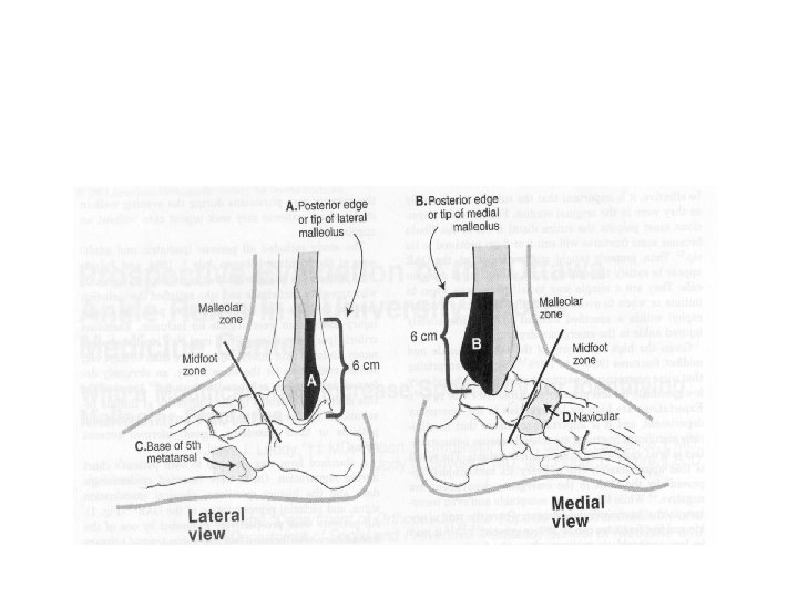
Ottawa - Buffalo Modification for Ankle Exam The incidence of ankle fractures in athletes involved in controlled sports activities is relatively low. However, the decision(s) on which ankles to study radiographically with xrays is not always easy. Not all ankle or foot injuries require immediate x-rays. The allied health practitioner can determine to a relatively accurate degree the need for futher study through a good clinical exam and by following the Ottawa Ankle Rules and with the use of the Buffalo Modification. (for more information, consult the AJSM, Vol 26, No 2. 1998)
Research has shown that in a hospital based E. R. of every 6 ankles approved for x-ray under the O. A. R. (Ottawa Ankle Rules), 5 have no radiographic findings. Steill, I. G. , JAMA, 269: 1127 -1132, 1993. • Clinical ankle exam If the patient needs x-rays, they will usually present with lateral fibular malleolus pain in the distal 6 cm, medial tibial malleolus pain in the distal 6 cm or pain to palpation over the proximal tip of the 5 th metatarsal or the Navicular. The inability to bear weight may indicate a lesion to the dome of the Talus or other associated trauma to the other structures of the ankle which may require x -ray study.
Squeeze test - check malleolus (2) Check tibia and fibula 7. Functional tests a) walking - check gait b) toe raises 1) both feet 2) one foot c) jump and land on both feet and then on one foot 8. Refer to doctor further evaluation and possible x-ray
Refer to physician for further evaluation and possible x-ray Non weight bearing x-rays Weight bearing x-rays (syndesmosis spreading) Stress x-rays for talar tilt and syndesmosis spreading Other associated ankle and lower leg tests Homan's Sign - patient is supine on the table, the knee is fully extended and the foot is dorsi flexed. Reproduction of pain with localized edema is considered a positive test for deep venous thrombophlebitis.
That’s All