Foot ANA 208 FOOT Ankle the narrowest and


- Slides: 21

Foot ANA 208

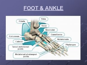
FOOT • Ankle: the narrowest and malleolar parts of the distal leg, proximal to the dorsum and heel of the foot, including the ankle joint. • Foot provides a platform for supporting the body when standing and has an important role in locomotion. • Skeleton of the foot consists of 7 tarsals, 5 metatarsals, and 14 phalanges. • Three anatomical and functional zones: - Hindfoot: talus and calcaneus. - Midfoot: navicular, cuboid, and cuneiforms. - Forefoot: metatarsals and phalanges. The part/region of the foot contacting the floor or ground is the sole. • Part superiorly is the dorsum of the foot or dorsal region of the foot. • Sole of the foot underlying the calcaneus is the heel, and the sole underlying the heads of the medial two metatarsals is the ball of the foot. • Great toe (L. hallux) is also the 1 st toe (digit of foot, and the little toe (L. digitus minimus) is also the 5 th toe. 2

SKIN AND SUBCUTANEOUS TISSUE • Skin of the dorsum of the foot is much thinner and less sensitive than skin on most of the sole. • Skin over the major weight-bearing areas of the sole —the heel is thick. • Fibrous septa—divide this tissue into fat-filled areas, making it a shock-absorbing pad, especially over the heel. • Skin ligaments anchor the skin to the plantar aponeurosis. • Skin of the sole is hairless and sweat glands are numerous 3

DEEP FASCIA OF FOOT • Deep fascia of the dorsum of the foot is thin where it is continuous proximally with the inferior extensor retinaculum. • Deep fascia is continuous with the plantar fascia which has a thick central part and weaker medial and lateral parts. • The thick, central part of the plantar fascia forms the strong plantar aponeurosis, which invests the central plantar muscles. • Plantar fascia holds the parts of the foot together, helps protect the sole from injury and to support the longitudinal arches of the foot. • Longitudinal bundles of the aponeurosis divide into five bands that become continuous with the fibrous digital sheaths that enclose the flexor tendons that pass to the toes. • At the anterior end of the sole, the aponeurosis is reinforced by transverse fibers forming the superficial transverse metatarsal ligament. 4

• In the midfoot and forefoot, vertical intermuscular septa forms the three compartments of the sole: • Medial compartment of the sole: contains the abductor hallucis, flexor hallucis brevis, the tendon of the flexor hallucis longus, and the medial plantar nerve and vessels. • Central compartment of the sole: contains the flexor digitorum brevis, the tendons of the flexor hallucis longus and flexor digitorum longus, quadratus plantae and lumbricals, and the adductor hallucis. The lateral plantar nerve. • Lateral compartment of the sole: contains the abductor and flexor digiti minimi brevis. 5

• Forefoot contains a fourth compartment interosseous compartment of the foot. - Contains the metatarsals, dorsal and plantar interosseous muscles, deep plantar and metatarsal vessels. • A fifth compartment, the dorsal compartment of the foot: contains extensors hallucis brevis and extensor digitorum brevis and neurovascular structures of the dorsum of the foot. 6

Muscles of Foot • 20 individual muscles of the foot: - 14: plantar aspect - 2: dorsal aspect • 4 - intermediate. • Muscles of the sole arranged in four layers within four compartments. • Plantar muscles function as a group during the support phase of stance, maintaining the arches of the foot. - They resist forces that tend to reduce the longitudinal arch as weight is received at 7

• The muscles of the foot are of little importance individually. Note that the: • Plantar interossei ADduct (PAD) and arise from a single metatarsal as unipennate muscles. • Dorsal interossei ABduct (DAB) and arise from two metatarsals as bipennate muscles. 8

9

10

11

12

13

• Two neurovascular planes between the muscle layers of the sole of the foot: - a superficial one between the 1 st and the 2 nd muscular layers - a deep one between the 3 rd and the 4 th muscular layers. • Tibial nerve divides posterior to the medial malleolus into the medial and lateral plantar nerves. • These nerves supply the intrinsic muscles of the plantar aspect of the foot. • The medial plantar nerve courses within the medial compartment of the sole between the 1 st and 2 nd muscle layers. • the lateral plantar nerve (and artery) run laterally between the muscles of the 1 st and 2 nd layers of plantar muscles. Their deep branches then pass medially between the muscles of the 3 rd and 4 th layers. • Two closely connected muscles on the dorsum of the foot are the extensor digitorum brevis (EDB) and extensor hallucis brevis (EHB). 14

15

16

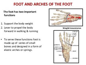
ARCHES OF FOOT • Foot is composed of bones connected by ligaments and absorb shock. • Tarsal and metatarsal bones are arranged in longitudinal and transverse arches which supports and add to the weightbearing capabilities and resiliency of the foot. • Distribute weight over the foot as shock absorbers and springboards during walking, running, and jumping. • Weight of the body is transmitted to the talus from the tibia, posteriorly to the calcaneus and anteriorly to the “ball of the foot” • Slightly flattened during standing and resume their curvature when body weight is removed. 17

Longitudinal arch of the foot • Medial and lateral parts which act as a unit • Transverse arch of the foot spreads weight in all directions. Medial longitudinal • Higher and more important than the lateral longitudinal arch. • Composed of the calcaneus, talus, navicular, three cuneiforms, and three metatarsals. • The talar head is the keystone • Supported by tibialis anterior and posterior and fibularis longus via their tendons. 18

Arches of the foot 19

Lateral longitudinal arch • Flatter than the medial part of the arch • Rests on the ground during standing. • Made up of the calcaneus, cuboid, and lateral two metatarsals. Transverse arch of the foot • Runs from side to side. • Formed by the cuboid, cuneiforms, and bases of the metatarsals. • Medial and lateral parts of the longitudinal arch serve as pillars for the transverse arch. • Tendons of the fibularis longus and tibialis posterior maintain the curvature of the transverse arch. 20

Integrity of the bony arches of the foot is maintained by: Passive factors: • Shape of the bones • Four layers of fibrous tissue that bowstring the longitudinal arch: v Plantar aponeurosis. v Long plantar ligament. v Plantar calcaneocuboid (short plantar) ligament. v Plantar calcaneonavicular (spring) ligament. Dynamic supports: v Active bracing action of intrinsic muscles of foot (longitudinal arch). v Active and tonic contraction of muscles with long tendons extending into foot: v Flexors hallucis and digitorum longus for the longitudinal arch. v Fibularis longus and tibialis posterior for the transverse arch. 21
 Ana208
 Anatomy and physiology of the foot
 Ankle foot orthoses
 Ankle foot orthosis project
 Example of narrowest graph
 Example of narrowest graph
 Narrowest graph example
 L
L Esophageal plexus
 The country with the narrowest population pyramid is
 Directional perm
 Ics 208
 Me gustan me encantan (p. 135) answers
 Pl 107-208
 Xkcd regexp
 Level 2 pp. 208-210 answers
 Memory express
 136xe isotope
 208.06 in standard notation
 208 huesos del cuerpo humano
 277/480 transformer bank
 Reaction injection molding
