DRUG REACTIONS Adverse drug reactions ADRs are a


![Bullous drug reactions (Stevens-Johnson syndrome [SJS] and toxic epidermal necrolysis [TEN]) Bullous drug reactions (Stevens-Johnson syndrome [SJS] and toxic epidermal necrolysis [TEN])](https://slidetodoc.com/presentation_image_h2/c0f8f53c843f8548c9caf8b9bfb0a660/image-13.jpg)
![S]S and TEN are considered by some as parts of a disease spectrum based S]S and TEN are considered by some as parts of a disease spectrum based](https://slidetodoc.com/presentation_image_h2/c0f8f53c843f8548c9caf8b9bfb0a660/image-14.jpg)


- Slides: 24

DRUG REACTIONS

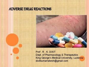
• Adverse drug reactions (ADRs) are a common cause of dermatologic consultation. • The risk for development of a drug eruption is related to the following factors: age, gender, dose, and the nature of the medication itself. • Females are 1. 3 -1. 5 times more likely to develop drug eruptions, except in children under the age of 3 where boys are more likely to be affected.

Evaluation 1. Previous general experience with the drug: • Has the suspected medication been reported to cause the reaction the patient is experiencing? • If so, how commonly? • Also ask the patient if he/she has had a previous reaction to any medications, as the current eruption may represent a crossreaction from a prior exposure.

• 2. Alternative etiologic candidates: • What are other possible causes of the patient's eruption? • An exanthem, could be an associated viral illness, not the medication. • 3. Timing of events: • When did the eruption appear relative to the administration of the suspected medication? • 4. Drug levels and evidence of overdose: • Certain reactions are known to be related to rate of administration or cumulative dose

• 5. Response to discontinuation (dechallenge): • Does the eruption clear when the suspected medication is stopped? • Because certain eruptions may clear in the face of continuation. • 6. Rechallenge: • If the offending medication reproduces the reaction on readministration, • this is strong evidence that the medication did indeed cause the reaction.

Pathogenesis • T cells, are felt to be important inducers of ADRs. These T cells act in two ways to induce reactions. • They can directly secrete biologically active molecules, resulting in direct tissue effects (epidermal necrosis, for example), or they can act by secreting chemokines that recruit the effector cells (eosinophils or neutrophils).

Clinical morphology • Cutaneous drug reaction can be described depend on their morphologic pattern. • In addition to the cutaneous eruption, some reactions may be associated with other systemic symptoms or findings. • The modifier "simple" is used to describe reactions without systemic symptoms or internal organ involvement. "Complex" reactions are those with systemic findings.

Exanthems are the most common form of adverse cutaneous drug eruption. They are characterized by erythema, often with small papules throughout. They tend to occur within the first 2 weeks of treatment but may appear later, or even up to 10 days after the medication has been stopped. Exanthems

Lesions tend to appear first proximally, especially in the groin and axilla, generalizing within 1 or 2 days. the face may be spared. Pruritusis usually prominent, helping to distinguish a drug eruption from a viral exanthem. Antibiotics, penicillins and trimethoprim are the most common causes of this reaction pattern.

drug reaction with eosinophilia and systemic symptoms (DRESS) 1. Rash developing late (more than 3 weeks) after the inciting medication is started. It often occurs with the first exposure to the medication. 2. Long-lasting symptoms (>2 weeks) after the discontinuation of the causative drug. Drug-induced hypersensitivity syndrome

3. Fever (over 3 S°C). 4. Multiorgan involvement. 5. Eosinophilia (>1500 absolute eosinophilia). 6. Lymphocyte activation (lymphocytosis, atypical lymphocytosis, lymphadenopathy). 7. Frequent reactivation of herpes viruses 1. anticonvulsants(phenobarbital, andphenytoin) 2. long-acting sulfonamides (sulfamethoxazole, sulfasalazine) 3. allopurinol 4. nevirapine 5. abacavir 6. dapsone 7. minocycline

The skin eruption typically morbilliform and can vary from faint and mild to severe with exfoliative erythroderma. Facial edema often accompanies the skin eruption, and may evolve to superficial pustules (especially on face). The internal organ involvement divided into two types: organ dysfunction occurring during or immediately associated with the acute episode; and late sequelae, possibly with an autoimmune basis. The first category includes colitis, intestinal bleeding, encephalitis, aseptic meningitis, hepatitis, interstitial nephritis, interstitial pneumonitis, respiratory distress syndrome, and myocarditis. Late sequelae include syndrome of inappropriate secretion of antidiuretic hormone (ADH), thyroiditis Graves' disease, and diabetes mellitus.
![Bullous drug reactions StevensJohnson syndrome SJS and toxic epidermal necrolysis TEN Bullous drug reactions (Stevens-Johnson syndrome [SJS] and toxic epidermal necrolysis [TEN])](https://slidetodoc.com/presentation_image_h2/c0f8f53c843f8548c9caf8b9bfb0a660/image-13.jpg)
Bullous drug reactions (Stevens-Johnson syndrome [SJS] and toxic epidermal necrolysis [TEN])
![SS and TEN are considered by some as parts of a disease spectrum based S]S and TEN are considered by some as parts of a disease spectrum based](https://slidetodoc.com/presentation_image_h2/c0f8f53c843f8548c9caf8b9bfb0a660/image-14.jpg)
S]S and TEN are considered by some as parts of a disease spectrum based on the following: 1. They are most commonly induced by the same medications. 2. Patients initially presenting with S]S may progress to extensive skin loss resembling TEN. 3. The histologic findings are indistinguishable. More than 100 medications have been reported to cause S]S and TEN. S]S has less than 10% body surface area (BSA) involved, cases with 10 -30% are S]S- TEN overlap cases, and more than 30% BSA erosion is called TEN.

In adults, common inciting medications 1. trimethoprim-sulfamethoxazole 2. nevirapine, lamotrigine, and carbamazepine. 3. Antibiotics (long acting sulfa drugs and penicillins), 4. Other anticonvulsants, (NSAIDs), and allopurinol. Fever and influenza-like symptoms often precede the eruption by a few days. Skin lesions appear on face and trunk and rapidly spread (usually within 4 days) Initial lesions are macular, followed by desquamation, or may form atypical targets with purpuric centers that coalesce, form bullae, then slough. In SJS, virtually always, two or more mucosal surfaces are also eroded, the oral mucosa and conjunctiva being most frequently affected. There may be photophobia, difficulty with swallowing, rectal erosions, painful urination, and cough

Fixed drug reactions (FDE) are common. Fixed drug eruptions are so named because they recur at the same site with each exposure to the medication. The time from ingestion of the offending agent to the appearance of symptoms is between 30 minutes and 8 hours, averaging 2 hours.

In most patients, six or fewer lesions occur, and frequently one. They may present anywhere on the body, but half occur on the oral and genital mucosa. Clinically, an FDE begins as a red patch that soon evolves to an iris or target lesion similar to erythema multiforme, and may eventually blister and erode.

Lesions of the genital and oral mucosa usually present as erosions. Most lesions are 1 to several cm in diameter, but larger plaques may occur Characteristically, prolonged or permanent postinflammatory hyperpigmentation results,

Urticaria/angioedema Medications may induce urticaria by immunologic and nonimmunologic mechanisms. In either case, clinically there are pruritic wheals or angioedema. Urticaria may be part of a more severe anaphylactic reaction with bronchospasm, laryngospasm, or hypotension.

Aspirin and the NSAIDs are the most common causes of nonimmunologic urticarial reactions Immunologic urticaria is most commonly associated with penicillin and related B~-lactam antibiotics. Angioedema is a known complication of the use of angiotensin-converting enzyme (ACE) inhibitors. Lisinopril and enalapril produce angioedema more commonly than captopril Angioedema typically occurs within a week of starting therapy, but may begin after months of treatment.

Photosensitivity reactions (photosensitive drug reactions) Medications may cause phototoxic, photoallergic, and lichenoid reactions Most medication related photosensitivity is triggered by radiation in the UVA range, partly for two reasons. First, most photosensitizing drugs have absorption spectra in the UVA and short-visible range (315 -430 nm), and second, UVA penetrates into the dermis where the photosensitizing drug is present.

NSAIDs, trimethoprim sulfamethoxazole, thiazide diuretics Phototoxic reactions are related to the dose of both the medication and the UV irradiation. appear from hours to days after exposure. present as immediate burning with sun exposure, exaggerated sunburn, Hyperpigmentation may complicate phototoxic reactions last for many months. Photoallergic reactions are typically eczematous and pruritic, and appear weeks to months after drug exposure. Treatment may include dose reduction and photoprotection, with a sunblock.

Drug-induced pigmentation Pigmentation of the skin may occur as a consequence of drug administration. The mechanism frequently is related to actual deposition of the offending drug in the skin. Minocycline induces many types of hyperpigmentation, Type I is a blue-black discoloration appearing in areas of prior inflammation, often acne or surgical scars.

The second type (type II) is the appearance of a similarcolored pigmentation on the normal skin of the anterior shins The least common type (type Ill)is generalized brown hyperpigmentation, accentuated in sun-exposed areas. In addition to the skin, minocycline type I and II pigmentation may also involve the sclera, conjunctiva, bone, thyroid, ear cartilage, nail bed, oral mucosa, and permanent teeth
 Antigentest åre
 Adverse food reactions
 Adr adverse drug reaction
 Chemical reactions section 3 reactions in aqueous solutions
 Section 2 classifying chemical reactions
 Chemistry unit 5 reactions balancing reactions worksheet
 How to write a balanced redox reaction
 Section 2 reinforcement classifying chemical reactions
 Methods of adulteration of crude drugs
 Mechanism of action of acetaminophen
 Ahca adverse incident reporting
 Definition of adverse event
 Mhmr of tarrant county
 Unanticipated problem vs adverse event
 Adverse reaction definition
 Alpha blocker
 Diuretic drugs classification
 What is adverse selection
 Serious adverse event reconciliation
 Adverse selection
 Adverse childhood experiences study
 Sentinel event and near miss
 Drugs that cross placenta mnemonic
 5.2 driving in adverse conditions assignment
 Dpp-4 inhibitors side effects


