Diagnosis of the Parasitic infections Mutah University Faculty


- Slides: 21

Diagnosis of the Parasitic infections Mutah University Faculty of Medicine General Microbiology 2019 -2020 Dr. Mohammad Odibate

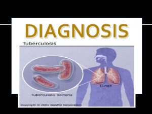
Diagnosis of Parasitic Infections 1. Clinical 2. Laboratory Purpose of laboratory diagnosis : – Confirmation of clinical suspicion. – Identification of unsuspected infection. 2

Specimens v Stool. v Blood. v Serum and plasma. v Others (anal swab, duodenal aspirate, sputum, urine, urogenital specimen). v Tissues and aspirates. 3

DIAGNOSIS DIRECT INDIRECT Urine Stool Sputum Biopsy Blood Aspirates IHAT LAT IFAT ELISA CFT MOLECULAR DNA probes PCR

Urine examination Parasites detected in the terminal drops of urine : Helminths: • Schistosoma haematobium eggs. • Enterobius vermicularis eggs in female patients. • Microfilaria of Wuchereria bancrofti. Protozoa: • Trichomonas vaginalis trophozoite in female patients. • Temporary stains, such as methylene blue is helpful to see T. Vaginalis. Note: Urine specimen should be centrifuged at 400 × g, the sediment mixed with a drop or two of saline, and examined by wet mount.

Urine examination

Urine examination Other techniques: • Membrane Filter technique: – For concentration of Schistosoma eggs. – Fill a syringe with urine, pass the urine through a filter. – Remove the filter and place it on the slide, and examine it microscopically.

Stool examination Sample collection: • Sample is collected in clean, dry container • Handled carefully • Samples in some cases fresh (amoeba, ciliates) • Liquid and soft stool examined within 15 min • Not mixed with urine or disinfectant (as they will kill trophozoites) Preservation of stool specimens: Aim: • To preserve protozoan morphology. • To prevent the continued development of some helminthic eggs and larvae. • The most common preservative used is 10% formalin. 8

Stool examination Microscopic Examination of Faecal Specimens: 1 - Direct Smears. 2 - Direct wet mount

Stool examination Direct Smears. Principle • To assess the worm burden of a patient • To provide a quick diagnosis of a heavily infected specimen • To check organism motility.

Stool examination Direct wet mount: • Small amount of faeces • Few drops of saline • Sometimes add lugol’s iodine (nuclear details, glycogen vacuole in cyst). • Protozoa (trophozoite), cyst, eggs and larva of helminths. 11

Stool examination Concentration methods • Used if parasites are scanty in the sample. • Two types: 1 - Floatation (eggs and cyst float , solution of high specific gravity) Saturated sodium chloride ii. Zinc sulphate centrifugation floatation (cyst, nematodes). i. 2 - Sedimentation (solution of formol ether Egg count in 1 gram 12 low specific gravity):

Stool examination Microscopic Examination of Faecal Specimens: Direct Smears Ascaris egg

Sputum examination üAbnormally, it is purulent, bloody, contains rusty brown particles (Paragonimus). Technique for examination: üAdd on a sputum sample equal volume of Na. OH to dissolve the mucus. üLeave this combination for a while, then centrifuge at 200 xg for 5 minutes, then examine the sediment. üThe specimen can be preserved in 10% formalin and a formalin-ethyl acetate

Sputum examination Parasitology Macroscopic Microscopic Appearance Concentration Bloody Take the sediment Rusty brown

Sputum examination Parasites that could be detected in sputum: 1. The inhabitant in the lung: ü Paragonimus 2. Migratory larvae: ü Ascaris ü Hook worm (Ancylostoma) ü Strongyloides. 3. Parasites causing pathology in the lung: üTrophozoites of Entamoeba histolytica. üHydatid sand due to rupture of hydatid cyst that could be present in the lung.

Blood examination • Fresh capillary blood of finger or ear lobe • Venous blood collected in EDTA (anticoagulant) Blood sample will be used for : – Microscopic examination (Thin Smear, Thick smear, Wet mount for microfilaria). • • Molecular diagnosis Detection of parasite antigen Isolation of organisms Special tests 17

Blood examination Two types of blood films can be made, thin and thick blood films: ü Thick films. üThin films.

Blood examination Thick blood film • Screen large amount of blood (light infection) • Can be stained latter 19 Thin blood film In malaria Parasitized red blood cells

Blood examination Parasites that could be detected in blood film: • Malaria • Trypanosoma (African and American). • Microfilaria of all types Filaria except Onchocerca volvulus. • Indian type of Leishmania donovani.

Examination of other Specimens 1. Lung and Liver • Aspiration from lung and liver could be examined for: üPneumocytosis üAmoebiasis Technique: The use of proteolytic enzymes is recommended to free the organisms from the aspirate material üHydatid Disease 2. Lymph nodes, Spleen, Liver, Bone Marrow and Spinal Fliud: Aspirated material may be examined for presence of trypanosomes, leishmanial forms and amoebae. 3. Cutaneous Ulcers : Leishmaniasis
 Nursing process definition
 Medical diagnosis and nursing diagnosis difference
 Second phase of nursing process
 Medical diagnosis and nursing diagnosis difference
 Perbedaan diagnosis gizi dan diagnosis medis
 Parasitic symbiosis
 Ascaris lumbricoides
 Parasitic ova
 Parasitic cone
 Parasitic cone
 Parasitic computing seminar report
 Rhizopus structure
 Cow parasitic twin
 Icd 10 morbus hansen
 Parasitic heterotrophs
 Insect name
 Saprophytic bacteria
 Plural of fungi
 Youtube
 Multualism
 Parasitic bacteria examples
 Parasitic fungi

