Degenerative scoliosis Considerations and indications for surgical treatment


- Slides: 34

Degenerative scoliosis Considerations and indications for surgical treatment Presenter’s name Arial 24 pt Meeting Arial 24 pt Presenter’s title Arial 20 pt City, Month, Year Arial 20 pt

Learning outcomes • Outline the treatment principles and options in relation to adult or degenerative spinal deformity • Recognize associated comorbidities and their influence on the outcome of surgery • Outline the principles in relation to restoration and maintenance of spinal balance • Perform assessment in relation to restoration and maintenance of spinal balance

Outline • Etiology • Relevant history • Significant examination findings • Indications for and interpretation of relevant investigations • Indications for surgery and surgical principles • Achieving spinal balance • Relevant complications

Etiology • Idiopathic scoliosis progressing into adulthood • Superimposed degeneration, usually worse in the concavity, leads to progression of deformity (typically over 50 years of age) • Usually, patients who have not had scoliosis as an adolescent develop a lumbar or thoracolumbar deformity due to symmetrical degeneration in later life

Natural history • Progression influenced by: • Magnitude of deformity • Spinal balance • Bone quality • Comorbidities • General fitness and muscle tone

Clinically (1/3) • Patient is usually over 60 years old • Presents with symptoms related to: • Degenerative disease (discs and/or facet joints) • Canal stenosis • Foraminal compromise • Spinal imbalance and muscle fatigue

Clinically (2/3) • Associated comorbidities • Hip, knee, and sacroiliac joint degeneration • Cardiac disease (BP, IHD, arrhythmias, anticoagulant use) • Respiratory disease • Diabetes • Smoking • Osteoporosis

Clinically (3/3) • Examination • Assess spinal balance and gait • Sagittal imbalance and loss of lumbar lordosis a common finding in patients presenting for surgical treatment • Neurological examination is often normal in the absence of nerve root compression (similar to lumbar canal stenosis patients) • Important to assess general medical condition of patient

Diagnosis • Identify origin of pain to determine most appropriate treatment

Degenerative scoliosis Pain from the scoliosis • Imbalance • Muscle fatigue

Degenerative scoliosis Pain from the scoliosis Degenerative LBP • Facet arthropathy • Disc degeneration

Degenerative scoliosis Pain from the scoliosis Degenerative LBP Claudicant pain

Degenerative scoliosis Pain from the scoliosis Degenerative LBP Claudicant pain Radicular pain

Diagnosis • Imaging should include: • Long, standing AP and lateral x-rays • MRI or CT myelography (if unable to have MRI) • Bending or traction x-rays may be indicated to assess flexibility of deformity

Red flags • Abnormal neurological findings (radicular or myelopathic) • Symptoms of neoplastic disease (nocturnal pain, constitutional upset, weight loss, etc) • Presence of a pathological fracture

Treatment (1/4) • Physical therapy and core stabilization • Hydrotherapy and regular swimming often useful in patients with this condition • Epidural or foraminal injections may provide relief of claudicant or radicular symptoms

Treatment (2/4) • Surgical intervention is usually major in this patient group • Localized foraminal or canal decompression may be tried for localized symptoms of neural compromise, but often develop recurrent symptoms and may aggravate back pain or lead to accelerated curve progression • Important for the patient to have appropriate expectations regarding the outcome of any surgery, which usually involves extensive fusion, often to the pelvis

Treatment (3/4) • Evaluate the risks and benefits of surgery in relation to: • Nature of the symptoms • Patient’s expectations • Chance of success • Risk of significant complications

Treatment (4/4) • Surgical principles • Achieve and maintain spinal balance • May need osteotomy to achieve this and important to consider pelvic parameters

Pelvic incidence (PI) (1/2) • The angle between the perpendicular to the sacral end plate at its midpoint and the line connecting this point to the midpoint of the femoral heads axis • This is a fixed anatomical relationship

Pelvic incidence (PI) (2/2) • The relationship of the sacrum to the pelvis is fixed • Average: • 53. 2°in men • 48. 2°in women

Pelvic tilt (PT) (1/4) • The angle between the vertical and the line joining the midpoint of the sacral end plate to the midpoint of the femoral head axis • Average: • 11. 9°Males • 10. 3°Females

Pelvic tilt (PT) (2/4) • Think about the pelvis rotating about an axis along a line joining the center of the femoral heads

Pelvic tilt (PT) (3/4) • Retroversion results in an increased pelvic tilt

Pelvic tilt (PT) (4/4) • Anteversion results in a reduced pelvic tilt

Sagittal vertical axis (SVA) (1/2) • The offset between the sagittal C 7 plumb line to the posterior superior corner of S 1 • A positive figure indicates anterior displacement of the plumb line relative to S 1

Sagittal vertical axis (SVA) (2/2) • Positive SVA > 5 cm is the most important and reliable radiographic predictor of health status • Increased pain, reduced function, and poor self-image

Treatment (1/2) • Surgical principles • Preoperative planning is essential to ensure surgical goals are understood and achievable • If normal disc and facets L 5–S 1 in younger patient, then avoid fusion to pelvis in first instance

Treatment (2/2) • Surgical principles • Degeneration of L 5–S 1 or oblique take-off of L 5 from pelvis requires fusion to pelvis • Fusion to pelvis requires A/P approach at L 5–S 1, and also generally at L 4/5 and above where accessible • Pelvic fixation may also be indicated • Important not to stop instrumentation at apex of kyphosis or scoliosis

Prognosis • For appropriately selected patients, clinical success and improved levels of activity and function are achieved in 80– 85% of patients • Good medical management is needed to minimize risks due to comorbidities

Potential complications • Cardiac or pulmonary complications: • Myocardial infarction, cardiovascular accident, pulmonary embolism • Persistent imbalance • Junctional breakdown • Degeneration, loss of fixation, or fracture • Nonunion and implant failure • Infection • Neurological complications • Persistent pain

Take-home messages • Degenerative scoliosis is not uncommon • Surgical intervention should be considered as a last resort in elderly patients, particularly those with significant comorbidities, due to associated risks • For well-selected patients with appropriate expectations, the outcome is generally good • Achieve and maintain spinal balance • Surgical planning is essential • Good medical management is needed to manage comorbidities

References 1. Aebi M. The adult scoliosis. Eur Spine J. 2005; 14(10): 925 -948. 2. Shapiro GS, Taira G Boachie-Adjei. Results of surgical treatment of adult idiopathic scoliosis with low back pain and spinal stenosis: a study of long-term clinical and radiographic outcomes. Spine. 2003; 28(4): 358 -363. 3. Ali RM, Boachie-Adjei O, Rowlins BA. Functional and radiographic outcomes after surgery for adult scoliosis using third-generation instrumentation techniques. Spine. 2003; 28(11): 1169 -1170. 4. Bradford DS, Tay BK, Hu SS. Adult scoliosis: surgical indications, operative management, complications, and outcomes. Spine. 1999; 24(24): 2617 -2629. 5. Ascani E, Bartolozzi P, Logroscino CA, et al. Natural history of untreated idiopathic scoliosis after skeletal maturity. Spine. 1986; 11(8): 784 -789. 6. Nachemson A. Adult scoliosis and back pain. Spine. 1979; 4(6): 513 -517.

Excellence in Spine
 Degenerative myelopathy wayne
 Fascetaria
 Vaginal pessary
 Rvad angle
 Scoliosis chiropractor seminole county
 Risser score scoliosis
 Scoliosis
 Scoliosis research society
 Wheelchair backrest for scoliosis
 Mri scoliosis protocol
 Thoracogenic scoliosis of thoracolumbar region
 Disorder of synovium and tendon
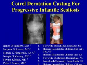
 Infantile scoliosis casting
 Costodesis
 Early onset scoliosis classification
 Rib hump scoliosis
 Coronal balance
 Dr abdullah sayegh
 Factors affecting traction
 Derotation cast
 Turnbuckle cast scoliosis
 Writing strategies and ethical considerations
 Ethical consideration in experimental research sample
 Pricing considerations and approaches
 What are the general considerations in machine design?
 Tax considerations for setting up a new business
 Software architecture of atm machine
 Three considerations of societal marketing concept
 Mechanical considerations of transmission line
 Database design considerations
 Contrasting acquisition
 Cloud delivery models
 Pre analytical considerations in phlebotomy
 Bioreactor considerations for animal cell culture
 Collaboration design considerations
