Craniosynostosis Cranial Vault Reconstruction Neil Anand MD Tzyy


 and AP (B) 3 -D Volume Rendered CT images")


 and AP (B) 3 -D Volume Rendered")

,")

 and Lateral (B) 3 -D Volume Rendered CT images")


 and Post surgical Bird’s")
 and Lateral (B) 3 -D Volume Rendered")

- Slides: 20
Craniosynostosis & Cranial Vault Reconstruction Neil Anand MD, Tzyy Shyang Chao MD, Scott Sosin DO, Neha Gowali MD, Jose Rios MD Ph. D Submission#: 1592 e. Ed. E#: 219 a
DISCLOSURE OF COMMERCIAL INTEREST Neither I nor my immediate family members have a financial relationship with a commercial organization that may have a direct or indirect interest on the content
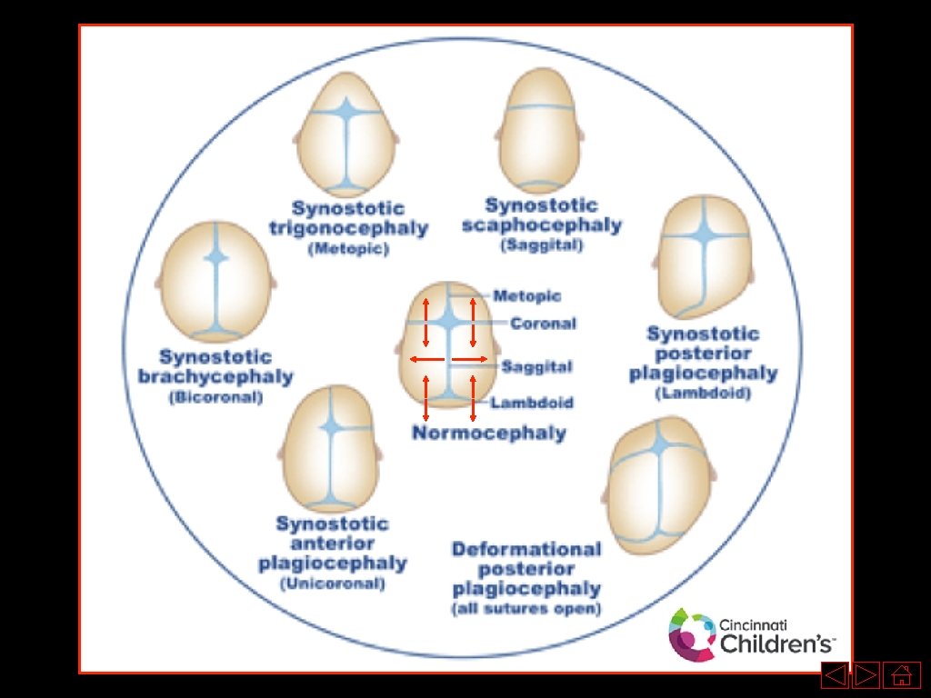
PURPOSE • Make radiologists and clinicians aware of the different disease entities, which involve craniosynostosis • Discuss the different sutures, which may be involved, with respect to their corresponding subtype of craniosynostosis • Educate radiologists and clinicians regarding the imaging characteristics, epidemiology, clinical presentation, differential diagnosis, associated syndromes, and treatment options (cranial vault reconstruction) of the varying types of craniosynostosis
Brachycephaly Background • Bi-parietal to fronto-occipital diameter approaches the 95 th percentile • Premature closure of the bi-coronal or bi-lambdoidal sutures Epidemiology • 30 -40% of all craniosynostosis • Slightly higher incidence of bilateral coronal synostosis in females • M: F = 1: 2 Clinical • Head may appear rounder than usual • Often associated with other syndromes • May lead to increased intracranial pressure or neurodevelopment delay from lack of brain growth • The incidence of associated anomalies is higher with bilateral coronal synostosis than with sagittal synostosis. Imaging • The ipsilateral frontal bone is flattened and the orbit is deformed with elevation of tits superior lateral (harlequin eyes) • Increased transverse diameter with decreased AP diameter • Abnormal calvarial widening in the transverse diameter • Increased cephalic index on antenatal ultrasound Prognosis and Treatment • Cranial remolding orthoses helmets for approximately 3 -4 months • Often have negative side effects such as skin irritation A AP view of the skull (A) in a patient with brachycephaly demonstrating increased bi-parietal diameter (red arrow).
Brachycephaly A B Lateral (A) and AP (B) 3 -D Volume Rendered CT images of the head in a patient with brachycephaly demonstrating partial premature fusion of the bilateral coronal sutures (A, B, red arrows). Note the decreased fronto-occipital diameter (A, green arrow).
Brachycephaly A AP view of the skull demonstrating premature closure of the left lateral aspect of the coronal suture (red arrow) leading to flattening of the calvarium in this region and a harlequin appearance to the left orbit.
Scaphocephaly / Dolichocephaly Background • Premature sagittal synostosis which results in diminished lateral growth of the skull while AP growth continues Epidemiology • Most common form of craniosynostosis, accounts for ~50% of cases • Male predominance 3. 5: 1 • Autosomal Dominant Clinical • May be associated with Marfan Syndrome • Narrow elongated skill • Near normal IQ • Often no hydrocephalus • May be secondary to certain hematological disorders, metabolic disorders, or bone dysplasia A Lateral view of the skull (A) in a patient with dolichocephaly demonstrating increased fronto. Imaging • Decreased transverse diameter, increased AP diameter with occipital diameter (red arrow). forehead bossing Prognosis and Treatment • Cranial vault reconstruction/cranioplasty is often implemented
Scaphocephaly / Dolichocephaly A B Lateral (A) and AP (B) 3 -D Volume Rendered CT images of the head in a patient with dolichocephaly demonstrating complete premature fusion of the sagittal suture (B, red arrow). Note the increased frontooccipital diameter (A, green arrow).
Non-positional Plagiocephaly Background • Asymmetric craniosynostosis with asymmetric coronal and/or lambdoid sutures premature closure Epidemiology • 20 - 30% Clinical • Do not confuse with positional plagiocephaly (results from external pressure and is not associated with premature suture synostosis). Imaging • Premature closure is often associated with harlequin eye deformity • May be divided into frontal (unilateral coronal suture) and occipital (unilateral lambdoid suture) plagiocephaly • Unilateral coronal: unilateral harlequin orbit, hemicalvarium shortened and pointed • Lambdoid synostosis: Trapezoid skull with ipsilateral posterior ear displacement and occipital flattening • Asymmetric growth results in displacement of the sagittal suture, nasal septum, and skull base to the affected side. Prognosis and Treatment • Cranial vault reconstruction has no utility • Cranial helmet therapy is the mainstay for unresolved cases
Non-positional Plagiocephaly Ant Post Ant Left Right A Post B C Bird’s Eye (A), AP (B), and Lateral (C) 3 -D Volume Rendered CT images of the head in a patient with nonpositional plagiocephaly demonstrating partial premature fusion of the right coronal suture (A, B, C, red arrows). Note the misshapen calvarium in (B, red circle).
Trigonocephaly Background • Premature fusion of the metopic suture. • Trigonon = triangle in greek Epidemiology • ~5% of cases • 1: 700 - 1: 15, 000 newborns. M: F = 2 -3. 3: 1 Clinical • Visible and palpable midline ridge may be present • Patients have a wedge shaped forehead and may demonstrate hypotelorism • May be associated with Jacobsen syndrome (intellectual disabilities, distinctive facial appearance, heart defects, and bleeding diathesis), Opitz syndrome, Muenke syndrome, Baller-Gerold syndrome, and Say. Meyer syndrome. Pathology • Metopic suture divides the frontal bones in the midline • Open at birth and fuses by 12 months • Premature fusion halts transverse growth of the forehead leading to a triangular shaped forehead Imaging • Premature fusion of the metopic suture leading to a beaked appearance • Teardrop shaped orbits angulated towards the midline of the forehead (‘surprised coon’ sign) in severe cases • Anterior curving of the metopic suture seen from the lateral view of the cranium • Often a normal cephalic index may be present • Bi-temporal shortening and bi-parietal broadening Prognosis and Treatment • Most compliant form of craniosynostosis for surgery (see slide: Cranial Vault Reconstruction).
Trigonocephaly A B Oblique (A) and Lateral (B) 3 -D Volume Rendered CT images of the head in a patient with Trigonocephaly demonstrating complete premature fusion of the Metopic suture (A, B red arrow). Note the frontal beaking (A, red circle).
Other Pachycephaly • Premature fusion of the lambdoid suture Oxycephaly • Multiple sutures involved • Coronal, sagittal, and lambdoid suture involvement • “Tower-like” skull
Apert Syndrome Background • Also known as Type 1 Acrocephalosyndrome • Craniosynostosis with syndactyly of fingers and toes Epidemiology • Increased paternal age • 1: 65 - 80, 000 Clinical • Midface hypoplasia, hypertelorism, syndactyly • Hearing loss in 90%, predominantly conductive hearing loss • Airway obstruction • Raised intracranial pressure Pathology • Autosomal dominant • Mutation in Fibroblast growth factor receptor 2 (FGFR 2) on chromosome 10 q 26 Imaging • Limb syndactyly, Tower shaped head • Coronal and/or squamous craniosynostoses • Widened sagittal and metopic sutures • EAC stenosis, cerebellar tonsillar ectopia, ventriculomegaly • Ear abnormalities such as EAC stenosis, dysmorphic ossicles, cochlear/vestibular malformations Prognosis and Treatment • Surgery: Le. Fort osteotomy and midface advancement • Strip craniotomy for release of sutures • Middle ear exploration, myringotomy, tympanostomy, etc…
Crouzon Syndrome Background • Premature fusion of multiple sutures, maxillary hypoplasia, shallow orbits Post Ant Epidemiology • Associated with Chiari I malformations, in up to 70% of cases. 1. 6 : 100, 000 Clinical • May have shallow orbits with exophthalmos, mid facial hypoplasia, and/or a bifid uvula. • Eyes may not point in the same direction Pathology • Autosomal dominant. Mutation in Fibroblast growth factor receptor 2 (FGFR 2) on chromosome 10 q 25 -26 Imaging • Cloverleaf skull/Kleeblattschadel in severe cases • Multiple sutures prematurely fused: bilateral coronal craniosynostosis A Right Left Prognosis and Treatment • Once treated: Normal life span. Without surgery, blindness and mental retardation often ensue • Surgery: Prevent closure of sutures. Open vault surgery followed by helmet prosthesis or strip craniotomy (<6 yo) can be performed Lateral (A) and AP (B) 3 -D Volume Rendered CT images of the head in a patient with Crouzon syndrome demonstrating a Kleeblattschadel configuration of the calvarium secondary to fusion of many cranial sutures, including the right coronal suture (B, red arrow), sagittal suture (B, green arrow), and left lambdoid suture (A, orange arrow). B
Cranial Vault Reconstruction Dolichocephaly • Most often include sagittal suturectomy with varying parietal and occipital osteotomies with or without frontal osteotomies and remodeling. • In older patients, surgical remodeling procedures may not re-ossify and may require a more comprehensive calvarial reconstruction leading to multiple interleaving osteotomies crossing the midline, in the coronal plane. Trigonocephaly • Standard approach consist of bi-frontal craniotomy, flap remodeling, and fronto-orbital advancement with recontouring. Anterior plagiocephaly • Standard fronto-orbital reconstruction • Surgical procedures have expanded from simple suturectomies to frontal-calvarial vault remodeling consisting of bi-frontal craniotomies and orbital frontal bandeau advancement Crouzon Syndrome • Initial treatment often requires cranio-orbital decompression, including bi-coronal suture release and osteotomies of the anterior cranial vault and upper orbits with reshaping and advancement
Cranial Vault Reconstruction A B Pre-surgical Bird’s eye view (A) and Post surgical Bird’s eye view (B) 3 -D Volume Rendered CT images of the head in the same patient demonstrate strip sagittal suturectomy (B, between green arrow) in a patient with premature fusion of the sagittal suture (A, red arrow). Additional osteotomies were noted along both coronal sutures as well as posteriorly within the parietal bones bilaterally (not shown). A AP (A) 3 -D Volume Rendered CT image of the head in a patient with Trigonocephaly demonstrating an osteotomy to the left of the region of the metopic suture (red arrow). Additional post-surgical changes were present along the coronal sutures, bilaterally (not shown)
Cranial Vault Reconstruction A B AP (A) and Lateral (B) 3 -D Volume Rendered CT images of the head in a patient who has undergone cranial vault reconstruction to treat underlying craniosynostosis. Multiple osteotomies along the anterior calvarium remain unfused. Note the underlying premature fusion of the bilateral coronal and sagittal sutures.
References 1. Badve, Chaitra A. et al. "Craniosynostosis: Imaging Review And Primer On Computed Tomography". Pediatric Radiology 43. 6 (2013): 728 -742. Web. 2. Brachycephaly | Radiology Reference Article | Radiopaedia. Org". Radiopaedia. org. N. p. , 2017. Web. 19 Feb. 2017. 3. Calandrelli, Rosalinda et al. "Identification Of Skull Base Sutures And Craniofacial Anomalies In Children With Craniosynostosis: Utility Of Multidetector CT". La radiologia medica 119. 9 (2014): 694 -704. Web. 4. Dixon, Andrew. "Trigonocephaly | Radiology Reference Article | Radiopaedia. Org". Radiopaedia. org. N. p. , 2017. Web. 19 Feb. 2017. 5. Gaillard, Frank. "Craniosynostosis | Radiology Reference Article | Radiopaedia. Org". Radiopaedia. org. N. p. , 2017. Web. 19 Feb. 2017. 6. Gaillard, Frank. "Scaphocephaly | Radiology Reference Article | Radiopaedia. Org". Radiopaedia. org. N. p. , 2017. Web. 19 Feb. 2017. 7. Glass, Ronald B. J. et al. "The Infant Skull: A Vault Of Information". Radio. Graphics 24. 2 (2004): 507 -522. Web. 8. Hinojosa, Jose. "Methods Of Cranial Vault Reconstruction For Craniosynostosis | Clinical Gate". Clinicalgate. com. N. p. , 2017. Web. 23 Feb. 2017. 9. Kapp-Simon, Kathleen A. et al. "Neurodevelopment Of Children With Single Suture Craniosynostosis: A Review". Child's Nervous System 23. 3 (2006): 269 -281. Web. 10. Kuang, Anna A. et al. "Benign Radiographic Coronal Synostosis After Sagittal Synostosis Repair". Journal of Craniofacial Surgery 24. 3 (2013): 937 -940. Web. 11. Massimi, Luca et al. "Isolated Sagittal Craniosynostosis: Definition, Classification, And Surgical Indications". Child's Nervous System 28. 9 (2012): 1311 -1317. Web. 12. St-Amant, Maxime. "Plagiocephaly | Radiology Reference Article | Radiopaedia. Org". Radiopaedia. org. N. p. , 2017. Web. 19 Feb. 2017. 13. "Statdx | Diagnostic Decision Support For Radiology". App. statdx. com. N. p. , 2017. Web. 19 Feb. 2017.