Chapter 6 An Introduction to Viruses Prions The


 (3) (5)")


 but may be single")


 Adsorption (2) Penetration (3) Uncoating (4) Synthesis")
 Adsorption and Host Range • Virus coincidentally collides with a susceptible host cell")
 Penetration • Flexible cell membrane is penetrated by the whole virus or its")
 Synthesis: Replication and Protein Production • Varies depending on whether the virus is")
 Release • Assembled viruses leave the host cell in one of two ways:")


- Slides: 32
Chapter 6 An Introduction to Viruses & Prions
The Search for the Elusive Virus • Louis Pasteur postulated that rabies was caused by a virus (1884) • Ivanovski and Beijerinck showed a disease in tobacco was caused by a virus (1890 s) • 1950 s virology was a multifaceted discipline – Viruses: noncellular particles with a definite size, shape, and chemical composition 2
The Position of Viruses in the Biological Spectrum • There is no universal agreement on how and when viruses originated • Viruses are considered the most abundant microbes on earth • Viruses played a role in the evolution of Bacteria, Archaea, and Eukarya • Viruses are obligate intracellular parasites 3
General Size of Viruses • Size range – most <0. 2 μm; requires electron microscope Copyright© The Mc. Graw-Hill Companies, Inc. Permission required for reproduction or display. BACTERIAL CELLS Rickettsia 0. 3 mm Viruses 1. Poxvirus 2. Herpes simplex 3. Rabies 4. HIV 5. Influenza 6. Adenovirus 7. T 2 bacteriophage 8. Poliomyelitis 9. Yellow fever Streptococcus 1 mm (1) (2) Protein Molecule 10. Hemoglobin molecule 250 nm 125 nm 110 nm 100 nm 75 nm 65 nm 30 nm 22 nm E. coli 2 mm long (10) (9) (8) 15 nm (7) (3) (4) (6) (5) 4 YEAST CELL – 7 mm
Viral Structure • Viruses bear no resemblance to cells – Lack protein-synthesizing machinery • Viruses contain only the parts needed to invade and control a host cell Copyright © The Mc. Graw-Hill Companies, Inc. Permission required for reproduction or display. Capsid Covering Virus particle Central core Envelope (not found in all viruses) Nucleic acid molecule(s) (DNA or RNA) Matrix proteins Enzymes (not found in all viruses) 5
General Structure of Viruses • Capsids – All viruses have capsids (protein coats that enclose and protect their nucleic acid) – The capsid together with the nucleic acid is the nucleocapsid – Some viruses have an external covering called an envelope made from the phospholipid bi-layer – Viruses lacking an envelope are naked Copyright © The Mc. Graw-Hill Companies, Inc. Permission required for reproduction or display. Capsid Nucleic acid (a) Naked Nucleocapsid Virus Envelope Spike Capsid Nucleic acid (b) Enveloped Virus 6
General Structure of Viruses Copyright © The Mc. Graw-Hill Companies, Inc. Permission required for reproduction or display. • Two structural capsid types: Capsomers 1. Helical - continuous helix (a) of capsomers forming a cylindrical nucleocapsid (b) Discs Nucleic acid Capsid begins forming helix. (c) 7
General Structure of Viruses Copyright © The Mc. Graw-Hill Companies, Inc. Permission required for reproduction or display. • Two structural capsid types: 2. Icosahedral - 20 sided with 12 corners (a) Capsomers Facet Capsomers Vertex Nucleic acid (b) Capsomers Vertex Fiber (c) 8 (d) © Dr. Linda Stannard, UCT/Photo Researchers, Inc.
General Structure of Viruses Copyright © The Mc. Graw-Hill Companies, Inc. Permission required for reproduction or display. • Viral envelope – Mostly animal viruses – Acquired when the virus leaves the host cell– taken from the host cell membrane – Exposed proteins on the outside of the envelope, called spikes, are essential for attachment of the virus to the host cell Are spikes considered a virulence factor? Capsid Nucleocapsid Nucleic acid © Dennis Kunkel/CNRI/Phototake (b) (a) Hemagglutinin spike Neuraminidase spike Matrix protein Lipid bilayer Nucleocapsid (c) 50 nm Spikes Nucleocapsid 9 (d) Dr. F. A. Murphy/CDC
Functions of Capsid/Envelope Copyright © The Mc. Graw-Hill Companies, Inc. Permission required for reproduction or display. • Protects the nucleic acid when the virus is outside of the host cell • Helps the virus bind to a cell surface and assists the penetration of the viral DNA or RNA into a suitable host cell Capsomers (a) © Dr. Linda Stannard, UCT/Photo Researchers, Inc. Fred P. Williams, Jr. /EPA Envelope Capsid DNA core 10 (b) © Eye of Science/Photo Researchers, Inc.
General Structure of Viruses • Complex viruses: atypical viruses – Poxviruses lack a typical capsid and are covered by a dense layer of lipoproteins – What are lipoproteins? – Some bacteriophages have a polyhedral nucleocapsid along with a helical tail and attachment fibers 240– 300 nm Core membrane Nucleic acid (a) Capsid head Collar Sheath 200 nm Outer envelope Soluble protein antigens Lateral body Nucleic acid Tail fibers Tail Base plate pins (c) 11 (b) © Bin Ni, Chisholm Lab, MIT
Types of Viruses B. Enveloped Viruses A. Complex Viruses Helical Icosahedral (1) (3) (5) (2) (4) (6) C. Nonenveloped Naked Viruses Helical Icosahedral (8) (7) (9) A. Complex viruses: (1) poxvirus, a large DNA virus (2) flexible-tailed bacteriophage B. Enveloped viruses: With a helical nucleocapsid: (3) mumps virus (4) rhabdovirus With an icosahedral nucleocapsid: (5) herpesvirus (6) HIV (AIDS) C. Naked viruses: Helical capsid: (7) plum poxvirus Icosahedral capsid: (8) poliovirus (9) papillomavirus
Concept Check: Copyright © The Mc. Graw-Hill Companies, Inc. Permission required for reproduction or display. How would you describe this virus? A. Icosahedral and Naked B. Helical and Naked C. Complex and Naked D. Icosahedral and Enveloped E. Helical and Enveloped F. Complex and Enveloped © Dennis Kunkel/CNRI/Phototake
Nucleic Acids • Viral genome – either DNA or RNA but never both • Carries genes necessary to invade host cell and redirect cell’s activity to make new viruses • Number of genes varies for each type of virus – few to hundreds • What is a gene? 14
Nucleic Acids • DNA viruses – Usually double stranded (ds) but may be single stranded (ss) – Circular or linear • RNA viruses – Usually single stranded, may be double stranded, may be segmented into separate RNA pieces – ss. RNA genomes ready for immediate translation are positive-sense RNA – ss. RNA genomes that must be converted into proper form are negative-sense RNA 15
Eukaryotic transcription & translation Viral transcription & translation • Viruses may have their own enzymes to carry out transcription & translation – Polymerases – DNA or RNA – Replicases – copy RNA – Reverse transcriptase – synthesis of DNA from RNA (AIDS virus) Eukaryotic cell machinery – RNA Polymerase for transcribing DNA to RNA – Ribosomes for translating RNA to Protein
Steps of Viral Replication General phases in animal virus multiplication cycle: 1. Adsorption – binding of virus to specific molecules on the host cell 2. Penetration – genome enters the host cell 3. Uncoating – the viral nucleic acid is released from the capsid 4. Synthesis – viral components are produced 5. Assembly – new viral particles are constructed 6. Release – assembled viruses are released by budding (exocytosis) or cell lysis What is exocytosis? Cell lysis? How do they differ? 17
Animal Virus Multiplication (1) Adsorption (2) Penetration (3) Uncoating (4) Synthesis
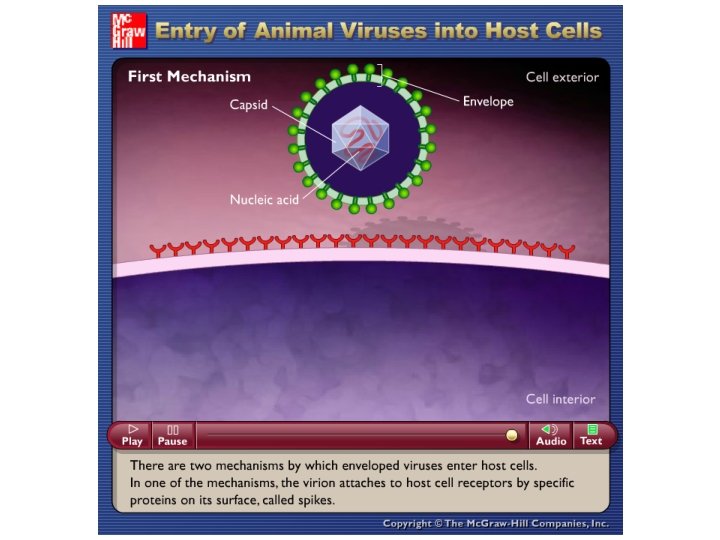
1) Adsorption and Host Range • Virus coincidentally collides with a susceptible host cell and adsorbs specifically to receptor sites on the membrane • Spectrum of cells a virus can infect – host range – Hepatitis B – human liver cells – Poliovirus – primate intestinal and nerve cells – Rabies – various cells of many mammals Copyright © The Mc. Graw-Hill Companies, Inc. Permission required for reproduction or display. Envelope spike Host cell membrane Capsid spike Receptor Host cell membrane Receptor 19 (a) (b)
2) Penetration • Flexible cell membrane is penetrated by the whole virus or its nucleic acid by: – Endocytosis – entire virus is engulfed and enclosed in a vacuole or vesicle – Fusion – envelope merges directly with membrane resulting in nucleocapsid’s direct entry into cytoplasm 21
4) Synthesis: Replication and Protein Production • Varies depending on whether the virus is a DNA or RNA virus • DNA viruses generally are replicated and assembled in the nucleus • RNA viruses generally are replicated and assembled in the cytoplasm – Positive-sense RNA contain the message for translation – Negative-sense RNA must be converted into positive-sense message 22
6) Release • Assembled viruses leave the host cell in one of two ways: – Budding – exocytosis; nucleocapsid binds to membrane which pinches off and sheds the viruses gradually; cell is not immediately destroyed – Lysis – nonenveloped and complex viruses released when cell dies and ruptures Copyright © The Mc. Graw-Hill Companies, Inc. Permission required for reproduction or display. (b) © Chris Bjornberg/Photo Researchers, Inc. Copyright © The Mc. Graw-Hill Companies, Inc. Permission required for reproduction or display. Host cell membrane Viral nucleocapsid Viral glycoprotein spikes Cytoplasm Capsid RNA Budding virion (a) Viral matrix protein Free infectious virion with envelope 23
Concept Check: Viruses commonly contain both DNA and RNA. A. True B. False
Viruses can damage the Host Cell Copyright © The Mc. Graw-Hill Companies, Inc. Permission required for reproduction or display. Cytopathic effects - virusinduced damage to cells 1. 2. 3. 4. 5. Changes in size and shape Cells fuse to form multinucleated cells Cell lysis Alter DNA Transform cells into cancerous cells Multiple nuclei Normal cell Giant cell (a) CDC Inclusion bodies 26 (b) © Massimo Battaglia, INe. MM CNR, Rome Italy
Persistent Infections • Persistent infections - cell harbors the virus and is not immediately lysed • Can last weeks or host’s lifetime; several can periodically reactivate – chronic latent state – Measles virus – may remain hidden in brain cells for many years – Herpes simplex virus – cold sores and genital herpes – Herpes zoster virus – chickenpox and shingles 27
Medical Importance of Viruses • Viruses are the most common cause of acute infections • Several billion viral infections per year • Some viruses have high mortality rates • Possible connection of viruses to chronic afflictions of unknown cause • Viruses are major participants in the earth’s ecosystem 28
Prions: Infectious Proteins Prions - misfolded proteins, contain no nucleic acid Extremely resistant to usual sterilization techniques Cause transmissible spongiform encephalopathies- fatal neurodegenerative diseases 29
Transmissible Spongiform Encephalopathies Common in animals: • Scrapie in sheep and goats • Bovine spongiform encephalopathies (BSE), a. k. a. mad cow disease • Wasting disease in elk • Humans – Creutzfeldt. Jakob Syndrome (CJS) Copyright © The Mc. Graw-Hill Companies, Inc. Permission required for reproduction or display. Brain cell Prion fibrils © James King-Holmes/Institute of Animal Health/Photo Researchers, Inc. (a) Dr. Art Davis/CDC (b) 30
Videos for Review Directions: copy the website link into your internet browser 1. Youtube: Prions: What are they? https: //www. youtube. com/watch? v=tfv 3 x. Aw 0 XOE 2. Youtube: Can chronic wasting disease jump from deer to humans? http: //youtu. be/Fu. Jgv 50 r. Uj. I 3. Youtube: Sheep with ataxia http: //youtu. be/-zll. YTt. HSks
Terms to Know • • • • • host range virus • endocytosis obligate intracellular parasites • uncoating capsid • budding nucleocapsid • exocytosis naked viruses • cytopathic effects helical capsids • persistent infections icosahedron • lysis enveloped viruses • plaques spikes • prions bacteriophages • transmissible spongiform genome encephalopathies adsorption penetration uncoating synthesis assembly release 32