Chapter 13 Viruses Viroids and Prions Adolf Mayer

 transmissible • Dimitri Iwanowski,")

![[INSERT FIGURE 13. 4]](https://slidetodoc.com/presentation_image/2d4d56857d2313bf8074d0bf631218ac/image-4.jpg "[INSERT FIGURE 13. 4]")


![[INSERT FIGURE 13. 5]](https://slidetodoc.com/presentation_image/2d4d56857d2313bf8074d0bf631218ac/image-14.jpg "[INSERT FIGURE 13. 5]")


![[INSERT FIGURE 13. 8]](https://slidetodoc.com/presentation_image/2d4d56857d2313bf8074d0bf631218ac/image-23.jpg "[INSERT FIGURE 13. 8]")


![Multiplication of RNA Virus [INSERT FIGURE 13. 13]](https://slidetodoc.com/presentation_image/2d4d56857d2313bf8074d0bf631218ac/image-32.jpg "Multiplication of RNA Virus [INSERT FIGURE 13. 13]")


![[INSERT FIGURE 13. 23] scrapie proteins accumulate in brain cells forming large vacuoles](https://slidetodoc.com/presentation_image/2d4d56857d2313bf8074d0bf631218ac/image-47.jpg "[INSERT FIGURE 13. 23] scrapie proteins accumulate in brain cells forming large vacuoles")
- Slides: 47
Chapter 13 Viruses, Viroids and Prions
• Adolf Mayer, 1886 – tobacco mosaic disease (TMD) transmissible • Dimitri Iwanowski, 1892 – Filtered sap still caused TMD – contagious fluid or filterable agent • Walter Reed, 1901 – Yellow Fever
• Felix d’Herelle, 1917 – Bacteriophage – Suggested phage therapy • 1930’s, term virus introduced and electron microscopy invented • Wendell Stanley, 1935 – Isolated tobacco mosaic virus
[INSERT FIGURE 13. 4]
• Viral Features – Obligate intracellular pathogen – Host range • highly specific or generalists – DNA or RNA – Protein coat – Surface proteins highly susceptible to mutations – Cause synthesis of specialized structures to transfer viral particles to other cells
• Virion – complete, fully developed, infectious viral particle – Living or non-living entities? ? ?
Viral Classification • Oldest system based on symptomology • International Committee on Taxonomy of Viruses (ICTV), 1966 – Describe viruses as elementary bio-systems – Classified into orders, families, genera and species – Over 1, 500 officially recognized species
• 3 primary means of classification: – Nucleic acid – Replication strategy – Morphology of protein coat (capsid)
Helical Viruses Polyhedral Viruses
• Viral Envelope – – Acquired from host cell Phospholipids and proteins Some glycoproteins are virally coded spikes Often play role in host recognition
Critical Swine Flu prevention tip: Don't DO this!
Complex Viruses
[INSERT FIGURE 13. 5]
Viral Taxonomy • Order –virales • Family –viridae • Genus – virus • Species – Common names – Subspecies designated by a number
Viral Taxonomy • Retroviridae– family – Lentivirus – genus • Human Immunodeficiency Virus– species • Herpesviridae – Simplexvirus • Human herpesvirus 1, HHV 2, HHV 3
Isolation and Cultivation of Viruses • Viruses must be grown in living cells – Cytopathic effects
• Animal viruses may be grown in living animals or in embryonated eggs
• Animal & plants viruses may be grown in cell cultures – Primary cell lines – Continuous cell lines (transformed cells )
Virus Identification • Serological tests – Detect antibodies against viruses in a patient • Nucleic acids – RFLPs – PCR
Multiplication of Bacteriophages • Lytic cycle Phage causes lysis and death of host cell • Lysogenic cycle Temperate phages incorporate DNA into host DNA (prophage)
Lytic Cycle • Attachment Phage attaches by tail fibers to host cell • Penetration Phage lysozyme opens cell wall, tail sheath contracts to force tail core and DNA into cell • Biosynthesis Production of phage DNA and proteins • Maturation Assembly of newly synthesized phage particles • Release Phage lysozyme breaks cell wall
[INSERT FIGURE 13. 8]
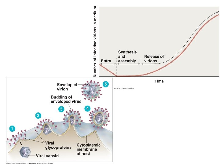
One-step Growth Curve
Lysogenic Cycle
• 3 Important results of lysogeny – Immunity to re-infection – Phage conversion – Specialized transduction
Specialized Transduction Prophage gal gene Bacterial DNA 1 Prophage exists in galactose-using host (containing the gal gene). Galactose-positive donor cell gal gene 2 gal gene Phage genome excises, carrying with it the adjacent gal gene from the host. 3 Phage matures and cell lyses, releasing phage carrying gal gene. 4 Phage infects a cell that cannot utilize galactose (lacking gal gene). Galactose-negative recipient cell 5 Along with the prophage, the bacterial gene becomes integrated into the new host’s DNA. 6 Lysogenic cell can now metabolize galactose. Galactose-positive recombinant cell
Multiplication of Animal viruses • • Attachment Penetration Uncoating Biosynthesis • Maturation • Release Viruses attaches to cell membrane By endocytosis or fusion By viral or host enzymes Production of nucleic acid and proteins Nucleic acid and capsid proteins assemble By budding (enveloped viruses) or rupture
• Attachment of animal viruses – Chemical attraction – No tails or tail fibers – Glycoprotein spikes or other attachment molecules
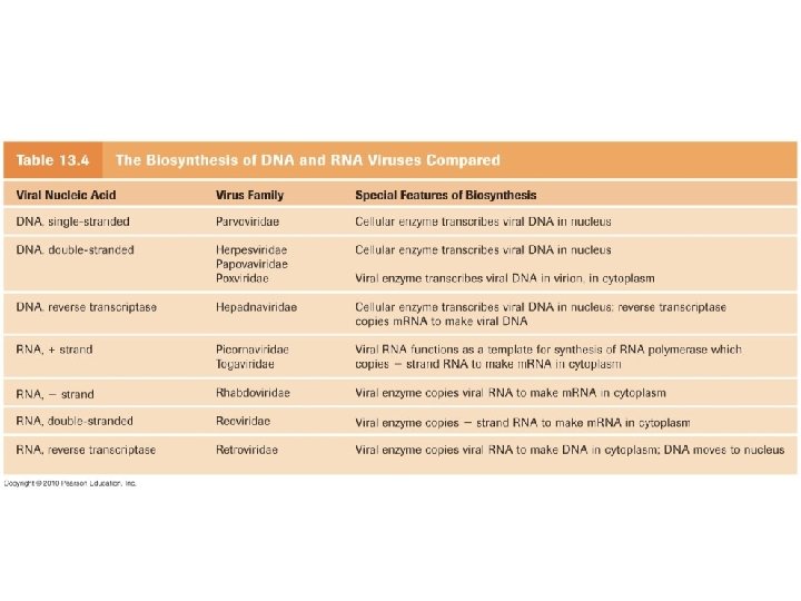
• Replication of Animal Viruses – Biosynthesis • Each virus requires different strategy depending on its nucleic acid • DNA viruses often enter the nucleus • RNA viruses typically replicate in cytoplasm • Must consider: – What serves as template for replication and how m. RNA is transcribed
Multiplication of DNA Virus Papovavirus 1 Virion attaches to host cell 7 Virions are released Host cell DNA Capsid DNA Cytoplasm 6 Virions mature 2 Virion penetrates cell and its DNA is uncoated Capsid proteins m. RNA 5 Late translation; capsid proteins are synthesized 4 Late transcription; DNA is replicated 3 Viral DNA penetrates host nucleus Early transcription and translation; enzymes are synthesized
Multiplication of RNA Virus [INSERT FIGURE 13. 13]
Multiplication of a Retrovirus Capsid Reverse transcriptase DNA Virus Two identical + stands of RNA 1 Retrovirus penetrates host cell. Host cell DNA of one of the host cell’s chromosomes 5 Mature retrovirus leaves host cell, acquiring an envelope as it buds out. Identical strands of RNA Viral proteins RNA Reverse transcriptase Viral RNA 2 Virion penetrates cell and its DNA is uncoated 4 Transcription of the provirus may also occur, producing RNA for new retrovirus genomes and RNA that codes for the retrovirus capsid and envelope proteins. Provirus 3 The new viral DNA is tranported into the host cell’s nucleus and integrated as a provirus. The provirus may divide indefinitely with the host cell DNA.
• Assembly and release of animal viruses • Most DNA viruses assemble in and are released from nucleus into cytosol • Most RNA viruses develop solely in cytoplasm • Enveloped viruses cause persistent infections • Naked viruses are released by exocytosis or may cause lysis and death of host cell
Viruses and Cancer • Oncogenes transform normal cells into tumor cells – Activated by mutagenic chemicals, radiation, viruses – Causes increased growth, loss of contact inhibition – Cells tend to be misshapen and exhibit chromosomal abnormalities
• Oncoviruses • Viral DNA integrated into host DNA • Induces tumors
Oncogenic Viruses • Oncogenic DNA Viruses – Adenoviridae – Herpesviridae – Poxviridae – Papovaviridae – Hepadnaviridae • Oncogenic RNA viruses – Retroviridae • DNA • HTLV 1 • HTLV 2
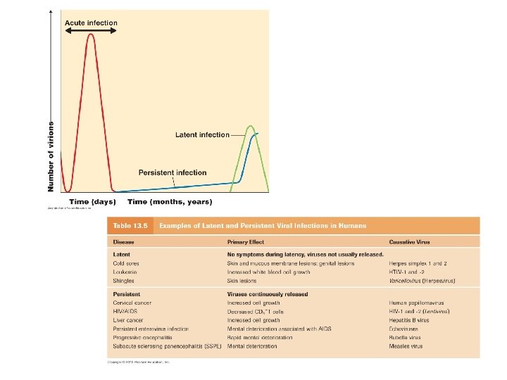
• Latent Viral Infections – Virus remains dormant in asymptomatic host cell for long periods • Cold sores, shingles • Persistent Viral Infections – Disease progresses slowly over a long period, generally fatal • Subacute sclerosing panencephalitis (measles virus)
• Plant Viruses – Plant viruses enter through wounds or via biting insects – May be transmitted in pollen • Viroids – infectious naked RNA Potato Spindle Tuber Viroid
Prions • Proteinaceous infectious particle • Inherited and transmissible diseases • Spongiform encephalopathies – Sheep scrapie, Creutzfeldt-Jakob disease, Gerstmann. Sträussler-Scheinker syndrome, fatal familial insomnia, mad cow disease
Pr. PC, normal cellular prion protein Pr. PSc, scrapie protein
[INSERT FIGURE 13. 23] scrapie proteins accumulate in brain cells forming large vacuoles