Chapter 13 Characterizing and Classifying Viruses Viroids and


 surrounding nucleic acid Nucleic acid and")
![[INSERT FIGURE 13. 1] Characteristics of Viruses](https://slidetodoc.com/presentation_image_h2/c33e52a29f7b9e0efb804eb3b4c3dd5b/image-4.jpg "[INSERT FIGURE 13. 1] Characteristics of Viruses")


![[INSERT FIGURE 13. 4] Size of Viruses](https://slidetodoc.com/presentation_image_h2/c33e52a29f7b9e0efb804eb3b4c3dd5b/image-7.jpg "[INSERT FIGURE 13. 4] Size of Viruses")


![[INSERT TABLE 13. 1]](https://slidetodoc.com/presentation_image_h2/c33e52a29f7b9e0efb804eb3b4c3dd5b/image-11.jpg "[INSERT TABLE 13. 1]")

![[INSERT TABLE 13. 2] - = negative sense: complimentary for m. RNA, cannot be](https://slidetodoc.com/presentation_image_h2/c33e52a29f7b9e0efb804eb3b4c3dd5b/image-13.jpg "[INSERT TABLE 13. 2] - = negative sense: complimentary for m. RNA, cannot be")

![[INSERT FIGURE 13. 8] Viral Replication of Bacteriophages](https://slidetodoc.com/presentation_image_h2/c33e52a29f7b9e0efb804eb3b4c3dd5b/image-15.jpg "[INSERT FIGURE 13. 8] Viral Replication of Bacteriophages")
![[INSERT FIGURE 13. 9] Viral Replication](https://slidetodoc.com/presentation_image_h2/c33e52a29f7b9e0efb804eb3b4c3dd5b/image-16.jpg "[INSERT FIGURE 13. 9] Viral Replication")

![[INSERT FIGURE 13. 11] Viral Replication - Bacteriophages](https://slidetodoc.com/presentation_image_h2/c33e52a29f7b9e0efb804eb3b4c3dd5b/image-18.jpg "[INSERT FIGURE 13. 11] Viral Replication - Bacteriophages")


![[INSERT FIGURE 13. 12] Viral Replication of Animal Viruses](https://slidetodoc.com/presentation_image_h2/c33e52a29f7b9e0efb804eb3b4c3dd5b/image-21.jpg "[INSERT FIGURE 13. 12] Viral Replication of Animal Viruses")


![[INSERT TABLE 13. 4] Viral Replication Comparison](https://slidetodoc.com/presentation_image_h2/c33e52a29f7b9e0efb804eb3b4c3dd5b/image-27.jpg "[INSERT TABLE 13. 4] Viral Replication Comparison")


![[INSERT TABLE 13. 5] Other Parasitic Particles: Viroids and Prions](https://slidetodoc.com/presentation_image_h2/c33e52a29f7b9e0efb804eb3b4c3dd5b/image-36.jpg "[INSERT TABLE 13. 5] Other Parasitic Particles: Viroids and Prions")

- Slides: 37
Chapter 13 Characterizing and Classifying Viruses, Viroids, and Prions
Cause many infections of humans, animals, plants, and bacteria Cannot carry out any metabolic pathway Neither grow nor respond to the environment Cannot reproduce independently Recruit the cell’s metabolic pathways to increase their numbers Cause most of the diseases that plague the industrialized world Virus – miniscule, acellular, infectious agent having one or several pieces of either DNA or RNA No cytoplasmic membrane, cytosol, organelles (with one exception) Have extracellular and intracellular state Characteristics of Viruses
Extracellular State Called virion Protein coat (capsid) surrounding nucleic acid Nucleic acid and capsid also called nucleocapsid Some have phospholipid envelope Outermost layer provides protection and recognition sites for host cells Intracellular State Capsid removed Virus exists as nucleic acid Characteristics of Viruses
[INSERT FIGURE 13. 1] Characteristics of Viruses
Genetic Material of Viruses Show more variety in nature of their genomes than do cells May be DNA or RNA, but never both Primary way scientists categorize and classify viruses Can be ds. DNA, ss. DNA, ds. RNA, ss. RNA May be linear and composed of several segments or single and circular Much smaller than genomes of cells Characteristics of Viruses
Hosts of Viruses Most viruses infect only particular host’s cells Due to affinity of viral surface proteins or glycoproteins for complementary proteins or glycoproteins on host cell surface May be so specific they only infect particular kind of cell in a particular host Generalists – infect many kinds of cells in many different hosts
[INSERT FIGURE 13. 4] Size of Viruses
Capsid Morphology Capsids – protein coats that provide protection for viral nucleic acid and means of attachment to host’s cells Capsid composed of proteinaceous subunits called capsomeres Some capsids composed of single type of capsomere; others composed of multiple types
Complex shape of a bacteriophage a. Notice helical capsid inside envelope b. Notice polyhedral capsid inside envelope Shapes of Viruses
The Viral Envelope Not all viruses have envelopes Acquired from host cell during viral replication or release; envelope is portion of membrane system of host Composed of phospholipid bilayer and proteins; some proteins are virally coded glycoproteins (spikes) Envelope’s proteins and glycoproteins often play role in host recognition
[INSERT TABLE 13. 1]
Classification of Viruses Based on type of nucleic acid, presence of envelope, shape, and size
[INSERT TABLE 13. 2] - = negative sense: complimentary for m. RNA, cannot be directly translated + = positive sense: equivalent to m. RNA
Dependent on hosts’ organelles and enzymes to produce new virions Replication cycle usually results in death and lysis of host cell → lytic replication Stages of lytic replication cycle Attachment Entry Synthesis Assembly Release Viral Replication of Bacteriophages
[INSERT FIGURE 13. 8] Viral Replication of Bacteriophages
[INSERT FIGURE 13. 9] Viral Replication
Lysogeny Modified replication cycle Infected host cells grow and reproduce normally for generations before they lyse Temperate phages Prophages – inactive phages Lysogenic conversion results when phages carry genes that alter phenotype of a bacterium
[INSERT FIGURE 13. 11] Viral Replication - Bacteriophages
Viral Replication Animation: Temperate Bacteriophages
Replication of Animal Viruses Same basic replication pathway as bacteriophages Differences result from Presence of envelope around some viruses Eukaryotic nature of animal cells Lack of cell wall in animal cells Attachment of animal viruses Chemical attraction Animal viruses do not have tails or tail fibers Have glycoprotein spikes or other attachment molecules that mediate attachment Viral Replication of Animal Viruses
[INSERT FIGURE 13. 12] Viral Replication of Animal Viruses
Replication of Animal Viruses Synthesis of animal viruses Each type of animal virus requires different strategy depending on its nucleic acid DNA viruses often enter the nucleus RNA viruses often replicated in the cytoplasm Must consider How m. RNA is synthesized? What serves as template for nucleic acid replication? Viral Replication of Animal Viruses
ds. DNA Viruses – similar to normal replication of host cellular DNA ss. DNA – in humans are paroviruses Host enzymes produce complimentary strand of DNA to make ds. DNA can act directly as m. RNA Use their reverse transcriptase to make DNA intermediate +ss. RNA – example is poliovirus Retroviruses - +ss. RNA but don’t use it as m. RNA, HIV -ss. RNA – rabies and flu Not recognized by host ribosome Has to use it’s RNA-dependent RNA transcriptase to make +RNA ds. RNA – rotavirus Unwinds, + strand acts immediately as m. RNA, +RNA is transcribed by RNA polymerase
Replication of Animal Viruses Assembly and release of animal viruses Most DNA viruses assemble in and are released from nucleus into cytosol Most RNA viruses develop solely in cytoplasm Number of viruses produced and released depends on type of virus and size and initial health of host cell Enveloped viruses cause persistent infections Are released by “budding”, do not lyse cell right away so cause long lasting infection Naked viruses are released by exocytosis or may cause lysis and death of host cell
Viral Replication Animation: Animal Viruses
Replication of Animal Viruses Latency of animal viruses Similar to lysogeny of bacteriophages but there are differences Some latent viruses do not become incorporated into host chromosome When provirus (latent virus) is incorporated into host DNA, condition is permanent, becomes physical part of host’s chromosome When animal viruses remain dormant in host cells May be prolonged for years with no viral activity, signs, or symptoms Viral Replication of Animal Viruses
[INSERT TABLE 13. 4] Viral Replication Comparison
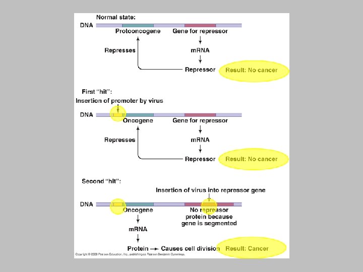
Normally, animal’s genes dictate that some cells can no longer divide and those that can divide are prevented from unlimited division Genes for cell division are “turned off” or genes that inhibit division are “turned on” Neoplasia – uncontrolled cell division in multicellular animal; mass of neoplastic cells is tumor Benign vs. malignant tumors Metastasis Cancers The Role of Viruses In Cancer
Environmental factors that contribute to the activation of oncogenes Ultraviolet light Radiation Carcinogens Viruses cause 20 -25% of human cancers in several ways Some carry copies of oncogenes as part of their genomes Some promote oncogenes already present in host Some interfere with tumor repression when they insert into host’s repressor gene Several specific DNA and RNA viruses are known to cause ~15% of human cancers Burkitt’s lymphoma Hodgkin’s disease Kaposi’s sarcoma Cervical cancer
Culturing Viruses in the Laboratory Has to be done using cells Live organisms – bacterial cultures, lab plants or animals Cell culture Bacteria, plant, animal Embryonated chicken eggs are great – large cell, free of microbes, contain nourishing yolk
Characteristics of Viroids Extremely small, circular pieces of RNA that are infectious and pathogenic in plants Similar to RNA viruses, but lack capsid May appear linear due to H bonding No animal diseases are known as of yet Other Parasitic Particles: Viroids and Prions
Characteristics of Prions Proteinaceous infectious agents Composed of single protein Pr. P All mammals contain gene that codes for primary sequence of amino acids in Pr. P Two stable tertiary structures of Pr. P Has role in activity of the brain but exaxt role still not known Normal functional structure with -helices called cellular Pr. P Disease-causing form with -sheets called prion Pr. P Prion Pr. P converts cellular Pr. P into prion Pr. P by inducing conformational change (acts enzymatically to do this) Results in fatal neurological degeneration Other Parasitic Particles: Viroids and Prions
Characteristics of Prions Normally, nearby proteins and polysaccharides force Pr. P into cellular shape Excess Pr. P production or mutations in Pr. P gene result in initial formation of prion Pr. P When prions are present, they cause newly synthesized cellular Pr. P to refold into prion Pr. P
Characteristics of Prions All prion diseases involve fatal neurological degeneration, deposition of fibrils in brain, and loss of brain matter Large vacuoles form in brain; characteristic spongy appearance Spongiform encephalopathies – BSE, CJD, kuru Prions only destroyed by incineration or autoclaving in 1 N Na. OH Normal cooking and sterilization procedures do not destroy them Other Parasitic Particles: Viroids and Prions
[INSERT TABLE 13. 5] Other Parasitic Particles: Viroids and Prions
Some scientists consider them complex pathogenic chemicals that lack the characteristics of life Other scientists consider them to be the least complex living entities because they Use sophisticated methods to invade cells Have the ability to take control of their host cell Are able to replicate themselves Are Viruses Alive?