
Liver 1 Function 1 Metabolic Glucose 2 Synthetic
Liver 1
• Function: 1 -Metabolic : Glucose 2 -Synthetic : Albumin, clotting factors …. . 3 -Detoxification : Drugs, hormones , NH 3 4 -Storage : Glycogen, TG, Fe, Cu, vit 5 -Excretory : Bile 2
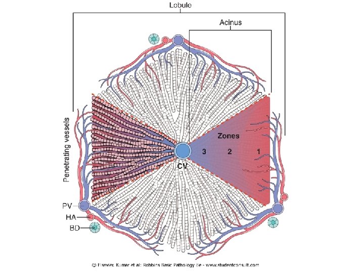
- Net wt. 1400 – 1600 gm - Blood supply: Portal v : 60 - 70% Hepatic a : 30 - 40% (2. 5% of body wt) - Microstructure - Hexagonal lobules → 6 acini - Acinus is divided into 3 zones: 1 -Zone 1 Periportal areas – closet to the vascular supply 2 -Zone 3 Pericentral area 3 -Zone 2 Inrermediate bet. Zone 1&2 3

Normal liver 5
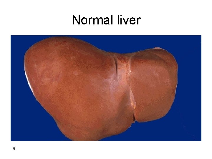
Normal liver 6
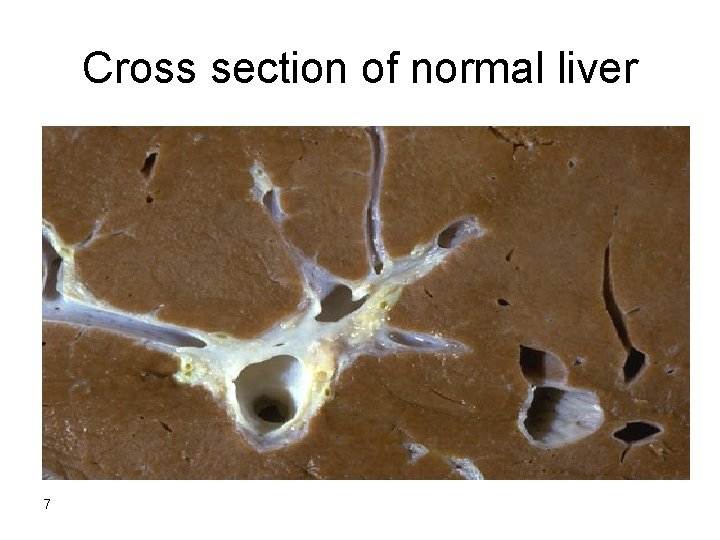
Cross section of normal liver 7
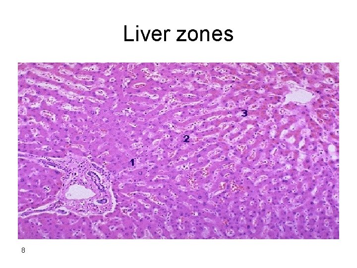
Liver zones 8
The parenbchyma is organized into plates of hepatocytes Hepatocytes are radially oriented around terminal hepatic vein ( central v. ) -Hepatocytes show only minimal variation in the overall size but nuclei may vary in size , number & ploidy esp. with advancing age -Vascular sinusoids present bet. cords of hepatocytes 9
Hepatic injury 1 -Inflammation (Hepatitis) 2 -Degeneration : ballooning degeneration feathery degeneration: retained biliary material accumulation of iron , copper 10
3 -Steatosis ( fatty change) microvesicular: ALD Reye syndrome acute fatty change of pregnancy macrovesicular: DM obesity 11

Fatty change 12
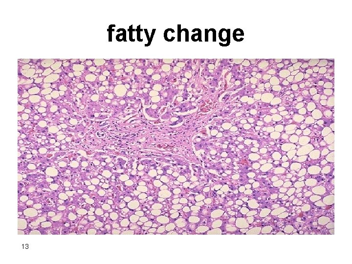
fatty change 13
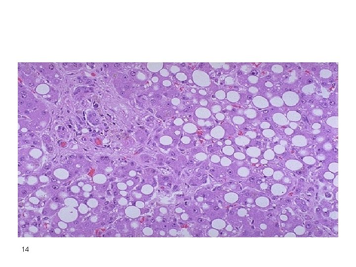
14
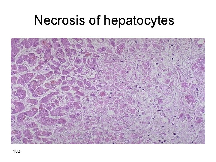
4 -Necrosis - Depending on the type: Coagulative necrosis Councilman bodies Lytic necrosis - Depending on the cause Ischemic Toxic 15
- Depending on location Centrilobular necrosis: Mid zonal : Periportal : interface hepatitis Focal: Piece meal necrosis bridging necrosis Diffuse: massive & submassive necrosis 16
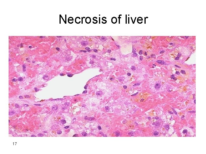
Necrosis of liver 17
5 -Regeneration -evidenced by increased mitosis or cell cycle markers. -the cells of the canal of Hering are the progenitor for hepatocytes & bile duct cells (oval cells ). 18
6 -Fibrosis bridging fibrosis 7 -Cirrhosis micronodular Macronodular 8 -Ductular proliferation 19
CLINICAL SYNDROMES • The major clinical syndromes of liver disease are: • 1 -hepatic failure • 2 -cirrhosis • 3 -portal hypertension • 4 -cholestasis. 20
liver failure • The alterations that cause liver failure fall into 3 categories: • 1 - Acute liver failure with massive hepatic necrosis • 2 - Chronic liver disease • 3 - Hepatic dysfunction without overt necrosis. 21
1 -Acute liver failure. • This is most often caused by drugs or fulminant viral hepatitis. • Acute liver failure denotes clinical hepatic insufficiency that progresses from onset of symptoms to hepatic encephalopathy within 2 to 3 weeks. • A course extending as long as 3 months is called subacute failure. 22
• The histologic correlate of acute liver failure is massive hepatic necrosis. • It is an uncommon but life-threatening condition that often requires liver transplantation. 23
2 -Chronic liver disease • This is the most common route to hepatic failure and is the end point of relentless chronic liver damage ending in cirrhosis. 24
3 -Hepatic dysfunction without overt necrosis. • Hepatocytes may be viable but unable to perform normal metabolic function: • 1 - Acute fatty liver of pregnancy (which can lead to acute liver failure a few days after onset) • 2 - Tetracycline toxicity • 3 - Reye syndrome 25
Clinical features 1 -Jaundice 2 -Hypoalbuminemia →edema 3 -Hyperammonemia 4 -hyperestrogenemia 5 -Spider angiomas 6 -Hypogonadism & gynecomastia 26
Complications: 1 -Multiple organ failure e. g lung 2 -Coagulopathy → bleeding def. factors II, VII, IX, X 3 -Hepatic encephalopathy 4 -Hepatorenal Syndrome 27
Alcoholic liver disease -Alcohol is most widely abused agent -Excessive ethanol consumption causes more than 60% of chronic liver disease in most Western countries and accounts for 40 -50% of deaths due to cirrhosis. -It is the 5 th leading cause of death in USA due to : 1. Accident 2. Cirrhosis 28
Pathogenesis • Short-term ingestion of as much as 80 gm of ethanol/d (8 beers or 7 ounces of 80 -proof liquor) generally produces mild reversible hepatic changes. • Chronic intake of 50 to 60 gm/day is considered a borderline risk for severe injury. • Women seem to be more susceptible to hepatic injury than are men because of low gastric metabolism of ethanol and differences in body composition. 29
-80– 100 mg/dl is the legal definition for driving under the influence of alcohol -44 ml of ethanol is required to produce this level in 70 kg person -In occasional drinkers, bl. Level of 200 mg/dl produces coma & death & resp. failure at 300 -400 mg/dl 30
- Habitual drinkers can tolerate levels up to 700 mg/dl without clinical effect due to metabolic tolerance explained by 5 -10 X induction of cytochrome P-450 system that includes enzyme CYP 2 E 1 which increases the metabolism of ethanol as well as other drugs as cocaine & acetominophen. 31

Forms of alcoholic liver disease 1 -Hepatic steatosis (90 -100% of drinkers) 2 -Alcoholic hepatitis ( 1 - 35% of drinkers) 3 -Cirrhosis ( 14% of drinkers) - Steatosis & hepatitis may develop independently 32

Hepatic steatosis -Can occur following even moderate intake of alcohol in form of microvesicular steatosis - initially centrilobular but in severe cases it may involve the entire lobule. -Chronic intake → diffuse steatosis -Liver is large ( 4 – 6 kg) soft yellow & greasy -Continued intake →fibrosis -Fatty change is reversible with complete absention from further intake of alcohol 33
Alcoholic hepatitis Characteristic findings : 1 -Hepatocyte swelling & necrosis -Accumulation of fat & water & proteins -Cholestasis -Hemosiderin deposition in hepatocytes & kupffer cells 2 -Mallory-hayline bodies -eosinoplilic cytoplasmic inclusions in degenerating hepatocytes formed of cytokeratin infermediate filaments & other proteins 34
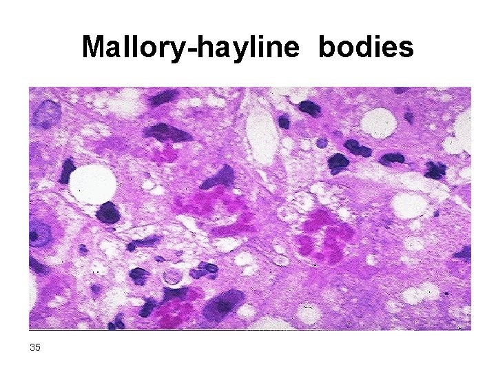
Mallory-hayline bodies 35
- Mallory-hayline inclusions are characteristic but not pathognomonic of alcoholic liver disease, they are also seen in : 1 -Primary biliary cirrhosis 2 -Wilson disease 3 -Chronic cholestatic syndromes 4 -Hepatocellular carcinoma 36
3 -Neutrophilic reaction 4 -Fibrosis -Sinusoidal & perivenular fibrosis -Periportal fibrosis 5 -Cholestasis 6 -Mild deposition of hemosiderin in hepatocytes & kupffer cells 37
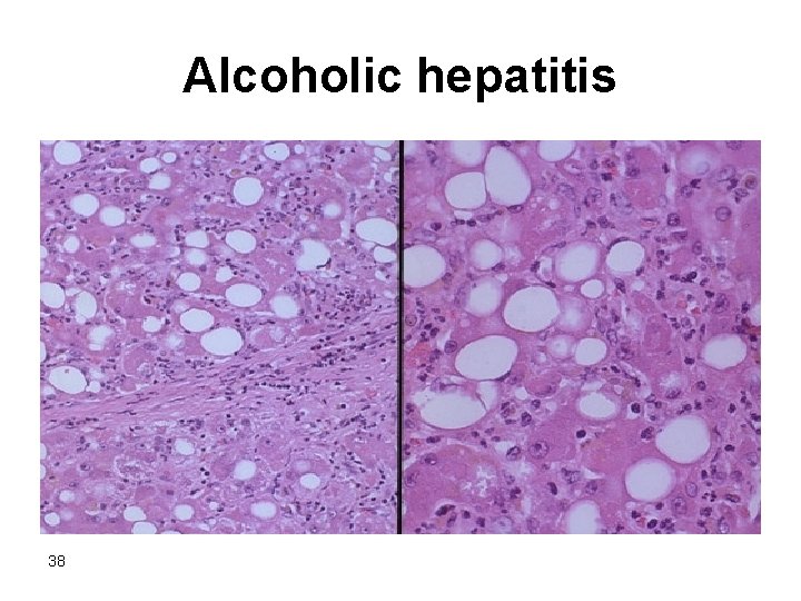
Alcoholic hepatitis 38
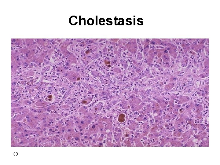
Cholestasis 39
Alcoholic cirrhosis -Usually it develops slowly -Initially the liver is enlarged yellow but over years it becomes brown shrunken non-fatty organ s. t < l kg in wt. -Micronodular → mixed micro & macronodular -Laennec cirrhosis = scar tissue -Bile stasis -Mallory bodies are only rarely evident at this stage -Irreversible -It can develop rapidly in the presence of alcoholic hepatitis (within 1 -2 yrs). 40
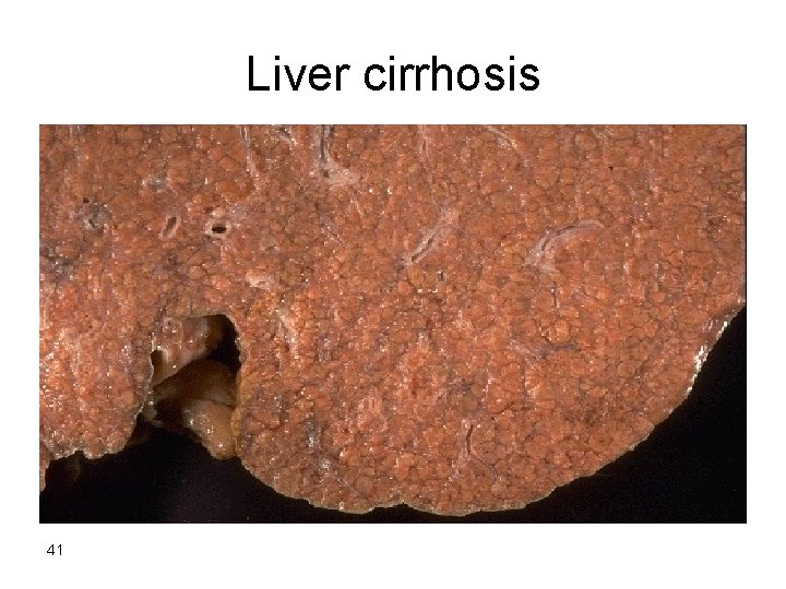
Liver cirrhosis 41
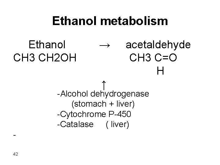
Ethanol metabolism Ethanol → acetaldehyde CH 3 CH 2 OH CH 3 C=O H ↑ -Alcohol dehydrogenase (stomach + liver) -Cytochrome P-450 -Catalase ( liver) 42
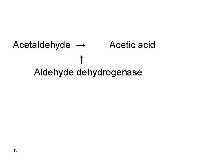
Acetaldehyde → Acetic acid ↑ Aldehyde dehydrogenase 43
- After absorption ethanol is distributed as Acetic acid in all tissues & fluid in direct proportion to blood level - Women have lower levels of gastric alcohol dehydrogenase activity than men & they may develop higher blood Levels than men after drinking the same quantity of ethanol. 44
- Less than 10% of absorbed ethanol is excreted unchanged in urine , sweat & breathe -There is genetic polymorphism in aldehyde dehydrogenase that affect ethanol metabolism e. g 50% of chinese , vietnamase & Japanese have lowered enzyme activity due to point mutation of the enzyme. → accumulation of acetaldehyde → facial flushing, tachycardia & hyperventilation. 45
Mechanism of ethanol toxicity 1 -Fatty change a-Shunting of lipid catabolism toward lipid bio-synthesis due to excess production of NADH over NAD in cystol & mitochondria b-Acetaldehyde forms adducts with tubulin & ↓ function of microtubules → ↓ in lipoprotein transport from liver c- ↑ peripheral catabolism of fat → ↑ FFA delivery to the liver d- ↓ sec. of lipoproteins from hepatocytes e. ↓ oxidation of FFA by mitochondria 2 -Induction of cytochrome P-450 enhances the metabolism of drugs to toxic metabolites (e. g acetominophen ) 46
3. ↑free radicals production due to activation of cytochrome P-4 so leads to membrane & protein damage 4. Alcohol directly affect microtubular & mitochondrial function & membrane fluidity 5. Acetaldehyde causes lipid peroxidation & antigenic alteration of hepatocytes → immune attack 6. Superimposed HCV infection causes acceleration of liver injury (HCV hepatitis occurs in 30% of alcoholics ) 47

7. Alcohol → release of bacterial endotoxins into portal circulation from the gut → inflammation of the liver 8. Alcohol → regional hypoxia in the liver due to release of endothelins which are potent vasoconstrictors → ↓ hepatic sinusoidal perfusion 9. Alteration of cytokine regulation TNF is a major effector of injury IL 6 IL 8 IL 18 48
Clinical features -Hepatic steatosis ( reversible ) • ↑ liver enz. • Severe hepatic dysfunction is unusual -Alcoholic hepatitis • 15 -20 yr. of excessive drinking • Non-specific symptoms, malaise, anorexia, wt. loss • Hepatosplenomegaly • ↑ LFT Each bout of hepatitis → 10 -20% risk of death → cirrhosis in 1/3 in few yrs. -Cirrhosis Portal hypertension 49
• Causes of death in alcoholic liver disease: 1 -hepatic failure 2 -Massive GI bleeding 3 -Infections 4 -Hepatorenal syndrome 5 -HCC in 3 -6% of cases 50
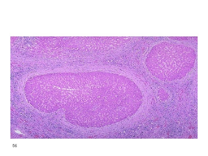
Cirrhosis • It is a diffuse process characterized by fibrosis & the conversion of liver parenchyma into nodules 51
• Main characteristics 1. Bridging fibrous septae 2. Parenchymal nodules encircled by fibrotic bands 3. Diffuse architecture disruption 52
• Types : Micronodules < 3 mm in diameter Macronodules > 3 mm in diameter 53
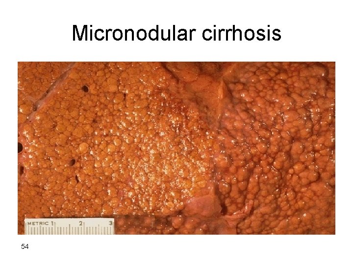
Micronodular cirrhosis 54
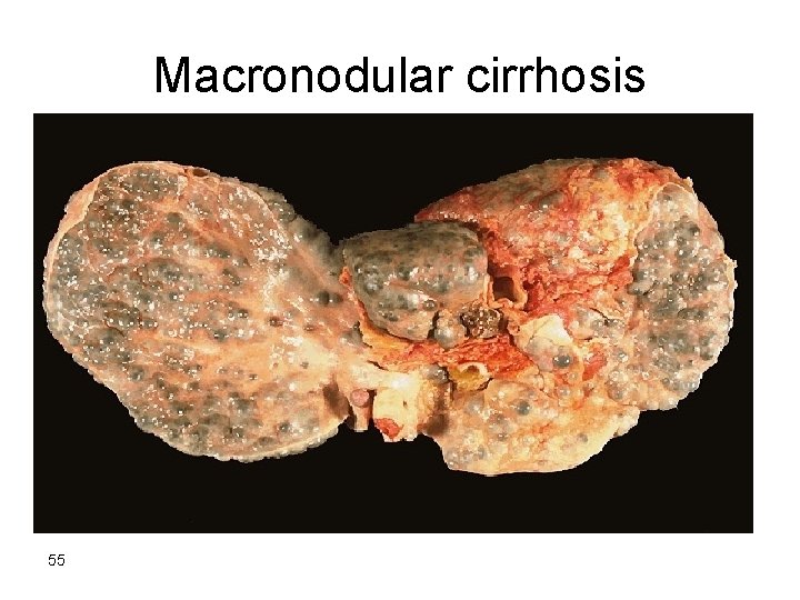
Macronodular cirrhosis 55
56
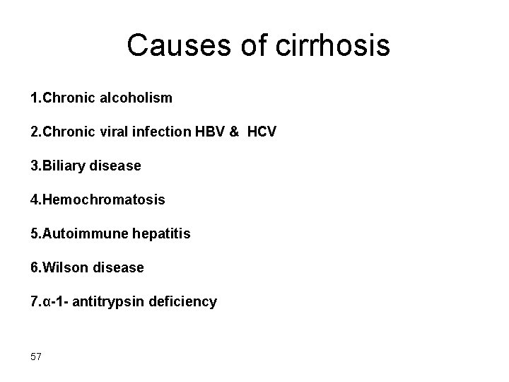
Causes of cirrhosis 1. Chronic alcoholism 2. Chronic viral infection HBV & HCV 3. Biliary disease 4. Hemochromatosis 5. Autoimmune hepatitis 6. Wilson disease 7. α-1 - antitrypsin deficiency 57
8. Rare causes Galactosemia Tyrosinosis Glycogen storage disease III &IV Lipid storage disease Hereditary fructose intolerance Drug induced e. g methyldopa 9. Cryptogenic cirrhosis 10% 58
Pathogenesis of cirrhosis -The mechanism of cirrhosis involves: 1 -Hepatocellular death 2 -Regeneration 3 -Progressive fibrosis 4 -Vascular changes 59
• The development of cirrhosis requires that cell death occur over long periods of time and be accompanied by fibrosis. • Fibrosis progresses to scar formation when the injury involves not only the parenchyma but also the supporting connective tissue. 60
-In normal liver the ECM collagen (types I, III, V & XI) is present only in : Liver capsule Portal tracts Around central vein 61
-delicate framework of type IV collagen & other proteins lies in space of Disse. -In cirrhosis types I & III collagen & others are deposited in the space of Disse. 62
• Vascular changes consisting of the loss of sinusoidal endothelial cell fenestrations and the development of portal vein-hepatic vein and hepatic artery-portal vein vascular shunts contribute to defects in liver function. 63
• Collagen deposition converts sinusoids with fenestrated endothelial channels that allow free exchange of solutes between plasma and hepatocytes to higher pressure fast-flowing vascular channels without such solute exchange. 64
• The movement of proteins (e. g. , albumin, clotting factors, lipoproteins between hepatocytes and the plasma is markedly impaired. • These functional changes are aggravated by the loss of microvilli from the hepatocyte surface which diminishes the transport capacity of the cell. 65
- The major source of collagen in cirrhosis is the perisinusoidal stellate cells (Ito cells) which lie in space of Disse - Ito cells are perisinusoidal stellate cells act normally as storage cells for vit A & fat. 66
• Activated stellate cells produce growth factors, cytokines, and chemokines that cause their further proliferation and collagen synthesis. • TGF-β is the main fibrogenic agent for stellate cells. 67
-The stimuli for the activation of stellate cells & production of collagen are : 1 -reactive oxygen species 2 -Growth factors 3 -cytokines TNF, IL-I, lymphotoxins 68
-Clinical features of cirrhosis : -Silent -Anorexia, wt loss, weakness -Complications : 1 -Progressive hepatic failure 2 -Portal hypertension 3 -Hepatocellular carcinoma 69
Portal hypertension - ↑ resistance to portal blood flow at the level of sinusoids & compression of central veins by perivenular fibrosis & parenchymal nodules - Arterial – portal anastomosis develops in the fibrous bands →increase the blood pressure in portal venous system 70
Causes of portal hypertension I. Prehepatic 1 -Portal vein thrombosis 2 -Massive splenomegaly II. Post hepatic 1 -Severe Rt. - sided heart failure 2 -Constrictive pericarditis 3 -Hepatic vein out flow obstruction III. Hepatic 1 -Cirrhosis 2 -Schistosomiasis 3 -Massive fatty change 4 -Diffuse granulomatosis as sarcoidosis, TB 5 -Disease of portal microcirculation as nodular regenerative hyperplasia 71
Clinical consequence of portal hypertension 1 -Ascites 2 -Portosystemic shunts 3 -Hepatic encephalopathy 4 -Splenomegaly 72
Ascites -Collection of excess fluid in peritoneal cavity -It becomes clinically detectable when at least 500 ml have accumulated -Features 1 -Serous fluid 2 -Contains as much as 3 g/ml of protein (albumin) 3 -It has the same concentration as blood of glucose, Na+, & K+ 4 -Mesothelial cells & lymphocytes 5 -Neutrophils = infection 6 -RBCs = DISSEMINATED CANCR 73
-Pathogenesis 1 -Sinusoidal ↑ Bp 2 -Hypoalbuminemia 3 -Leakage of hepatic lymph into the peritoneal cavity N- thoracic duct lymph flow is 800 -1000 ml/d in cirrhosis it may approach 20 L /day 4 -Renal retention of Na+ & water due to 2 ry hyperaldosteronism 74
Portosystemic shunt -Because of ↑portal venous pressure bypasses develop wherever the systemic & portal circulation share capillary beds. -Sites: 1 -Around & within the rectum (Hemorrhoids) 2 -Gastroesophageal junction (varicies ) 3 -Retroperitoneum 4 -Falciform ligament of the liver (periumbilical & abdominal wall collaterals ) → caput medusae 75
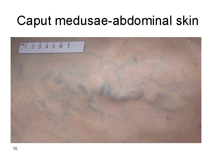
Caput medusae-abdominal skin 76
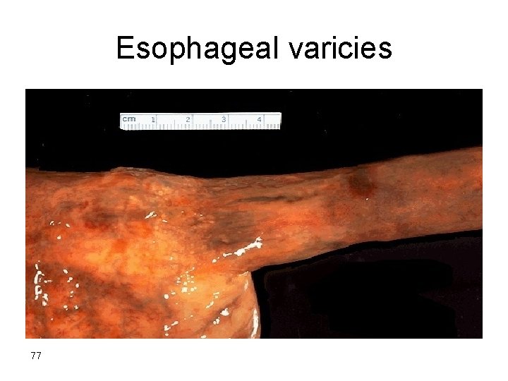
Esophageal varicies 77
- Gastroesophageal varicies appear in 65% of pts. with advanced cirrhosis & cause death in 50% of then due to UGI bleeding.
Splenomegaly -Usu. 500 -1000 gms (N <300 gms) -Not necessarily correlated with other features of portal ↑Bp -May result in hypersplenism 79

Splenomegaly 80
Hepatic encephalopthy -It is a complication of acute & chronic hepatic failure -Disturbance in brain function ranging from behavioural changes to marked confusion & sutpor to deep coma & death -The changes may progress over hrs. or days 81
-Neurological signs: Rigidity Hyper-reflexia Non – specific EEG Seizures Asterixis ( non-rhythmic rapid extension flexion movements of head & extremities). -Brain shows edema & astrocytic reaction
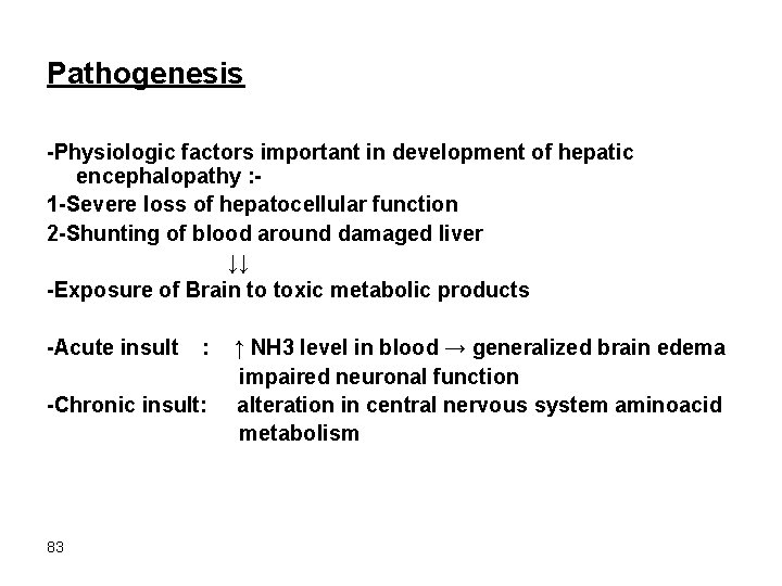
Pathogenesis -Physiologic factors important in development of hepatic encephalopathy : 1 -Severe loss of hepatocellular function 2 -Shunting of blood around damaged liver ↓↓ -Exposure of Brain to toxic metabolic products -Acute insult : -Chronic insult: 83 ↑ NH 3 level in blood → generalized brain edema impaired neuronal function alteration in central nervous system aminoacid metabolism
Hepatorenal Syndrome • appears in individuals with severe liver disease. • consists of the development of renal failure without primary abnormalities of the kidneys themselves. 84
• Excluded by this definition are concomitant damage to both liver and kidney, as may occur with exposure to CCL 4 and certain mycotoxins and the copper toxicity of Wilson disease. • Also excluded are instances of advanced hepatic failure in which circulatory collapse leads to acute tubular necrosis & acute renal failure. 85
• Kidney function promptly improves if hepatic failure is reversed. • The exact cause is unknown. • Systemic vasoconstriction leading to severe reduction of renal blood flow particularly to the cortex. 86
• Onset of this syndrome is typically by a drop in urine output associated with rising BUN and creatinine values. • The renal failure may increase the risk of death in the patient with acute fulminant or advanced chronic hepatic disease. 87
Drug – lnduced liver disease -Drug reactions: 1 -Predictable (intrinsic) 2 -Unpredictable (idiosyncratic) 88
• Predictable drug reactions may occur in anyone who accumulates a sufficient dose (dose-dependent). • Unpredictable reactions depend on idiosyncrasies of the host: • 1 -the host's propensity to mount an immune response to the antigenic stimulus. • 2 -the rate at which the host metabolizes the agent.

• The injury may be immediate or take weeks to months to develop. • drug-induced chronic hepatitis is clinically and histologically indistinguishable from chronic viral hepatitis or autoimmune hepatitis and hence serologic markers of viral infection are critical for making the distinction.
Predictable drugs: Acetaminophen Tetracycline Antineoplastic agents CCL 4 Alcohol Unpredictable drugs Chlorpromazine Halothane Sulfonamides Methyldopa Allopurinol
-Mechanism of drug injury : 1 -Direct toxic damage e. g acetaminophen CCl 4 mushroom toxins 2 -Immune-mediated damage 92
-Patterns of injury 1 -Hepatocellular necrosis 2 -Cholestasis 3 -Steatosis 4 -Steatohepatitis 5 -Fibrosis 6 -Vascular lesions 7 -Granuloma 8 -Neoplasms benign & malignant 93
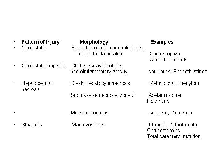
• Pattern of Injury Morphology Examples • Cholestatic Bland hepatocellular cholestasis, without inflammation Contraceptive Anabolic steroids • Cholestatic hepatitis Cholestasis with lobular necroinflammatory activity Antibiotics; Phenothiazines • Hepatocellular Spotty hepatocyte necrosis Methyldoya, Phenytoin necrosis Submassive necrosis, zone 3 Acetaminophen Halothane • Massive necrosis Isoniazid, Phenytoin • Steatosis Macrovesicular Ethanol, Methotrexate Corticosteroids Total parenteral nutrition
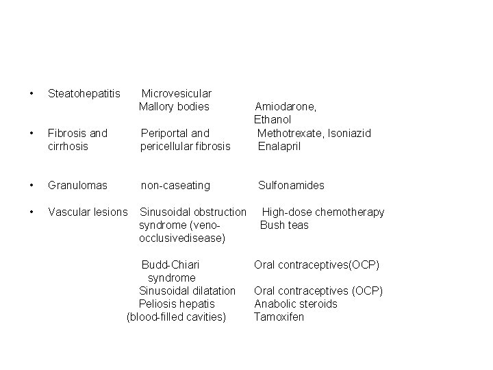
• Steatohepatitis Microvesicular Mallory bodies Amiodarone, Ethanol • Fibrosis and Periportal and Methotrexate, Isoniazid cirrhosis pericellular fibrosis Enalapril • Granulomas non-caseating Sulfonamides • Vascular lesions Sinusoidal obstruction High-dose chemotherapy syndrome (veno- Bush teas occlusivedisease) Budd-Chiari Oral contraceptives(OCP) syndrome Sinusoidal dilatation Oral contraceptives (OCP) Peliosis hepatis Anabolic steroids (blood-filled cavities) Tamoxifen
• Neoplasms Hepatic adenoma OCP Anabolic steroids HCC Thorotrast Cholangiocarcinoma Thorotrast Angiosarcoma Thorotrast, Vinyl chloride

Drugs that may cause acute liver failure 1 -Acetaminophen 2 -Halothane 3 -Antituberculosis drugs (rifampin, isoniazid) 4 -Antidepressant monoamine oxidase inhibitors 5 -Toxins as CCL 4 & mushroom poisoning 97
• The most common cause (46% of cases of acute liver failure) is acetaminophen intoxication. • 60% of these are a consequence of accidental overdosage.
Morphology: Massive necrosis → 500 – 700 gm liver Submassive necrosis Patchy necrosis 99
• Patient survival for more than a week permits regeneration of surviving hepatocytes. • Regeneration is initially in the form of strings of ductular structures which mature into hepatocytes. • If the parenchymal framework is preserved liver architecture is restored. • With massive destruction of lobules leads to formation of nodular masses of liver cells. • Scarring may occur in patients with a protracted course of submassive or patchy necrosis representing a route for developing so-called macronodular cirrhosis
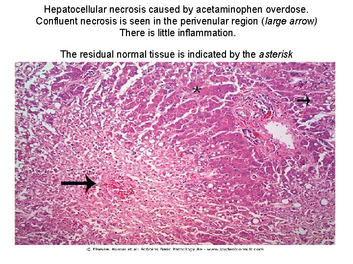
Hepatocellular necrosis caused by acetaminophen overdose. Confluent necrosis is seen in the perivenular region (large arrow) There is little inflammation. The residual normal tissue is indicated by the asterisk
Necrosis of hepatocytes 102
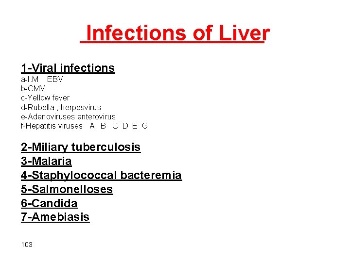
Infections of Liver 1 -Viral infections a-I. M EBV b-CMV c-Yellow fever d-Rubella , herpesvirus e-Adenoviruses enterovirus f-Hepatitis viruses A B C D E G 2 -Miliary tuberculosis 3 -Malaria 4 -Staphylococcal bacteremia 5 -Salmonelloses 6 -Candida 7 -Amebiasis 103
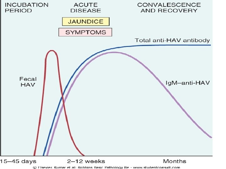
Hepatitis A virus • Hepatitis A ("infectious hepatitis") is a benign, self-limited disease. • incubation period of 15 to 50 days (average 28 days). • HAV does not cause chronic hepatitis or a carrier state and only rarely causes fulminant hepatitis. • Fatality rate is 0. 1%
-Transmission : Feco-oral rout -Endemic in developing countries with low hygiene & sanitation → anti-HAV Abs by the age of 10 yrs. → 50% by the age of 50 yrs.
-Clinically the disease is mild to asymptomatic affecting children of school age & rare thereafter -The virus is shed in bile & feces -The virus is shed is the stool 2 -3 wks before & 1 wk after the onset of jaundice -HAV is not shed in saliva, urine, or semen -HAV viremia is transient & b. I. Donors are not screened for the virus 106
• Waterborne epidemics may occur in developing countries where people live in overcrowded, unsanitary conditions. • Among developed countries, sporadic infections may be contracted by the consumption of raw or steamed shellfish (oysters, mussels, clams), which concentrate the virus from seawater contaminated with human sewage. • Ingestion of raw green onions contaminated with HAV caused outbreaks of the disease in the United States in 2003
Serelogic dx Anti HAV Ig. M: at the onset of symptoms → ↓ in few months Anti HAV Ig. G: appears later & persists for life -HAV vaccine is effective
Hepatitis B Virus • carrier rate of approximately 400 million. • About 80% of all chronic carriers live in Asia and the Western Pacific rim, where prevalence of chronic hepatitis B is more than 10%. • In the United States there approximately 185, 000 new infections per year.
-HBV is a hardy virus can withstand extremes of temperature & humidity -Prolonged IP 4 -26 wks -Prolonged viremia HBV remains in blood during the last stages of incubation period and during active episodes of acute and chronic hepatitis -Present in all body fluids as tears, saliva, sweat, breast milk, vaginal sec. , semen & pathological body fluids except stool 111
• vertical transmission from mother to child during birth constitutes the main mode of transmission. • horizontal transmission via: • 1 - transfusion • 2 - blood products • 3 - dialysis • 4 - needle-stick accidents among health care workers • 5 -IV drug abuse • 6 -sexual transmission (homosexual or heterosexual) • 7 -In 1/3 of patients the source of infection is unknown.
• HBV infection in adults is mostly cleared, but vertical transmission produces a high rate of chronic infection.
-Phases of infection : 1. Proliferative phase 2. Integrative phase 114
HBV antigens : 1. HBc Ag(hepatitis B core antigen) - hepatocytes 2. HBe Ag(pre-core protein) -blood 3. HBs Ag -blood -hepatocytes 4. DNA polymerase (HBV-DNA) (reverse transcriptase activity) 5. HBx protein ( transcriptional transactivator ) required for viral infectivity and may have a role in the causation of hepatocellular carcinoma by regulating p 53 degradation and expression 115
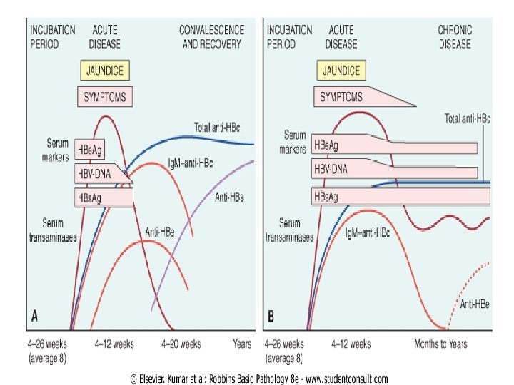
• HBs. Ag appears before the onset of symptoms, peaks during overt disease, and then declines to undetectable levels in 3 to 6 months. • Anti-HBs antibody does not rise until the acute disease is over and is usually not detectable for a few weeks to several months after the disappearance of HBs. Ag. • Anti-HBs may persist for life conferring protection • HBV-DNA, and DNA polymerase appear in serum soon after HBs. Ag, and all signify active viral replication
• Persistence of HBe. Ag is an important indicator of continued viral replication, infectivity, and probable progression to chronic hepatitis. • The appearance of anti-HBe Abs shortly after the disappearance of HBe. Ag indicates the end of the infection. • Ig. M anti-HBc becomes detectable in serum shortly before the onset of symptoms • Over a period of months the Ig. M anti-HBc antibody is replaced by Ig. G anti-HBc.
• Anti – HBs Ig. G: rise after the acute phase is over & remains detectable after wks or months after disappearance of HBs. Ag • Hepatitis B can be prevented by vaccination and by the screening of donor blood, organs, and tissues 118
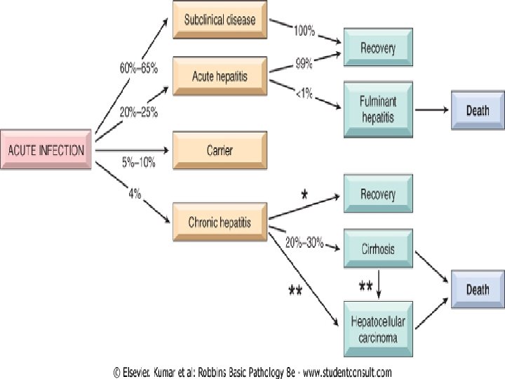
Clinical syndromes associated with HBV infection 1 -Acute hepatitis with recovery 2 -Nonprogressive chronic hepatitis 3 -Progressive chronic hepatitis ending in cirrhosis 4 -Fulminant hepatitis with massive liver necrosis 5 -Asymptomatic carrier state 120
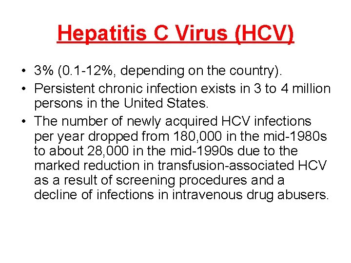
Hepatitis C Virus (HCV) • 3% (0. 1 -12%, depending on the country). • Persistent chronic infection exists in 3 to 4 million persons in the United States. • The number of newly acquired HCV infections per year dropped from 180, 000 in the mid-1980 s to about 28, 000 in the mid-1990 s due to the marked reduction in transfusion-associated HCV as a result of screening procedures and a decline of infections in intravenous drug abusers.
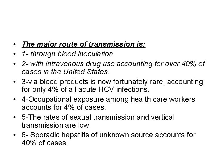
• The major route of transmission is: • 1 - through blood inoculation • 2 - with intravenous drug use accounting for over 40% of cases in the United States. • 3 -via blood products is now fortunately rare, accounting for only 4% of all acute HCV infections. • 4 -Occupational exposure among health care workers accounts for 4% of cases. • 5 -The rates of sexual transmission and vertical transmission are low. • 6 - Sporadic hepatitis of unknown source accounts for 40% of cases.
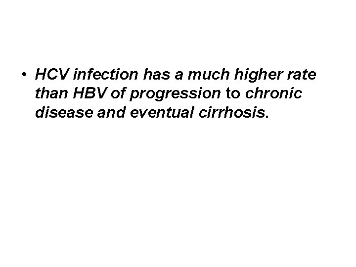
• HCV infection has a much higher rate than HBV of progression to chronic disease and eventual cirrhosis.
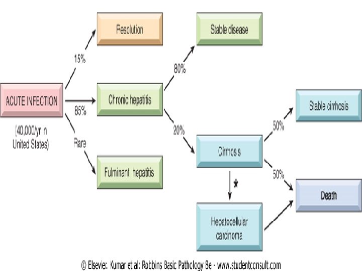
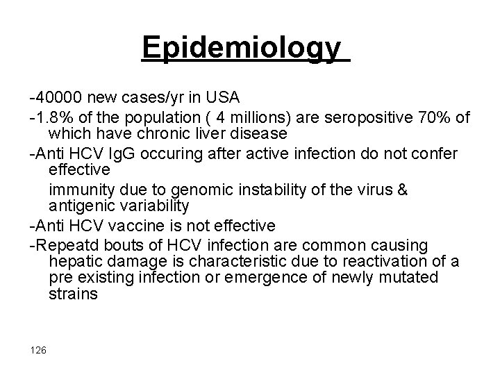
Epidemiology -40000 new cases/yr in USA -1. 8% of the population ( 4 millions) are seropositive 70% of which have chronic liver disease -Anti HCV Ig. G occuring after active infection do not confer effective immunity due to genomic instability of the virus & antigenic variability -Anti HCV vaccine is not effective -Repeatd bouts of HCV infection are common causing hepatic damage is characteristic due to reactivation of a pre existing infection or emergence of newly mutated strains 126
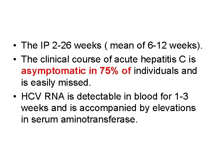
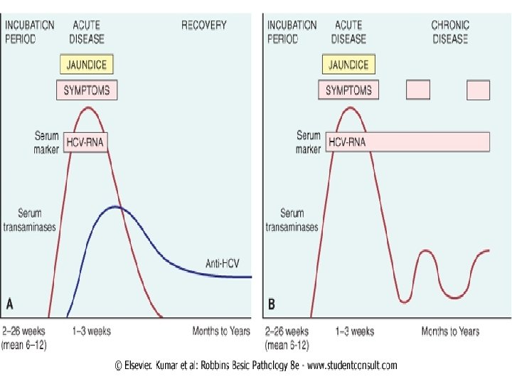
• The IP 2 -26 weeks ( mean of 6 -12 weeks). • The clinical course of acute hepatitis C is asymptomatic in 75% of individuals and is easily missed. • HCV RNA is detectable in blood for 1 -3 weeks and is accompanied by elevations in serum aminotransferase.
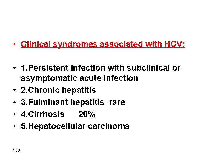
• Clinical syndromes associated with HCV: • 1. Persistent infection with subclinical or asymptomatic acute infection • 2. Chronic hepatitis • 3. Fulminant hepatitis rare • 4. Cirrhosis 20% • 5. Hepatocellular carcinoma 128
Serological diagnosis • HCV RNA is detectable in bl. For 1 – 3 wks peak coincides with ↑ in serum transaminases • Anti HCV Abs detected in 50 – 70% of patients during symptomatic acute infection • In 30 – 50% of patients the anti HCV Abs emerge after 3 – 6 wks • In chronic HCV infection circulating HCV-RNA persists despite the presence of Abs in many patients ( > 90%) 129
Hepatitis D Virus -Hepatitis delta virus -Replication defective virus -Causes infection only when it is encapsulated by HBs. Ag -I. P 4 – 7 wks in superinfection 131
• 8% among HBs. Ag carriers in southern Italy to as high as 40% in Africa and the Middle East. • HDV infection is uncommon in Southeast Asia and China, areas in which HBV infection is endemic. • In the United States HDV infection is largely restricted to drug addicts and individuals receiving multiple transfusions (e. g. hemophiliacs who have prevalence rates of 1% to 10%).
• Delta hepatitis arises in two settings: • (1) acute coinfection after exposure to serum containing both HDV and HBV • (2) superinfection of a chronic carrier of HBV with a new inoculum of HDV. • Most coinfected individuals can clear the viruses and recover completely. • in superinfected individuals there is an acceleration of hepatitis, progressing to more severe chronic hepatitis 4 to 7 weeks later.
• Routes of transmission: • Parenteral (close personal contact)
• HDV Ag are detectable in the blood and liver just before and in the early days of acute symptomatic disease. • Ig. M anti-HDV antibody is the most reliable indicator of recent HDV exposure, but its appearance is transient. • acute coinfection by HDV and HBV is best indicated by detection of Ig. M against both HDV Ag and HBc. Ag • With HDV superinfection, HBs. Ag is present in serum; and anti-HDV antibodies (Ig. M and Ig. G) persist in low titer for months or longer.
Serologic diagnosis. HDV-RNA is detectable in blood & liver just prior to & in early days of acute symptomatic disease . Anti HDV Ig. M = recent HDV infection. Anti HDV Ig. M appears late & freq. short-lived. Coinfection : Ig. M against HDV Ag & HBV Ag. Superimposed infection: anti HDV Ig. M & HBs. Ag 136
Hepatitis E virus • HEV hepatitis is an enterically transmitted, waterborne infection occurring primarily beyond the years of infancy. • HEV is endemic in India • Prevalence rates of anti-HEV Ig. G antibodies approach 40% in the Indian population. • Sporadic infection seems to be uncommon & occurs mainly in travelers and accounts for more than 50% of cases of sporadic acute viral hepatitis in India.
- Water-borne infection Young – middle – aged adults Rare in children Endemic infection in India, Africa, Mexico…… Sporadic infection is uncommon & occurs mainly in travelers - Self-limiting mild disease except in pregnant women with high mortality rate (20%) - I. P: 6 wks ( range 2 -8 wks) - No chronic liver disease or carrier state 138
Serelogy -HEV-RNA can be detected in stool & liver before the onset of clinical symptoms -Anti HEV-Ig. M appears during acute illness & replaced by Ig. G when symptoms resolve (ie in 2 – 4 wks) 139
Clinicopathologic Syndromes 1 -Acute asymptomatic : serologic evidence only A B C D E 2 -Acute symptomatic hepatitis icteric or anicteric A B C D E 3 -Chronic hepatitis with or without progression to cirrhosis B &C 4 -Fulminant hepatitis with massive or submassive hepatic necrosis B, D A & C very rare 5 -Chronic carrier state B, C 140
Acute asymptomatic infection with recovery -Minimally ↑ serum tranaminases -HAV & HBV infections are freq. subclinical in childhood period -HCV infection is subclinical in 75% of the cases 141
Acute symptomatic infection with recovery -Can be caused by any hepatotropic viruses although it is uncommon in HCV infection -Phases: 1 -Incubation period 2 -Symptomatic preicteric phase . Malaise. General fatigability. Nausea. Loss of appetite. Fever, headaches, muscle pain, diarrhea. 10% of pts. Develop serum sickness-like synd. esp. with HBV infection (fever, rash, arthralgia ) due to circulating immune complexes 142
3 -Symptomatic icteric phase. Usual in adults but not children with HAV. Absent in 50% of cases of HBV & the majority of HCV. Conj. hyperbilirubinemia, dark colored urine , dark stool, pruritus . Prolonged PT, hyperglobulinemia, ↑ serum alkaline phosphatase 143
• 1 - diffuse swelling (ballooning degeneration) • 2 - cholestasis, with bile plugs in canaliculi and brown pigmentation of hepatocytes. • 3 -Fatty change is mild and is unusual except with HCV infection. • 4 - HBV infection may generate "ground-glass" hepatocytes • a finely granular, eosinophilic cytoplasm shown by electron microscopy to contain massive quantities of HBs. Ag in the form of spheres and tubules. • Other HBV-infected hepatocytes may have "sanded" nuclei, resulting from abundant intranuclear HBc. Ag.
• 5 - patterns of hepatocyte death are seen. • 6 -confluent necrosis of hepatocytes may lead to bridging necrosis • 7 -lobular disarray • 8 -Inflammation.
• 9 - Kupffer cells undergo hypertrophy and hyperplasia and are often laden with lipofuscin pigment caused by phagocytosis of hepatocellular debris. • 10 -The portal tracts are usually infiltrated with a mixture of inflammatory cells. • 11 -interface hepatitis). • 12 -bile duct proliferation
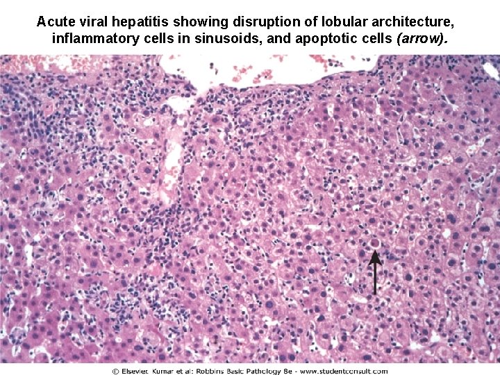
Acute viral hepatitis showing disruption of lobular architecture, inflammatory cells in sinusoids, and apoptotic cells (arrow).
Fulminant hepatitis • Hepatic insufficiency that progresses from onset of symptoms to hepatic escepholopathy in 2 -3 wks • Subfulminant ( up to 3 mon) 148
Causes : 1 -Viral hepatitis 50 – 65% B, C, E HBV 2 x > HCV 2 -Drugs & chemical 25 - 50% e. g Isoniazid , halothane , methyldopa & acetominophen 3 -Obstruction of hepatic vein 4 -Wilson’s disease 5 -Acute fatty change of pregnancy. 6 -Massive tumor infiltration 7 -Reactivation of chronic hepatitis B 8 -Acute immune hepatitis
• Morphology -↓ liver size ( 500 – 700 gm) -Necrosis of hepatocytes -Collapsed reticulin tissue -Inflammatory infillrate -Regenerative activity of hepatocytes -Fibrosis 150
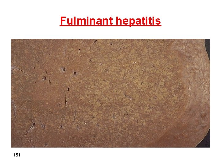
Fulminant hepatitis 151
Chronic Hepatitis • Symptomatic, biochemical or serelogic evidence of continuing or relapsing hepatic disease for more than 6 months with histologically documented inflammation and necrosis. • Progressive or non progressive • HBV , HCV, HBV-HDV. 152
Morphology of chronic hepatitis • Mild to severe 1. Protal inflammation 2. Lymphoid aggregate 3. Necrosis of hepatocytes-councilman bodies 4. Bile duct damage 5. Steatosis 6. Interface hepatitis 7. Bridging necrosis & fibrosis 8. Fibrosis 9. Ground-glass appearance 10. Sanded nuclei 11. Lobular disarray 153
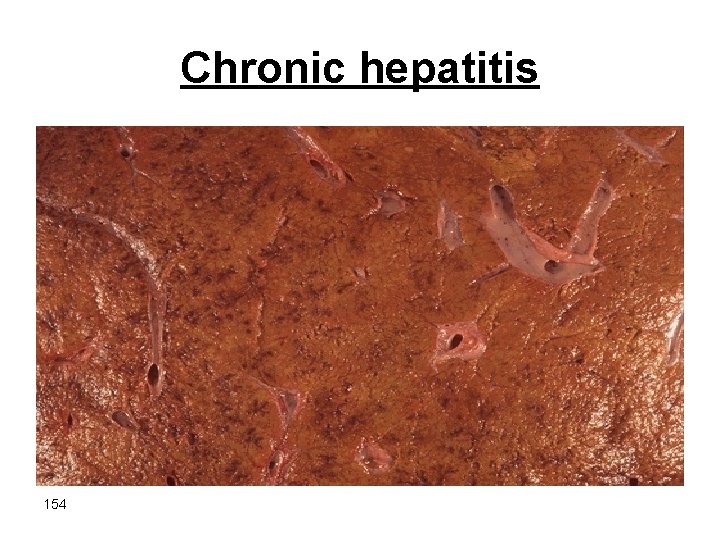
Chronic hepatitis 154

Chronic hepatitis 155
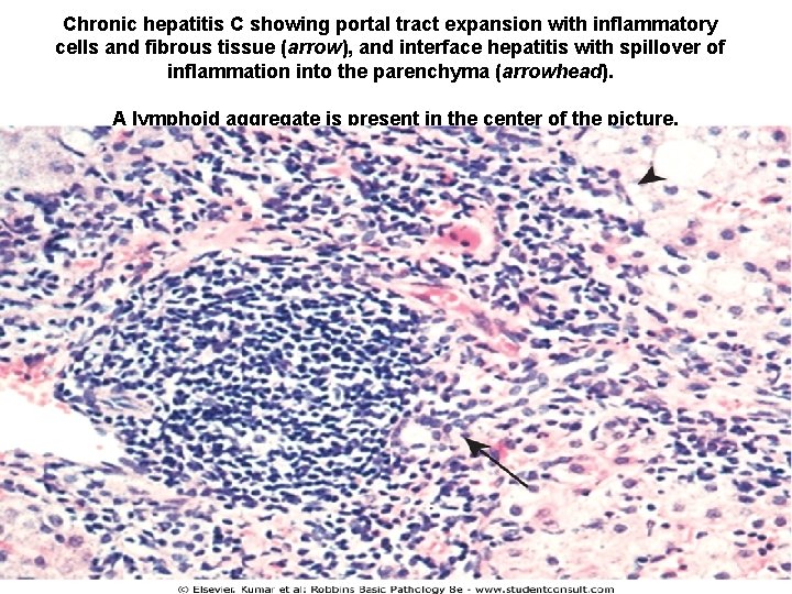
Chronic hepatitis C showing portal tract expansion with inflammatory cells and fibrous tissue (arrow), and interface hepatitis with spillover of inflammation into the parenchyma (arrowhead). A lymphoid aggregate is present in the center of the picture.
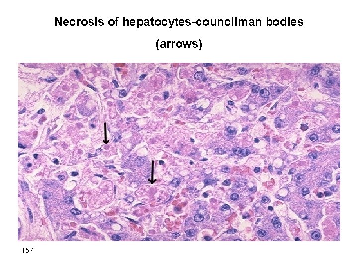
Necrosis of hepatocytes-councilman bodies (arrows) 157
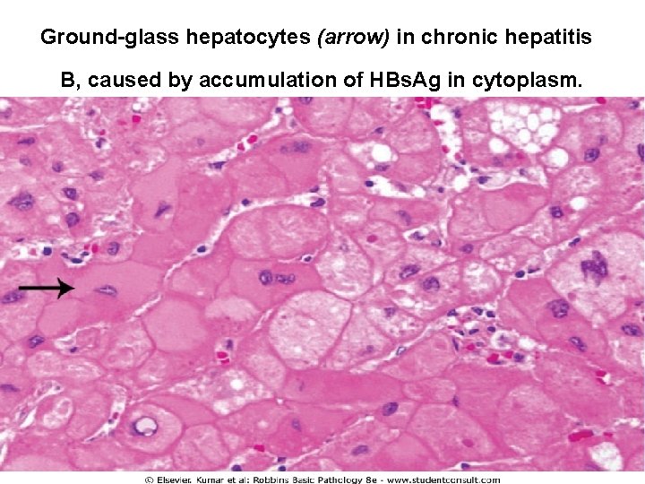
Ground-glass hepatocytes (arrow) in chronic hepatitis B, caused by accumulation of HBs. Ag in cytoplasm.
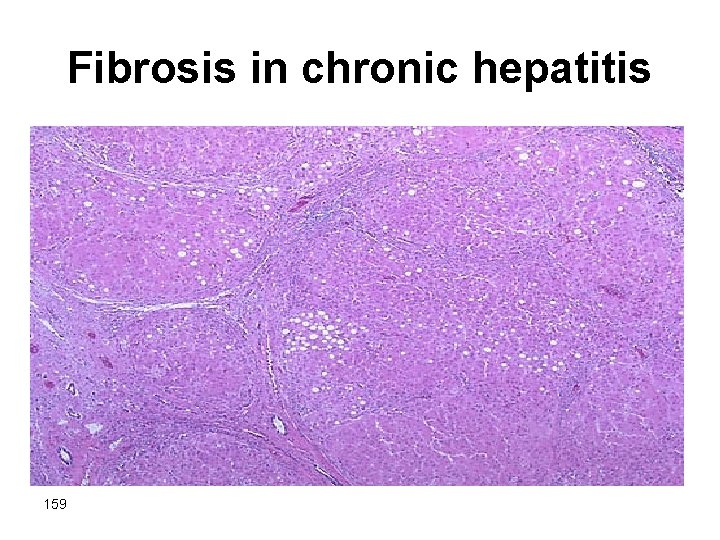
Fibrosis in chronic hepatitis 159
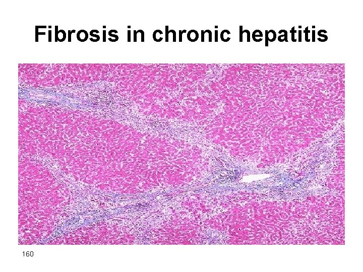
Fibrosis in chronic hepatitis 160
Carrier state • Carriers are: • (1) those who harbor one of the viruses but are suffering little or no adverse effects • (2) those who have nonprogressive liver damage but are essentially free of symptoms or disability • Both constitute reservoirs of infection. 161
Predisposing factors • 1 -HBV infection early in life, particularly through vertical transmission during childbirth, produces a carrier state 90 -95% of the time. • only 1 -10% of HBV infections acquired in adulthood yield a carrier state. • 2 -impaired immunity • 3 -HBV, HCV, ? HDV
- Slides: 162