Caloric Restriction and Resveratrol Reverse a Highfat Diet

, based on low calorie intake, is")


 Ki")
 NC HF CR R 400 R 50 Insulin")


 NC CR HF R 400 R 50")


 NC CR HF R 400 R")


- Slides: 23
Caloric Restriction and Resveratrol Reverse a High-fat Diet Induced Diabetes and Improve Islet β cell Dysfunction in Mice Jiaoyue Zhang Ph. D Endocrinology Department, Union hospital, Huazhong University of Science & Technology, Wuhan 430022, China
Caloric Restriction and Resveratrol n Calorie restriction (CR), based on low calorie intake, is basic for therapy of diabetes n SIRT 1 is the NAD+-dependent deacetylase, and controls the activities of genes that regulate circadian rhythm, apoptosis. n Resveratrol (Res), a type of natural phenol, is the potent natural SIRT 1 activator. It has been found to lower plasma glucose in streptozotocin-induced diabetic rats.
SIRT 1 is involved in the activities of β cell n n n SIRT 1 was preferentially expressed in pancreatic beta cells while not in exocrine cells In beta cell-specific Sirt 1 -overexpressing (BESTO) transgenic mice,GSIS ↑; SIRT 1 reduced by si. RNA in beta cel lines (INS, MIN 6) , GSIS↓ Our hypothesis: SIRT 1 could possibly play an important role in the effect of CR and Res on beta cell Bordone L, et al. PLo. S Biol, 2006, 4: e 31 Moynihan KA, et al. Cell Metab, 2005, 2: 105 -17
Aim of the research n Explore the anti-hyperglycaemic role and mechanisms of CR and Res on beta cell protection
male C 57 BL/6 J mice 15 standard chow × 24 w control 65 High fat diet× 8 w model a high fat diet × 16 w standard chow +60%CR 15只 Normal control NC High fat diet HF Caloric restriction CR a high fat diet +Res 400 mg/kg/d a high fat diet +Res 50 mg/kg/d IPGTT Large dosage of Res R 400 Low dosage of Res R 50 Caloric intake、BW、lipid IPGTT、insulin、ITT Obesity index=(total fat weight)/body weight× 100
Caloric intake Body weight Obesity index Blood FFA #▲ #▲ #▲ *▲ Compared to NC, *P<0. 05,#P<0. 01;compared to HF, △P<0. 05,▲P<0. 01
FBG 8 w IPGTT curve AUC # * 24 w # ▲ ▲ *△ # ▲ ▲ #▲
Fasting blood insulin HOMA-IR # # #▲ #▲ ▲ ▲ Insulin tolerance (ITT) Ki # #▲ #▲ #▲
Islet morphology HE stainig (× 200) NC HF CR R 400 R 50 Insulin protein expressions by immunohistochemistry (× 400) NC HF CR R 400 R 50
Β cell mass Insulin content in pancreas # #△ ▲ *▲ △ △
Insulin stimulatory index ΔI 30/ΔG 30 #▲ #▲ ▲ *▲ #▲ # △ * A B C
Apoptosis by TUNEL (× 200) NC CR HF R 400 R 50
SIRT 1 protein SIRT 1 m. RNA #▲ UCP 2 m. RNA #▲ # △ # Marker NC △ * HF CR R 400 ▲ # * R 50 SIRT 1 #▲ Marker NC HF CR R 400 ▲ NC HF CR R 400 R 50 SIRT 1 UCP 2
PDX-1 m. RNA Bax m. RNA Bcl-2 m. RNA # ▲ ▲ Marker NC HF CR R 400 R 50 PDX-1 GAPDH Marker NC HF CR ▲ R 400 ▲ #▲ #▲ # R 50 GAPDH Bax Marker NC HF CR R 400 R 50 GAPDH Bcl-2
Mitochondrial changes in islets ( electron microscopy ) NC CR HF R 400 R 50
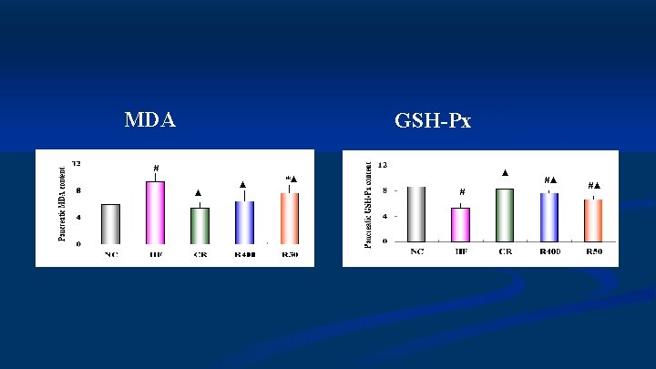
n CR and Res administration rendered the animals resistant to diet-induced obesity and insulin resistance, which was supported by previous studies. Importantly, the functions of GSIS in vivo and in vitro were improved robustly in HR group compared with NC and HF group in our experiment. n Compared with CR,Res does not affect caloric intake and sorts of food, less weight loss, lower blood glucose and less insulin sensitivity.
The role of beta cell protection n Long term CR intervetion can inhibit compensary changes of islet β cell, and improve increased basal insulin secretion and impaired GSIS. It might be related to improved lipotoxicity and apoptosis. n Res also has β cell protection, especially in the capacity of insulin secretion. Res 400 mg/kg/d is more effective than 50 mg/kg/d.
SIRT 1 as a mediator of CR 热卡限制 Visceral fat, liver, kidney and brain NAD+/NADH Anti-aging SIRT 1 islet NAD+/NADH SIRT 1 凋亡 UCP 2 ATP GSIS
Res impact on SIRT 1 pathway n Res increased SIRT 1 expression on β cell only in higher dosage, but inhibited UCP 2 in both groups. n The above role is still unclear especially in different tissues. n The impact on UCP 2 of Res might induce increased insulin secretion, and the role is independent on the expression of SIRT 1. Baur JA, et al. Nature, 2006, 444(7117): 337 -42 Lagouge M, et al. Cell, 2006, 127(6): 1109 -22
Conclusions n Both CR and Res can reverse diabetes induced by a highfat diet. CR is more effective on insulin resistance. Res is more potent on blood glucose while not affecting caloric intake and body weight. n Both can improve beta cell dysfunction in secretion and morphology, and Res is more potent than CR.
Conclusions n SIRT 1 pathway might mediate the protective role of CR and Res on β cell,but the effects on SIRT 1 and UCP 2 expressions are different. n CR and Res do not affect PDX-1 m. RNA expression,but might improve Bcl-2/Bax imbalance and oxidative damage, which induce improved mitochondrial damage and apoptosis