Appraoch to a child with anaemia Dr Pushpa
- Slides: 35
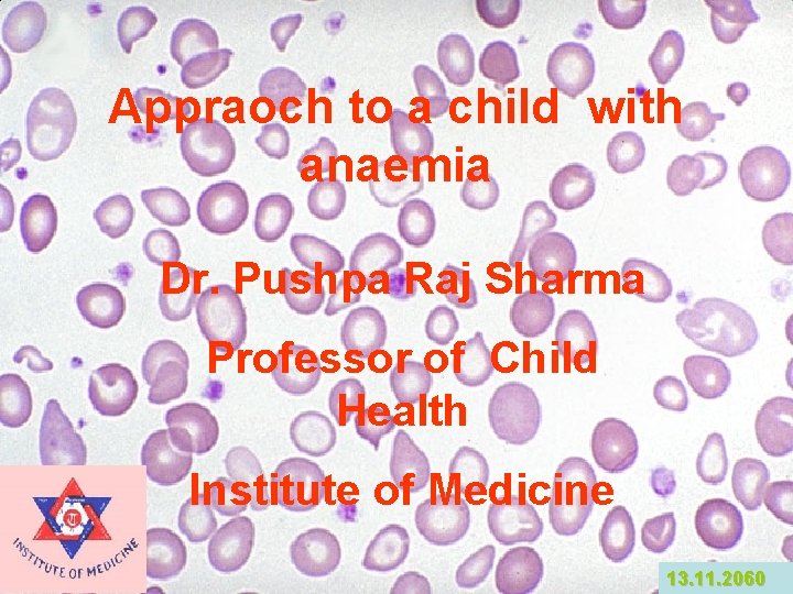
Appraoch to a child with anaemia Dr. Pushpa Raj Sharma Professor of Child Health Institute of Medicine 13. 11. 2060
Definition of anaemia • The condition of having too few red blood cells. • It is a reduction of the red cell volume or hemoglobin concentration below -2 SD for age, race and sex. • Microscopic: normocytic, microcytic, normochromic macrocytic or specific abnormalities (spherocyte, sickle cell, target cell)
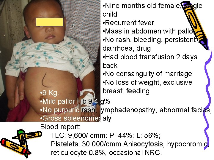
Case • • Nine months old “Rai” female, single child Recurrent fever Mass in abdomen with pallor No history of rash, bleeding, persistent diarrhoea, drug, jaundice, pica, Had blood transfusion 2 days back No consanguity of marriage No family history of gall stones, jaundice. No loss of weight, exclusive breast feeding
Summary of examination finding • 9 Kg. • Mild pallor • No purpuric rash, lymphadenopathy, abnormal facies, • Gross spleenomegaly
Diagnostic Approach-History • Age : Iron def rare without blood loss before 6 mo in term infants. • Family Hist & Genetics: (1)X-linked: G 6 PD def (2)Aut dominant: Spherocytosis (3)Aut recessive: Sickle cell, Fanconi anemia (4)Family member with early age of cholecystectomy/splenectomy (5)Ethnicity: Thalassemia; G 6 PD def
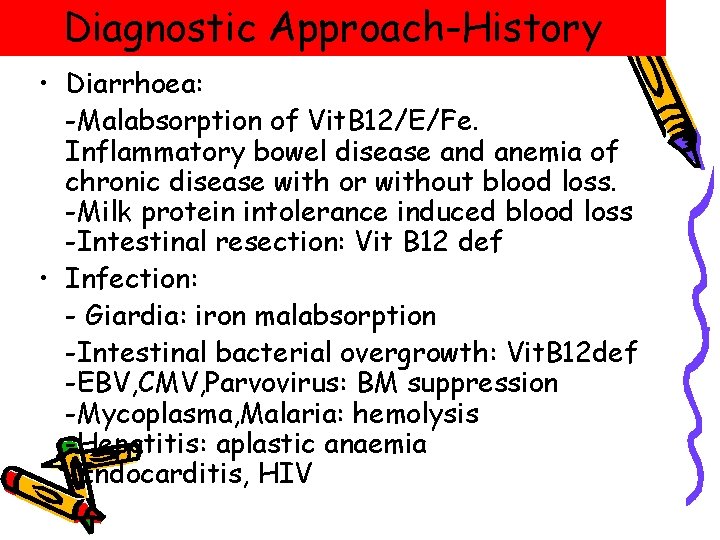
Diagnostic Approach-History • Diarrhoea: -Malabsorption of Vit. B 12/E/Fe. Inflammatory bowel disease and anemia of chronic disease with or without blood loss. -Milk protein intolerance induced blood loss -Intestinal resection: Vit B 12 def • Infection: - Giardia: iron malabsorption -Intestinal bacterial overgrowth: Vit. B 12 def -EBV, CMV, Parvovirus: BM suppression -Mycoplasma, Malaria: hemolysis -Hepatitis: aplastic anaemia -Endocarditis, HIV
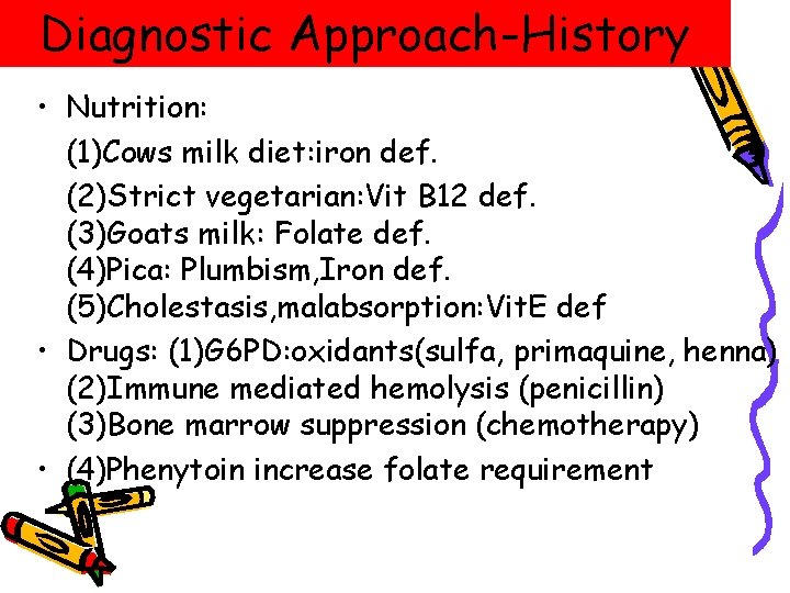
Diagnostic Approach-History • Nutrition: (1)Cows milk diet: iron def. (2)Strict vegetarian: Vit B 12 def. (3)Goats milk: Folate def. (4)Pica: Plumbism, Iron def. (5)Cholestasis, malabsorption: Vit. E def • Drugs: (1)G 6 PD: oxidants(sulfa, primaquine, henna) (2)Immune mediated hemolysis (penicillin) (3)Bone marrow suppression (chemotherapy) • (4)Phenytoin increase folate requirement
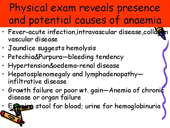
Physical exam reveals presence and potential causes of anaemia • Fever-acute infection, intravascular disease, collagen vascular disease • Jaundice suggests hemolysis • Petechia&Purpura—bleeding tendency • Hypertension&oedema-renal disease • Hepatosplenomegaly and lymphadenopathy— infiltrative disease • Growth failure or poor wt. gain—Anemia of chronic disease or organ failure • Examine stool for blood; urine for hemoglobinuria
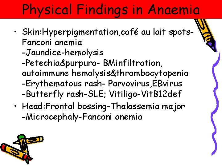
Physical Findings in Anaemia • Skin: Hyperpigmentation, café au lait spots. Fanconi anemia -Jaundice-hemolysis -Petechia&purpura- BMinfiltration, autoimmune hemolysis&thrombocytopenia -Erythematous rash- Parvovirus, EBvirus -Butterfly rash-SLE; Vitiligo-Vit. B 12 def • Head: Frontal bossing-Thalassemia major -Microcephaly-Fanconi anemia
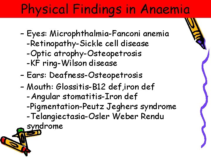
Physical Findings in Anaemia – Eyes: Microphthalmia-Fanconi anemia -Retinopathy-Sickle cell disease -Optic atrophy-Osteopetrosis -KF ring-Wilson disease – Ears: Deafness-Osteopetrosis – Mouth: Glossitis-B 12 def, iron def -Angular stomatitis-Iron def -Pigmentation-Peutz Jeghers syndrome -Telangiectasia-Osler Weber Rendu syndrome
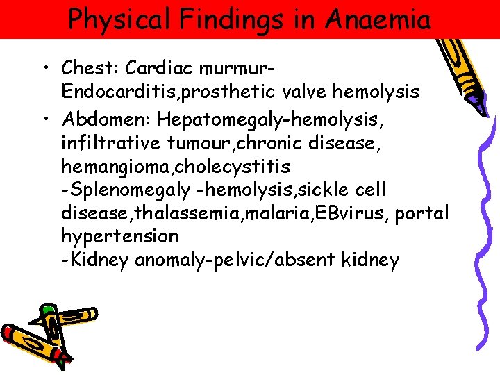
Physical Findings in Anaemia • Chest: Cardiac murmur. Endocarditis, prosthetic valve hemolysis • Abdomen: Hepatomegaly-hemolysis, infiltrative tumour, chronic disease, hemangioma, cholecystitis -Splenomegaly -hemolysis, sickle cell disease, thalassemia, malaria, EBvirus, portal hypertension -Kidney anomaly-pelvic/absent kidney
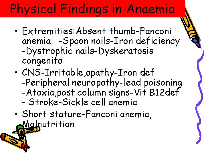
Physical Findings in Anaemia • Extremities: Absent thumb-Fanconi anemia -Spoon nails-Iron deficiency -Dystrophic nails-Dyskeratosis congenita • CNS-Irritable, apathy-Iron def. -Peripheral neuropathy-lead poisoning -Ataxia, post. column signs-Vit B 12 def - Stroke-Sickle cell anemia • Short stature-Fanconi anemia, Malnutrition
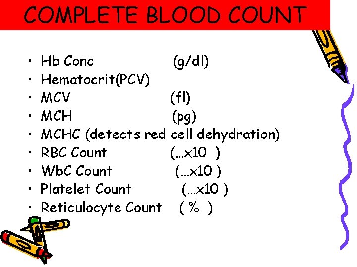
COMPLETE BLOOD COUNT • • • Hb Conc (g/dl) Hematocrit(PCV) MCV (fl) MCH (pg) MCHC (detects red cell dehydration) RBC Count (…x 10 ) Wb. C Count (…x 10 ) Platelet Count (…x 10 ) Reticulocyte Count ( % )
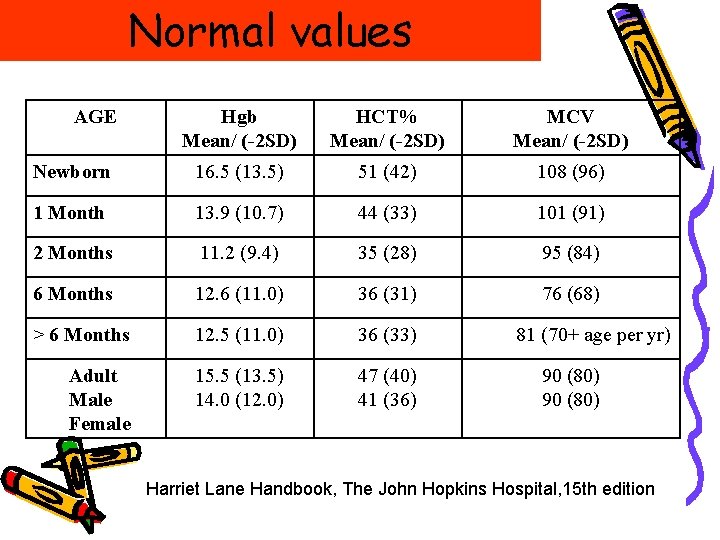
Normal values AGE Hgb Mean/ (-2 SD) HCT% Mean/ (-2 SD) MCV Mean/ (-2 SD) Newborn 16. 5 (13. 5) 51 (42) 108 (96) 1 Month 13. 9 (10. 7) 44 (33) 101 (91) 2 Months 11. 2 (9. 4) 35 (28) 95 (84) 6 Months 12. 6 (11. 0) 36 (31) 76 (68) > 6 Months 12. 5 (11. 0) 36 (33) Adult Male Female 15. 5 (13. 5) 14. 0 (12. 0) 47 (40) 41 (36) 81 (70+ age per yr) 90 (80) Harriet Lane Handbook, The John Hopkins Hospital, 15 th edition
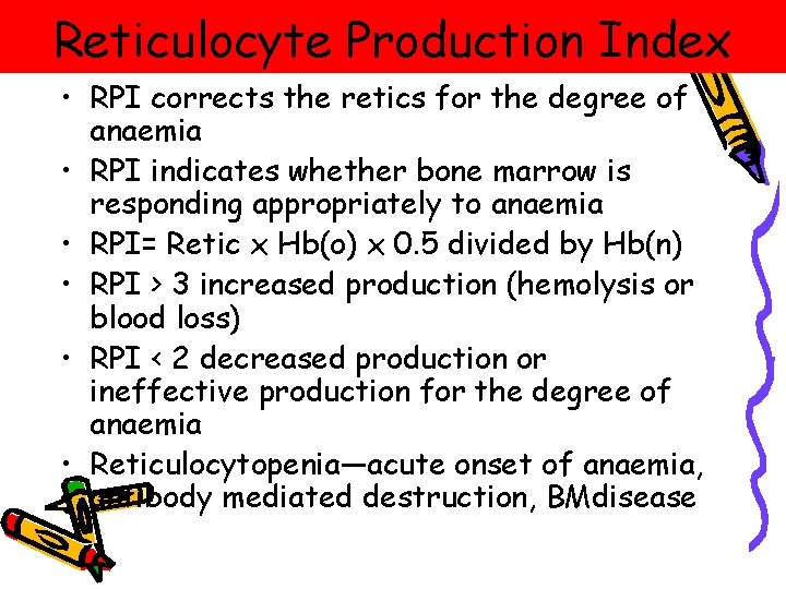
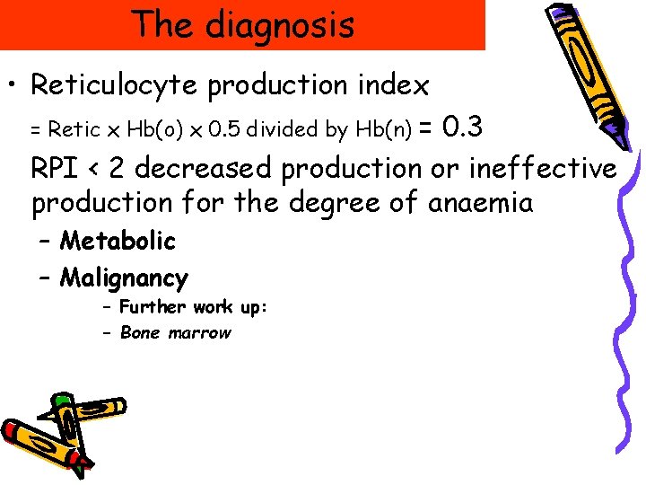
Reticulocyte Production Index • RPI corrects the retics for the degree of anaemia • RPI indicates whether bone marrow is responding appropriately to anaemia • RPI= Retic x Hb(o) x 0. 5 divided by Hb(n) • RPI > 3 increased production (hemolysis or blood loss) • RPI < 2 decreased production or ineffective production for the degree of anaemia • Reticulocytopenia—acute onset of anaemia, antibody mediated destruction, BMdisease
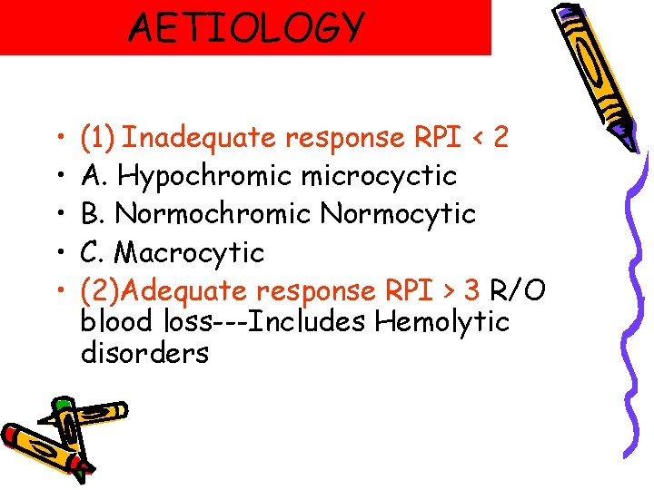
AETIOLOGY • • • (1) Inadequate response RPI < 2 A. Hypochromic microcyctic B. Normochromic Normocytic C. Macrocytic (2)Adequate response RPI > 3 R/O blood loss---Includes Hemolytic disorders
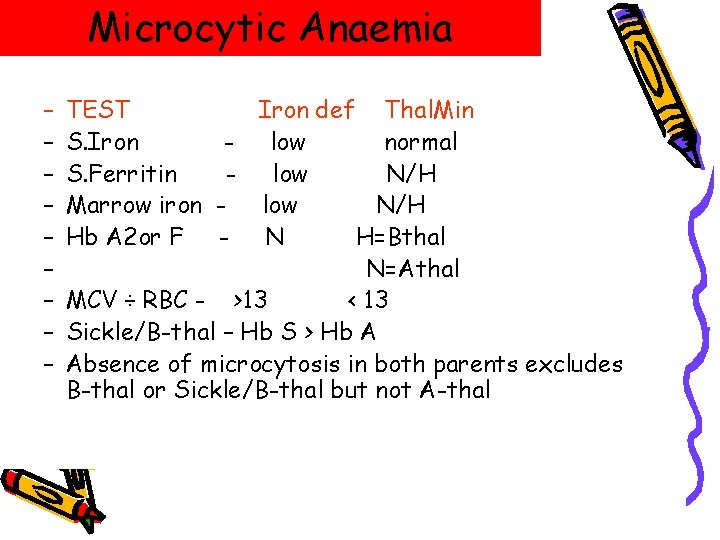
Microcytic Anaemia – – – – – TEST Iron def Thal. Min S. Iron - low normal S. Ferritin - low N/H Marrow iron - low N/H Hb A 2 or F - N H=Bthal N=Athal MCV ÷ RBC - >13 < 13 Sickle/B-thal – Hb S > Hb A Absence of microcytosis in both parents excludes B-thal or Sickle/B-thal but not A-thal
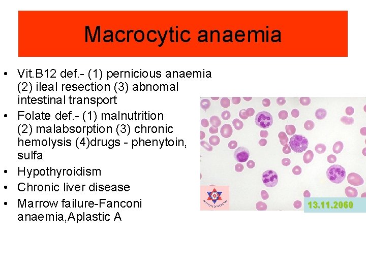
Macrocytic anaemia • Vit. B 12 def. - (1) pernicious anaemia (2) ileal resection (3) abnomal intestinal transport • Folate def. - (1) malnutrition (2) malabsorption (3) chronic hemolysis (4)drugs - phenytoin, sulfa • Hypothyroidism • Chronic liver disease • Marrow failure-Fanconi anaemia, Aplastic A 13. 11. 2060
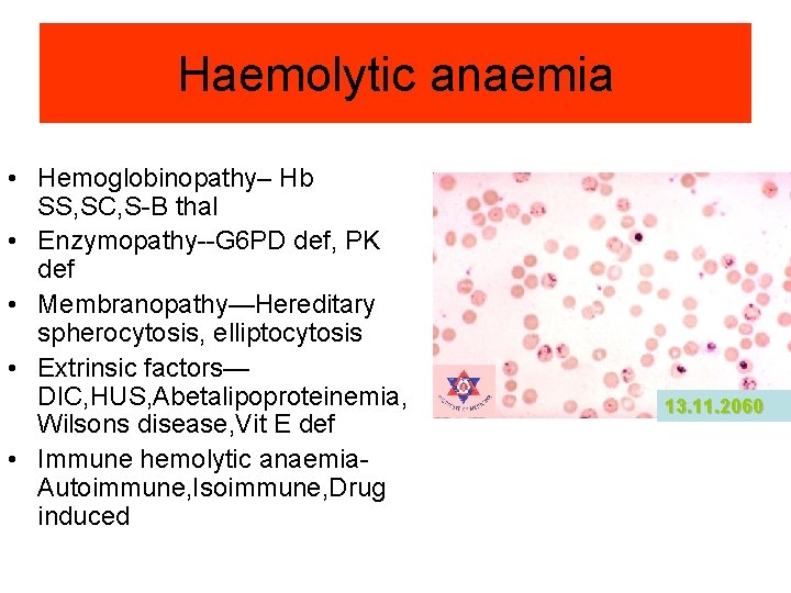
Haemolytic anaemia • Hemoglobinopathy– Hb SS, SC, S-B thal • Enzymopathy--G 6 PD def, PK def • Membranopathy—Hereditary spherocytosis, elliptocytosis • Extrinsic factors— DIC, HUS, Abetalipoproteinemia, Wilsons disease, Vit E def • Immune hemolytic anaemia. Autoimmune, Isoimmune, Drug induced 13. 11. 2060
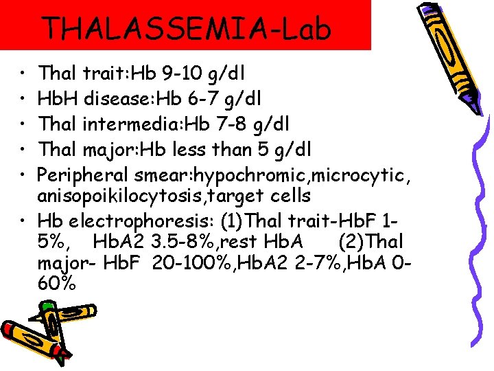
THALASSEMIA-Lab • • • Thal trait: Hb 9 -10 g/dl Hb. H disease: Hb 6 -7 g/dl Thal intermedia: Hb 7 -8 g/dl Thal major: Hb less than 5 g/dl Peripheral smear: hypochromic, microcytic, anisopoikilocytosis, target cells • Hb electrophoresis: (1)Thal trait-Hb. F 15%, Hb. A 2 3. 5 -8%, rest Hb. A (2)Thal major- Hb. F 20 -100%, Hb. A 2 2 -7%, Hb. A 060%
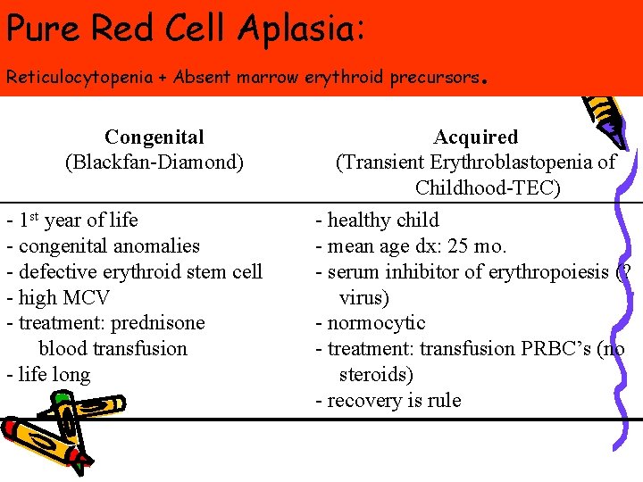
Pure Red Cell Aplasia: Reticulocytopenia + Absent marrow erythroid precursors Congenital (Blackfan-Diamond) - 1 st year of life - congenital anomalies - defective erythroid stem cell - high MCV - treatment: prednisone blood transfusion - life long . Acquired (Transient Erythroblastopenia of Childhood-TEC) - healthy child - mean age dx: 25 mo. - serum inhibitor of erythropoiesis (? virus) - normocytic - treatment: transfusion PRBC’s (no steroids) - recovery is rule
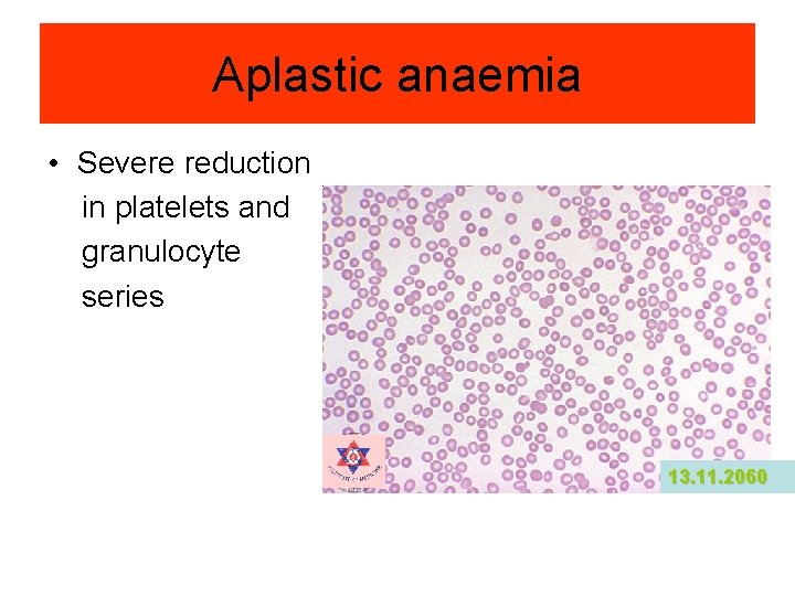
Aplastic anaemia • Severe reduction in platelets and granulocyte series 13. 11. 2060
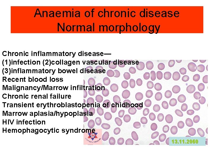
Anaemia of chronic disease Normal morphology Chronic inflammatory disease— (1)infection (2)collagen vascular disease (3)inflammatory bowel disease Recent blood loss Malignancy/Marrow infiltration Chronic renal failure Transient erythroblastopenia of chidhood Marrow aplasia/hypoplasia HIV infection Hemophagocytic syndrome 13. 11. 2060
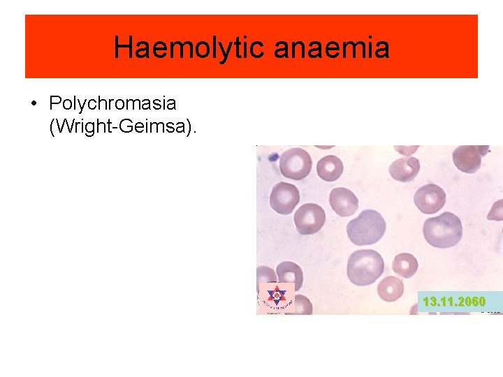
Haemolytic anaemia • Polychromasia (Wright-Geimsa). 13. 11. 2060
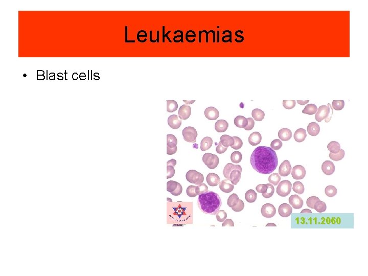
Leukaemias • Blast cells 13. 11. 2060
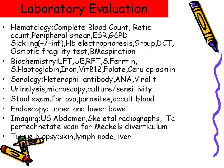
Laboratory Evaluation • Hematology: Complete Blood Count, Retic count, Peripheral smear, ESR, G 6 PD Sickling(+/-inf), Hb electrophoresis, Group, DCT, Osmotic fragility test, BMaspiration • Biochemistry: LFT, UE, RFT, S. Ferrtin, S. Haptoglobin, Iron, Vit. B 12, Folate, Ceruloplasmin • Serology: Heterophil antibody, ANA, Viral t • Urinalysis, microscopy, culture/sensitivity • Stool exam. for ova, parasites, occult blood • Endoscopy: upper and lower bowel • Imaging: US Abdomen, Skeletal radiographs, Tc pertechnetate scan for Meckels diverticulum • Tissue biopsy: skin, lymph node, liver
• Nine months old female, single child • Recurrent fever • Mass in abdomen with pallor • No rash, bleeding, persistent diarrhoea, drug • Had blood transfusion 2 days back • No consanguity of marriage • No loss of weight, exclusive breast feeding • 9 Kg. • Mild pallor Hb: 9. 4 g% • No purpuric rash, lymphadenopathy, abnormal facies, • Gross spleenomegaly Blood report: TLC: 9, 600/ cmm: P: 44%: L: 56%; Platelets: 30. 000/cmm Anisocytosis, hypochromic, reticulocyte 0. 8%, occasional NRC.
The diagnosis • Reticulocyte production index = Retic x Hb(o) x 0. 5 divided by Hb(n) = 0. 3 RPI < 2 decreased production or ineffective production for the degree of anaemia – Metabolic – Malignancy – Further work up: – Bone marrow
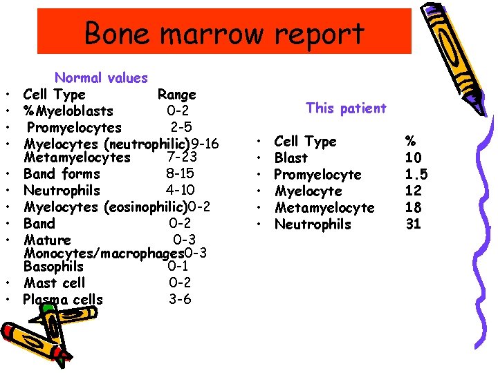
Bone marrow report • • • Normal values Cell Type Range %Myeloblasts 0 -2 Promyelocytes 2 -5 Myelocytes (neutrophilic)9 -16 Metamyelocytes 7 -23 Band forms 8 -15 Neutrophils 4 -10 Myelocytes (eosinophilic)0 -2 Band 0 -2 Mature 0 -3 Monocytes/macrophages 0 -3 Basophils 0 -1 Mast cell 0 -2 Plasma cells 3 -6 This patient • • • Cell Type Blast Promyelocyte Metamyelocyte Neutrophils % 10 1. 5 12 18 31
Final diagnosis • Malignancy – Juvenile Chronic Myelogenous Leukemia
The commonest cause • Iron def. is common in children 9 mo-3 yr • Iron def. anemia in a child over 3 yr should prompt consideration of occult blood loss. • Infants less than 6 months generally do not develop iron def. ; the exception to this rule is premature infants, who are at risk of iron def. at 4 mo, if iron supplementation is not given.
Iron deficiency Anemia – Dietary iron def is the usual cause – Manifestations of anemia – CNS abnormalities: apathy, irritability, poor conc, cognitive deficits – Poor muscle endurance – GIT dysfunction – Impaired WBC and T-cell function
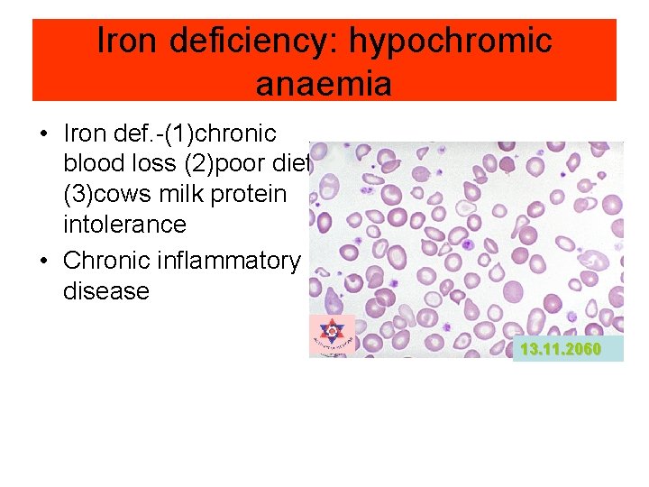
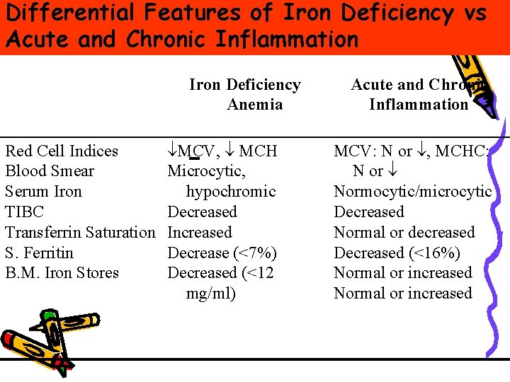
Iron deficiency: hypochromic anaemia • Iron def. -(1)chronic blood loss (2)poor diet (3)cows milk protein intolerance • Chronic inflammatory disease 13. 11. 2060
Differential Features of Iron Deficiency vs Acute and Chronic Inflammation Iron Deficiency Anemia Red Cell Indices Blood Smear Serum Iron TIBC Transferrin Saturation S. Ferritin B. M. Iron Stores ¯MCV, ¯ MCH Microcytic, hypochromic Decreased Increased Decrease (<7%) Decreased (<12 mg/ml) Acute and Chronic Inflammation MCV: N or ¯, MCHC: N or ¯ Normocytic/microcytic Decreased Normal or decreased Decreased (<16%) Normal or increased
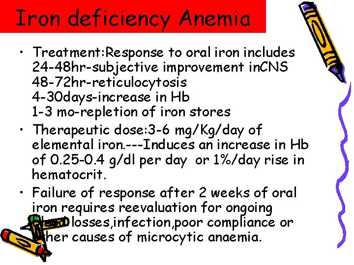
Iron deficiency Anemia • Treatment: Response to oral iron includes 24 -48 hr-subjective improvement in. CNS 48 -72 hr-reticulocytosis 4 -30 days-increase in Hb 1 -3 mo-repletion of iron stores • Therapeutic dose: 3 -6 mg/Kg/day of elemental iron. ---Induces an increase in Hb of 0. 25 -0. 4 g/dl per day or 1%/day rise in hematocrit. • Failure of response after 2 weeks of oral iron requires reevaluation for ongoing blood losses, infection, poor compliance or other causes of microcytic anaemia.