
Approach to child with heart disease Pushpa Raj


- Slides: 30
Approach to child with heart disease Pushpa Raj Sharma Professor of Child Health Institute of Medicine
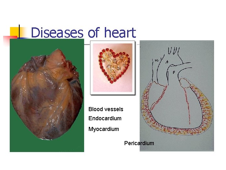
Diseases of heart Blood vessels Endocardium Myocardium Pericardium
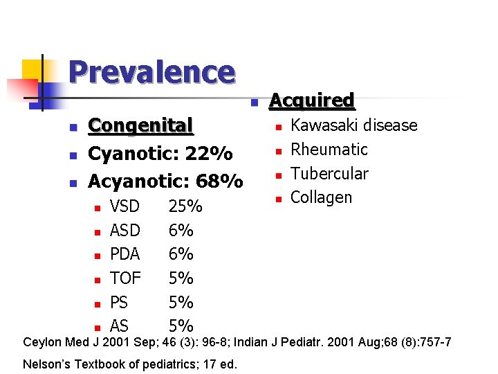
Prevalence n n Congenital Cyanotic: 22% Acyanotic: 68% n n n VSD ASD PDA TOF PS AS 25% 6% 6% 5% 5% 5% Acquired n n Kawasaki disease Rheumatic Tubercular Collagen Ceylon Med J 2001 Sep; 46 (3): 96 -8; Indian J Pediatr. 2001 Aug; 68 (8): 757 -7 Nelson’s Textbook of pediatrics; 17 ed.
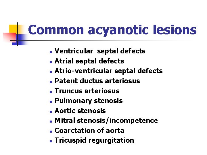
Common acyanotic lesions n n n n n Ventricular septal defects Atrial septal defects Atrio-ventricular septal defects Patent ductus arteriosus Truncus arteriosus Pulmonary stenosis Aortic stenosis Mitral stenosis/incompetence Coarctation of aorta Tricuspid regurgitation
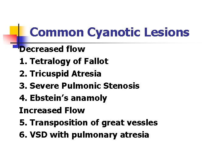
Common Cyanotic Lesions Decreased flow 1. Tetralogy of Fallot 2. Tricuspid Atresia 3. Severe Pulmonic Stenosis 4. Ebstein’s anamoly Increased Flow 5. Transposition of great vessles 6. VSD with pulmonary atresia
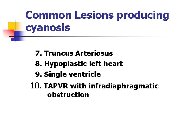
Common Lesions producing cyanosis 7. Truncus Arteriosus 8. Hypoplastic left heart 9. Single ventricle 10. TAPVR with infradiaphragmatic obstruction
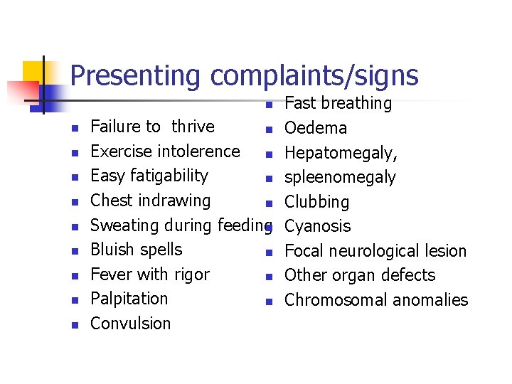
Presenting complaints/signs Fast breathing Failure to thrive n Oedema Exercise intolerence n Hepatomegaly, Easy fatigability n spleenomegaly Chest indrawing n Clubbing Sweating during feedingn Cyanosis Bluish spells n Focal neurological lesion Fever with rigor n Other organ defects Palpitation n Chromosomal anomalies Convulsion n n
Cyanosis: is it a cardiac cause or lung cause n Hyperoxia test n Neonates with cyanotic congenital heart disease usually do not have significantly raised arterial Pao 2 during administration of 100% oxygen.
Ventricular Defect n Small VSD n n n Asymptomatic A loud, harsh, or blowing holosystolic murmur. Large VSD dyspnea, feeding difficulties, poor growth, profuse perspiration, recurrent pulmonary infections, and cardiac failure in early infancy. Syndromes associated with this condition n 80%
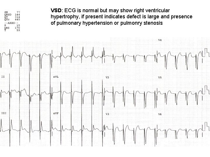
VSD: ECG is normal but may show right ventricular hypertrophy, if present indicates defect is large and presence of pulmonary hypertension or pulmonry stenosis
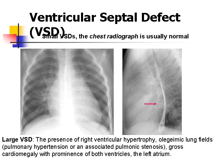
Ventricular Septal Defect (VSD) Small VSDs, the chest radiograph is usually normal Large VSD: The presence of right ventricular hypertrophy, olegeimic lung fields (pulmonary hypertension or an associated pulmonic stenosis), gross cardiomegaly with prominence of both ventricles, the left atrium.
Ventricular Septal defects n n 30– 50% of small defects close spontaneously, most frequently during the 1 st 2 yr of life. Small muscular VSDs are more likely to close (up to 80%) than membranous VSDs are (up to 35%). infants with large defects have repeated episodes of respiratory infection and heart failure despite optimal medical management. Surgical repair prior to development of an irreversible increase in pulmonary vasculalr resistance (usually prior to the patient's second birthday).
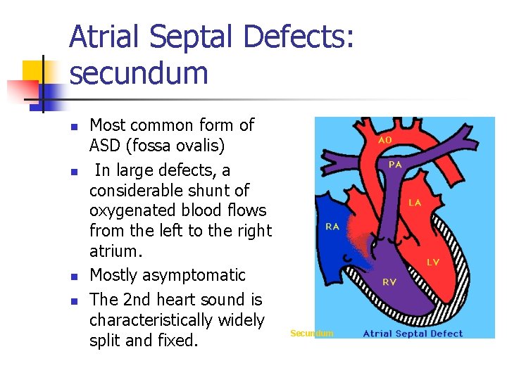
Atrial Septal Defects: secundum n n Most common form of ASD (fossa ovalis) In large defects, a considerable shunt of oxygenated blood flows from the left to the right atrium. Mostly asymptomatic The 2 nd heart sound is characteristically widely split and fixed. Secundum
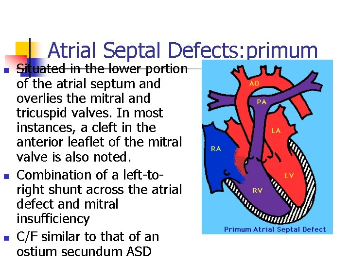
Atrial Septal Defects: primum n n n Situated in the lower portion of the atrial septum and overlies the mitral and tricuspid valves. In most instances, a cleft in the anterior leaflet of the mitral valve is also noted. Combination of a left-toright shunt across the atrial defect and mitral insufficiency C/F similar to that of an ostium secundum ASD
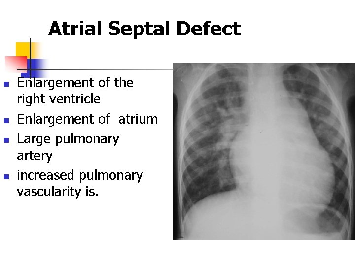
Atrial Septal Defect n n Enlargement of the right ventricle Enlargement of atrium Large pulmonary artery increased pulmonary vascularity is.
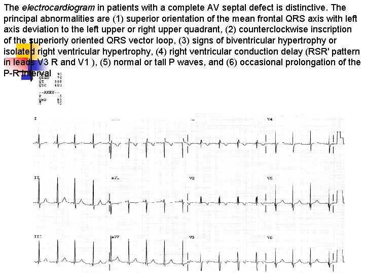
The electrocardiogram in patients with a complete AV septal defect is distinctive. The principal abnormalities are (1) superior orientation of the mean frontal QRS axis with left axis deviation to the left upper or right upper quadrant, (2) counterclockwise inscription of the superiorly oriented QRS vector loop, (3) signs of biventricular hypertrophy or isolated right ventricular hypertrophy, (4) right ventricular conduction delay (RSR′ pattern in leads V 3 R and V 1 ), (5) normal or tall P waves, and (6) occasional prolongation of the P-R interval
Atrial Septal Defects n n Secundum ASDs are well tolerated during childhood. Antibiotic prophylaxis for isolated secundum ASDs is not recommended. Surgery or transcatheter device closure is advised for all symptomatic patients and also for asymptomatic patients with a Qp: Qs ratio of at least 2: 1. Ostium primum defects are approached surgically
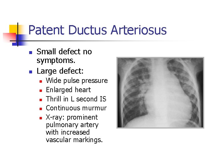
Patent Ductus Arteriosus n n Small defect no symptoms. Large defect: n n n Wide pulse pressure Enlarged heart Thrill in L second IS Continuous murmur X-ray: prominent pulmonary artery with increased vascular markings.
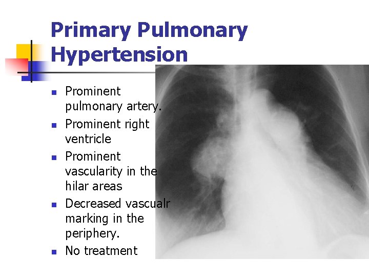
Primary Pulmonary Hypertension n n Prominent pulmonary artery. Prominent right ventricle Prominent vascularity in the hilar areas Decreased vascualr marking in the periphery. No treatment
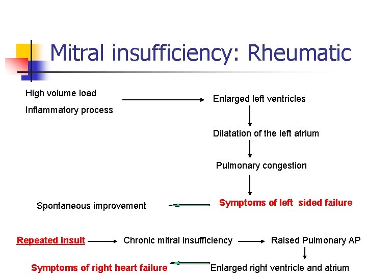
Mitral insufficiency: Rheumatic High volume load Enlarged left ventricles Inflammatory process Dilatation of the left atrium Pulmonary congestion Spontaneous improvement Repeated insult Symptoms of left sided failure Chronic mitral insufficiency Symptoms of right heart failure Raised Pulmonary AP Enlarged right ventricle and atrium
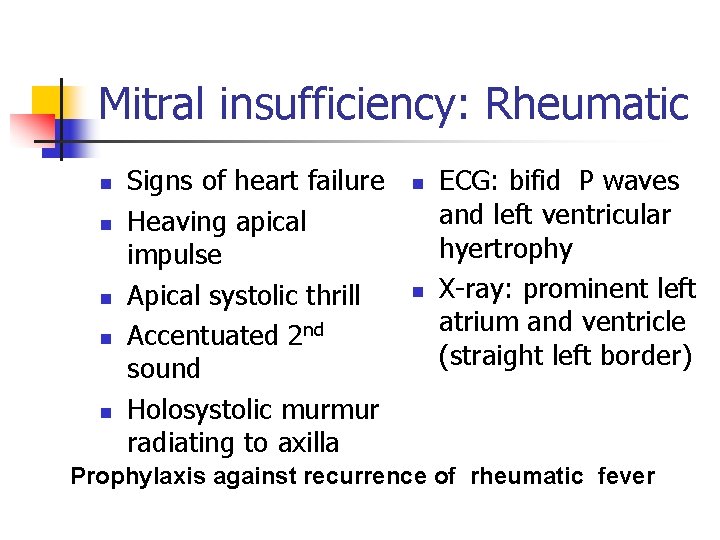
Mitral insufficiency: Rheumatic n n n Signs of heart failure Heaving apical impulse Apical systolic thrill Accentuated 2 nd sound Holosystolic murmur radiating to axilla n n ECG: bifid P waves and left ventricular hyertrophy X-ray: prominent left atrium and ventricle (straight left border) Prophylaxis against recurrence of rheumatic fever
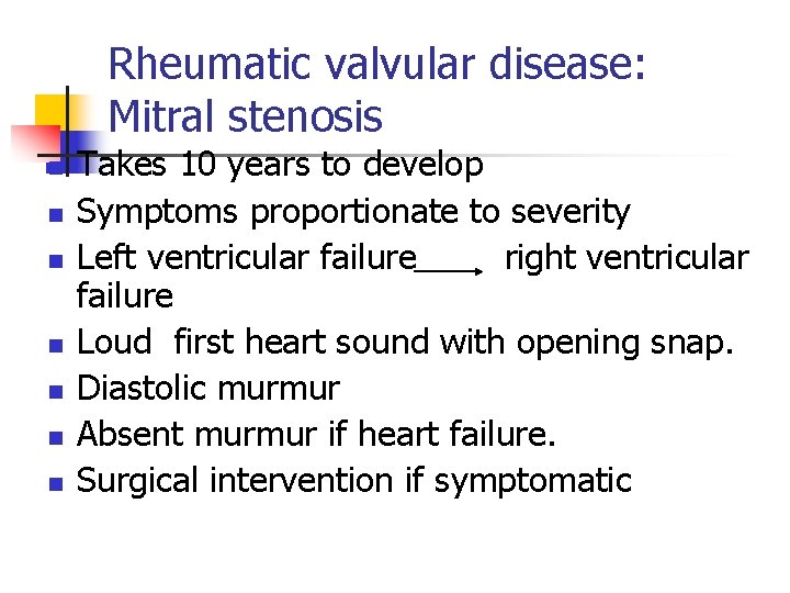
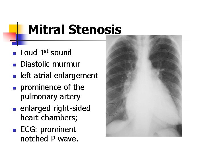
Rheumatic valvular disease: Mitral stenosis n n n n Takes 10 years to develop Symptoms proportionate to severity Left ventricular failure right ventricular failure Loud first heart sound with opening snap. Diastolic murmur Absent murmur if heart failure. Surgical intervention if symptomatic
Mitral Stenosis n n n Loud 1 st sound Diastolic murmur left atrial enlargement prominence of the pulmonary artery enlarged right-sided heart chambers; ECG: prominent notched P wave.
Pericardial Effusion n Presenting complaint n n n n Precordial pain Cough Dyspnoea Abdominal pain Vomiting Fever Other organs involvement n Signs: n n n n Position: leaning forward. Puffy face Friction rub Absent apical impulse Muffled heart sounds Pulsus paradoxus Distended neck veins Low QRS complex, T inversion
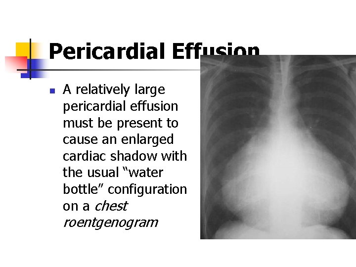
Pericardial Effusion n A relatively large pericardial effusion must be present to cause an enlarged cardiac shadow with the usual “water bottle” configuration on a chest roentgenogram
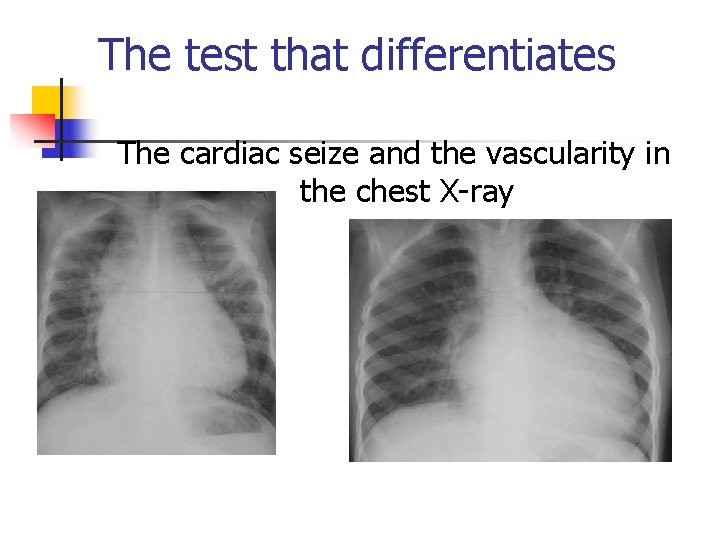
The test that differentiates The cardiac seize and the vascularity in the chest X-ray
Cardiac disease with normal/decreased vasculature n n n n Viral myocarditis Tetralogy of Fallot Pulmonary atresia Tricuspid atresia Endocardial fibroelastosis Aberrant left coronary artery Cystic medial necrosis Diabetic mother
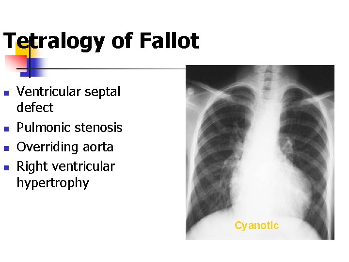
Tetralogy of Fallot n n Ventricular septal defect Pulmonic stenosis Overriding aorta Right ventricular hypertrophy Cyanotic
Cardiac disease with increased vasculature n n n n Atrioventricular septal defects Congestive cardiac failure Transposition of great arteries with VSD Total anomalous pulmonary venous drainage Truncus arteriosus Single ventricle without pulmonary stenosis Hypoplastic left heart syndrome
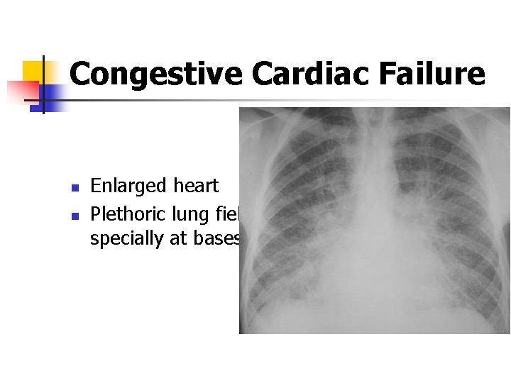
Congestive Cardiac Failure n n Enlarged heart Plethoric lung fields specially at bases