
Multiple pregnancy antenatal care for twin and triplet














- Slides: 99
Multiple pregnancy: antenatal care for twin and triplet pregnancies professor Dr Esraa AL-Maini
Introduction The incidence of twinning ranges from 4 per 1000 births in Japan to as frequent as 54 per 1000 in some regions of Nigeriais 1‐ more prevalent in pregnancies with advancing maternal age (strongest at 30– 39 years) (presumed to be secondary to the rise in follicle‐stimulating hormone concentrations ( spontaneous dizygous) 2‐ Familial predisposition to multiple ovulations (usually dizygous twinning) may occur and is presently best explained by an autosomal dominant inheritance pattern 3‐monozygotic constant incidence of approximately 3. 9 per 1000. The incidence of multiple births has risen in the last 30 years. The combined effects of delayed childbearing and high uptakes of assist reproductive technologies at advanced maternal age have been responsible for this rise.
Up to 24% of successful IVF procedures result in multiple pregnancies. increase is due to assisted reproductive technologies, such as superovulation (using antioestrogens or gonadotrophins)and in vitro fertilization (IVF) with embryo transfer. There is evidence that the number of multiple pregnancies is influenced by the number of embryo transfers and, as such, the number of multiple pregnancies associated with IVF has reduced since the recommendation that embryo transfer numbers be reduced. In addition, epidemiological evidence suggests that assisted reproduction techniques increase the incidence of monozygous twinning by up to eightfold. This is particularly associated with the techniques of ‘blastocyst hatching’. Monochorionic twins comprise 20% of spontaneous and 5% of iatrogenic twin gestations.
maternal mortality associated with multiple births is 2. 5 times that for singleton births. This high perinatal fetal loss and morbidity is largely attributed to : the increased risk of prematurity and also intrauterine growth restriction (IUGR) with associated iatrogenic prematurity (irrespective of chorionicity. Cerebral palsy is approximately three times more common in twins and 10 times more common in triplets compared with singletons. Multiple pregnancy may be classified according to: Number of fetuses: twins, triplets, quadruplets, etc. Number of fertilized eggs: zygosity. Number of placentae: chorionicity. Number of amniotic cavities: amnionicity.
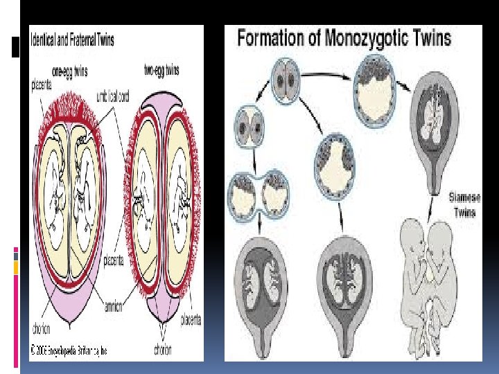
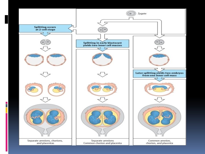
Twin pregnancy may be dizygotic (70%) or monozygotic (30%). Dizygotic twins (non‐identical) occur from ovulation and subsequent fertilization of two oocytes. This results in dichorionic diamniotic twins, where each fetus has its own placenta and amniotic cavity. Although they always have two functionally separate placentae (dichorionic), the placentae can become anatomically fused together and appear to the naked eye as a single placental mass. They always have separate amniotic cavities (diamniotic) and the two cavities are separated by a thick three‐layer membrane (fused amnion in the middle with chorion on either side; The fetuses can be either same‐ sex or different‐sex pairings.
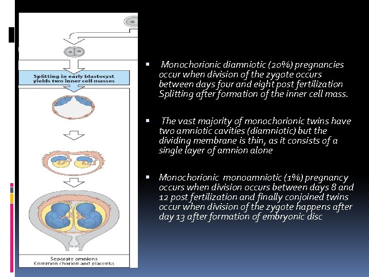
Monozygotic (identical) pregnancies result from fertilization of a single ovum with subsequent division of the zygote. If the zygote splits shortly after fertilization with in 3 days , the twins will each have a separate placenta and thus will be dichorionic diamniotic in 10%
Monochorionic diamniotic (20%) pregnancies occur when division of the zygote occurs between days four and eight post fertilization Splitting after formation of the inner cell mass. The vast majority of monochorionic twins have two amniotic cavities (diamniotic) but the dividing membrane is thin, as it consists of a single layer of amnion alone Monochorionic monoamniotic (1%) pregnancy occurs when division occurs between days 8 and 12 post fertilization and finally conjoined twins occur when division of the zygote happens after day 13 after formation of embryonic disc
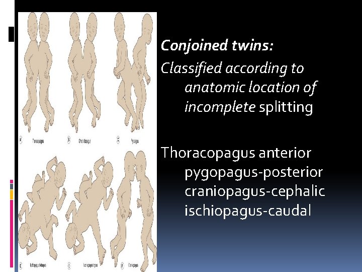
Conjoined twins: Classified according to anatomic location of incomplete splitting Thoracopagus anterior pygopagus-posterior craniopagus-cephalic ischiopagus-caudal
Maternal homeostatic responses All the normal physiological adaptations, such as increased cardiac output, glomerular filtration rate and renal blood flow, are further increased in a multiple pregnancy. Women with twins increase their plasma volume by one‐third more than women with singletons. Red cell mass increases approximately 300 m. L more than in singleton pregnancies but because this is disproportionately less than the increase in plasma volume, haemoglobin and haematocrit values fall.
. Maternal iron stores are diminished in 40% of women with twins routine haematinic supplementation is recommended (usually as combined iron sulfate and folic acid supplementation) . Hyperemesis gravidarum is more common in multiple pregnancies and is managed as in singleton pregnancies. Severe cases may respond to maternal steroid therapy and require pyroxidine (B 6) supplementation. The majority of minor pregnancy complications such as backache, symphysis pubis dysfunction, oedema, varicose veins, haemorrhoids and stria are all increased as a result of both the physical effects of greater uterine size and greater placental hormone production. Hypertensive disease of pregnancy and pre‐eclampsia are up to 10 times more common in multiple compared with singleton pregnancies but are managed once diagnosed on standard principles (as in singletons).
Consideration should be given to low‐dose aspirin prophylaxis but there are no national/international recommendations to this effect. Maternal pregnancy‐related hypertension remains a significant cause of maternal morbidity (and mortality) in multiple pregnancies and a significant cause of iatrogenic preterm delivery, increasing perinatal morbidity and mortality. This occurs in 15– 20% of twin pregnancies, 25% of triplets and up to 60% of higher‐order multiple pregnancies
Postnatally, the physical difficulties and socioeconomic impact of coping with the demands of two or more babies are considerable. Postnatal depression is more common in women nursing twins than singletons. With the high perinatal loss rates there are often associated problems of postnatal grieving and bereavement. Families of women who give birth to babies after a multiple pregnancy may require additional social support
Woman‐centred care Treatment and care should take into account women's needs and preferences. Women with twin and triplet pregnancies should have the opportunity to make informed decisions about their care and treatment, in partnership with their healthcare professionals. If women do not have the capacity to make decisions, healthcare professionals should follow the Department of Health's advice on consent Good communication between healthcare professionals and women is essential.
General care 1‐Information and emotional support 2‐Explain sensitively the aims and possible outcomes of all screening and 3‐diagnostic tests to women with twin and triplet pregnancies to minimise their anxiety. 4‐Diet, lifestyle and nutritional suppleme Give women with twin and triplet pregnancies the same advice about diet, lifestyle and nutritional supplements as in routine antenatal care 5‐Be aware of the higher incidence of anaemia in women with twin and triple pregnancies compared with women with singletone 6‐ Perform a full blood count at 20– 24 weeks to identify women with twin and 7‐triplet pregnancies who need early supplementation with iron or folic acid, and repeat at 28 weeks as in routine antenatal care
Special Clinical care for women with twin and triplet pregnancies should be provided by a multidisciplinary team consisting of: a core team of named specialist obstetricians, specialist midwives and ultrasonographers, all of whom have experience and knowledge of managing twin and triplet pregnancies an enhanced team for referrals, which should include: a perinatal mental health professional a women's health physiotherapist an infant feeding specialist a dietitian.
Coordinate clinical care for women with twin and triplet pregnancies to: minimise the number of hospital visits provide care as close to the woman's home as possible provide continuity of care within and between hospitals and the community. The core team should offer information and emotional support specific to twin and triplet pregnancies at their first contact with the woman and provide ongoing opportunities for further discussion and advice including: antenatal and postnatal mental health and wellbeing antenatal nutrition the risks, symptoms and signs of preterm labour and the potential need for corticosteroids for fetal lung maturation likely timing and possible modes of delivery breastfeeding parenting.
1 -Determining gestational age and chorionicity ‐women with a twin pregnancy should be offered an ultrasound examination between 11 +0 weeks and 13+6 weeks of gestation (crown–rump length 45‐ 84 mm) to assess; 1‐ Fetal viability 2‐Gestational age 3‐Chorionicity 4‐ Exclude major congenital malformations. Chorionicity should be determined at the time the twin pregnancy is detected by ultrasound based upon ( 90%100%accuracy ) 1‐The number of placental masses, 2‐The appearance of the membrane attachment to the( placenta twin peak sign) 3‐The membrane thickness. (no. of layers , thick or thin) This scan is best performed before 14 weeks of gestation.
If transabdominal ultrasound scan views are poor because of 1 -A retroverted uterus 2 - A high body mass index (BMI), use a transvaginal ultrasound scan to determine chorionicity. If a woman with a twin or triplet pregnancy presents after 14 weeks 0 days, determine chorionicity at the earliest opportunity by ultrasound using all of the following: 1‐The number of placental masses 2‐The lambda or T‐sign 3‐Membrane thickness 4‐Discordant fetal sex (16‐ 20 weeks)for medical reasons
‐A photographic (thermal copy) record should be taken and placed in the patients notes documenting the ultrasound appearance of the membrane attachment to the placenta( may need another opinion , ) as chorionicity is best determined before 14 weeks of gestation. ‐On ultrasound, the fetuses in twin pregnancies should be assigned nomenclature (i. e. upper and lower, or left and right) and this should be clearly documented in the womans case notes ‐Women should informed monochorionic twin pregnancies have higher fetal loss rates than dichorionic twin pregnancies, mainly due to second trimester loss and, overall, may have a higher risk of associate neurodevelopmental morbidity
This is because chorionicity is relevant to: ●● counselling parents in relation to the risk of perinatal morbidity and mortality ●● counselling parents in relation to their risk of genetic and structural abnormalities; ●● invasive testing and the management of discordant congenital anomaly; ●● feasibility of multiple fetal pregnancy reduction; ●● risk of complications that may occur in a multiple pregnancy and potential sequelae ; ●● early detection and management of feto‐fetal transfusion syndrome.
ZYGOSITY: Monochorionic twins by definition are monozygotic, Discordant‐sex twins are dizygous. while in dichorionic concordant‐sex twins the chance of dizygosity is 75– 80% In the remaining 50%, zygosity cannot be determined without DNA fingerprinting, such as the polymerase chain reaction Technique Not only are parents curious but knowledge of zygosity influences the twins’ rearing, their sense of identity, their genetic risks and their transplantation compatibility. However, it is not routine to offer this in current practice in UK. Rarely there may be indications for zygosity testing in utero on invasive
2‐Miscarriage Twins have a high incidence of spontaneous early pregnancy loss. 12% of human conceptions start as twins , are found at least twice as commonly in the first trimester as at birth. First‐trimester early pregnancy loss and resorption of one previously indefinable twin on ultrasound is known as the ‘vanishing twin’ phenomenon and is estimated to occur in up to 20% of twin pregnancies Spontaneous first‐trimester loss of one or more fetuses in high‐order pregnancies is estimated to occur approximately 50% of the time. (up to 13 weeks ) When one twin dies in utero in mid‐trimester (13‐ 20), a papyraceous fetus (the squashed paper‐like remains of the baby) may be found alongside the placenta after delivery. In some cases, this is only identifiable histopathologically
In dichorionic twins, the second or third trimester after 20 weeks intrauterine death of one fetus may be associated with the onset of labour, although in some cases the pregnancy may continue uneventfully and even result in delivery at term. Careful fetal and maternal monitoring is required. By contrast, fetal death of one twin in monochorionic twins may result in immediate complications in the survivor. These include death or brain damage with subsequent neurodevelopmental handicap. Acute hypotensive episodes, secondary to placental vascular anastomoses between the two fetuses, result in haemodynamic volume shifts from the live to the dead fetus. The acute release of vasoactive substances into the survivor’s circulation may also play a role. Death or handicap of the co‐twin occurs in up to 30% of cases.
3‐Screening for Down’s syndrome ‐Should be done by health care professional with experience with care for twin and counseling. ‐Inform women about the complexity of decisions they may need to make depending on the outcomes of screening, including different options according to the chorionicity of the pregnancy - map the fetal positions
Women with dizygous twins can be counselled that the chance of their pregnancy producing a child with Down’s syndrome is theoretically double their age‐related risk, where as women with monozygous twins simply have their age‐related risk that both twins will be aneuploid. Serum screening is, in general, inapplicable to multiple pregnancies
second trimester serum screening for Down’s syndrome in triplet pregnancies not used Offer women with twin and triplet pregnancies who have a high risk of Down’s syndrome referral to a fetal medicine specialist in a tertiary level fetal medicine centre of women with a multiple pregnancy
Women with twins who wish to have aneuploidy screening should be offered use the combined screening test (nuchal translucency, beta‐human chorionic gonadotrophin, pregnancy‐ associated plasma protein‐A) at approximately 11 weeks 14) calculate the risk of Down’s syndrome per pregnancy In dichorionic twins, the risk of aneuploidy is that of each of the individual fetuses. In monochorionic twin pregnancies, the risk of aneuploidy is the average between the twins
In women with twin pregnancies who miss or who have unsuccessful first trimester screening for aneuploidy, second trimester screening by the quadruple test(alpha feto protein , B HCG , E 2 Inhibin 14‐ 21) should be offered (Result is awaited) Down’s syndrome in twins, but the results of large prospective studies are awaited. Screening for Down’s syndrome using a maternal plasma sample to perform cell‐free DNA sequencing also known as non‐invasive prenatal testing (NIPT), IN monochorionic twins should be equally reliable as in singletons. In monochorionic twin pregnancies, higher failure rate and lower accuracy compared with MONO small study
Invasive test consider second trimester serum screening and explain to the woman the potential problems of such screening. These include the increased likelihood of pregnancy loss associated with double invasive testing (amniocentesis) because the risk of Down's syndrome cannot be calculated separately for each baby Invasive procedures in twins and other higher‐order multiple pregnancies are potentially complex techniques and should only be performed in fetal medicine referral centres The in utero topography (placental and membranes) is mapped using ultrasound. The location of the fetuses, the placental site(s) and the plane of the dividing septum in three dimensions should be noted and recorded. Such is a prerequisite for interpretation of discordant results and for selective termination of pregnancy.
In monochorionic twins, sample only one fetus by either amniocentesis or chorionic villous sampling (CVS). However, rare cases of heterokaryotypic monochorionic twins may be missed (occurring in <1%). So, amniocentesis on both amniotic sacs is worthy of consideration if monochorionic twins are 1‐discordant for structural anomalies 2‐nuchal translucencies or growth.
In dichorionic twins, there has been controversy CVS is less desirable than amniocentesis for performing karyotyping. Because of problems with contamination 2% miss dsaignosis So limited to CVS to high‐risk cases such as ‐ monogenic disease or ‐there is an aneuploidy risk of greater than 1 in 50. RCOG guidelines amniocentesis as the preferred option for karyotyping in dichorionic twins. to be weighed against the increased risks of selective reduction at increased gestational ages.
When performing fetal blood sampling, the intrahepatic vein may be sampled to avoid confusing the cord origins in twins. The overall pregnancy‐loss rate was 2‐ 3%
4‐Screening for structural abnormalities Monozygous twins have a 50% increase in structural abnormalities per baby. In particular, they have twice the frequency of congenital heart disease (a fourfold increase per pregnancy). monochorionic twins should undergo a routine detailed ultrasound scan between 18 and 20+6 weeks of gestation which includes extended views of the fetal heart anatomy (as recommended in the Fetal Anomaly Screening Programme screening of a singleton fetus). Consider scheduling ultrasound scans in twin and triplet pregnancies at a slightly later gestational age than in singleton pregnancies and be aware that the scans will take longer to perform. 30‐ 45 minut
In dichorionic twins discordant for fetal anomaly, Selective termination of pregnancy by the induction of asystole using an abortifacient is associated with an 8% loss rate, with lower rates if the procedure is performed before 16 weeks’ gestation Selective termination of monochorionic twins cannot be performed using injection of an abortifacient as this would lead to death of the healthy twin due to sharing of the circulation along vascular anastomoses. However, a variety of cord occlusion techniques has been developed to render selective termination feasible, associated increased risk of co‐twin demise and co‐twin morbidity when these Survival rates of the co‐twin vary between 70 and 80% in reported single‐centre series
What is the optimum ultrasound regimen for twin pregnancies? uncomplicated dichorionic twin pregnancies at least eight antenatal appointments with a healthcare professional from the core team. At least two of these appointments should be with the specialist obstetrician every 4 weeks from 16 to 36 weeks , Offer additional appointments without scans at 16 and 34 weeks. Fetal ultrasound assessment should take place every 2 weeks in uncomplicated monochorionic pregnancies from 16 +0 weeks onwards until delivery at least nine antenatal appointments (30‐ 34 escape ) Offer women with uncomplicated monochorionic triamniotic and dichorionic triamniotic triplet pregnancies at least 11 antenatal trichorionic triamniotic triplet pregnancies at least seven antenatal appointments. Offer an additional appointment scan at 16 weeks. Women with twin and triplet pregnancies involving a shared amnion should be offered individualised care from a consultant in a tertiary level fetal medicine centre
Monitoring for intrauterine growth restriction Ultrasound is the primary tool for monitoring growth in multiple pregnancies for several reasons. The risk of IUGR (~25%) is higher than in singleton pregnancy in two‐thirds of cases growth will be discordant (affecting one twin only). In addition, abdominal palpation andsymphysis–fundal height measurement are unreliable as indices of growth in individual fetuses as they reflect total intrauterine growth. There is no proven agreement on the ideal frequency of ultrasound examination. However, it is common policy to scan dichorionic twins at up to 4‐weekly intervals from 24 weeks’ gestation with or without Doppler measurements as indicated.
Monochorionic twins are often scanned more frequently, at 2‐weekly intervals, from 16 weeks onwards, (significant overlap in diagnosis between early twin–twin transfusion syndrome and selective IUGR. ) singleton biometric charts should be used. Many centres use the measurement of discordancy in estimated fetal weight Consider a 25% or greater difference in size between twins or triplets as a clinically important indicator of intrauterine growth restriction and offer referral to a tertiary level fetal medicine centre. This parameter has some predictive value in monochorionic twins for bad outcome in feto‐fetal transfusion syndrome and stillbirth but not in dichorionic twin. Do not use umbilical artery Doppler ultrasound to monitor for intrauterine growth restriction or birth weight differences in twin or triplet pregnancies.
Indications for referral to a tertiary level fetal medicine centre Seek a consultant opinion from a tertiary level fetal medicine centre for: monochorionic monoamniotic twin pregnancies monochorionic monoamniotic triplet pregnancies monochorionic diamniotic triplet pregnancies dichorionic diamniotic triplet pregnancies complicated by any of the following: discordant fetal growth fetal anomaly discordant fetal death feto‐fetal transfusion syndrome
The standard principle of management of IUGR (i. e. delivery when the risks of continued intrauterine life outweigh those of extrauterine existence) needs modification in twin pregnancy to account for the risks to both twins. The latency between absent end‐diastolic flow velocity in IUGR in twins is longer before ‘pre‐terminal’ factors precipitate delivery than in singleton pregnancies. this latency is longest in monochorionic twins. cessation of fetal growth with pre‐terminal arterial cerebral, peripheral and intracardia carterial and venous Doppler studies may warrant deliveryat 26 weeks in a singleton fetus.
Discordant IUGR at such an early gestation in dichorionic twins might better be managed by allowing the severely affected IUGR fetus to die in utero, sparing the healthy fetus the risks of iatrogenic prematurity. In monochorionic twins, such decisions are even more complex. There is some evidence that the presence or absence of umbilical artery Doppler flow during diastole is indicative of prognosis. Positive end‐diastolic velocities indicate the best prognostic group when there is significant discordancy between monochorionic twins in terms of growth.
Absent end‐diastolic velocity indicates an intermediate risk group and so‐called intermittent absent end‐diastolic velocity indicates the worst prognostic group ( perinatal morbidity ) monochorionic the ‘larger’ twin may have the highest morbidity in terms of neurodevelopmental sequelae. In some cases (of early onset) it may be necessary to consider selective cord occlusion rather than delivery of the whole pregnancy(are difficult decisions )evoking Management in a tertiary centre
Maternal complications Hypertension Measure blood pressure and test urine for proteinuria to screen for hypertensive disorders at each antenatal appointment in twin and triplet pregnancies as in routine antenatal care Advise women with twin and triplet pregnancies that they should take 75 mg of aspirin 1 daily from 12 weeks until the birth of the babies if they have one or more of the following risk factors for hypertension: first pregnancy , age 40 years or older , pregnancy interval of more than 10 years BMI of 35 kg/m 2 or more at first visit family history of pre‐eclampsi , Preterm labour 52. 2% of multiple births deliver before 37 weeks, The median gestational ages at delivery in twins and triplets is 37 and 34 weeks, respectively and This is the major cause of neonatal death in multiple pregnancies:
mortality rates are up to seven times higher in twins than singleton pregnancies; in triplets and higher‐order pregnancies, rates of nearly 40 per 1000 live births have been recorded. Parents should be informed of the symptoms and signs of threatened preterm labour and the advisability of early presentation.
The risk is not clearly defined. 1‐Certainly (as in polyhydramnios) there is increased uterine distension 2‐There is also focus on the potential maternal–fetal endocrine interaction, which may predispose to this increased risk Identification of twin pregnancies at risk of preterm birth may improve outcome if effective interventions are used. More reliable than screening
Management of preterm labour in multiple Prediction One of the most promising methods of prediction of spontaneous labour in twins is the measurement of maternal cervical length using transvaginal ultrasound. Between 23 and 24 weeks gestation, the mean maternal cervical length is similar to that of singleton pregnancies (38 mm). At this gestation, a cervical length of 25 mm or less will have a positive for delivery prior to 34 weeks. Do not use fetal fibronectin testing nor home uterine activitity nor cervical length (with or without fetal fibronectin) routinely to predict the risk of spontaneous preterm birth in twin or triplet pregnancies.
Prevention Do not use the following interventions (alone or in combination) routinely to prevent spontaneous preterm birth in twin or triplet pregnancies: bed rest at home or in hospital intramuscular or vaginal progesterone cervical cerclage oral tocolytics.
here is no evidence that prophylactic measures, either physical fitting of an Arabin cervical pessary, or pharmacological, prevent spontaneous preterm labour in multiple pregnancies. There is no specific preventive measure (aside from fetal reduction in higher‐order multiple pregnancies
Management 1‐The use of β 2‐sympathomimetic infusions in multiple pregnancy, along with steroids and fluid overload, are known risk factors for the rare but potentially fatal complication of pulmonary oedema( active tocolysis has been abandoned) 2‐equally use of oral nifedipine or intravenous atosiban (an oxytocin receptor antagonist) only leads to a relatively modest prolongation of gestation data is sparse.
3‐use of such therapy is usually only advocated to allow prophylaxis with corticosteroids. 4‐ The relative ‘resistance’ of multiple pregnancies to pulmonary surfactant maturation compared with singleton pregnancies has been postulated and has raised the possibility that a larger dose is required in multiple pregnancies but this remains to be tested
It is certainly theoretically possible that this could do more harm than good, as such therapy would potentially need to be administered on a weekly basis and there is no evidence that repeat doses of steroids are of increased value. Furthermore, and not with standing the safety of steroids in follow‐up studies, there remain concerns about the potential adverse fetal effects of repeated steroid courses on glial formation and hippocampal development in children exposed in utero. Do not use single or multiple untargeted (routine) courses of corticosteroids in twin or triplet pregnancies.
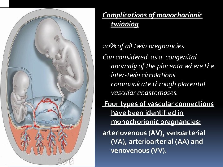
Complications of monochorionic twinning 20% of all twin pregnancies Can considered as a congenital anomaly of the placenta where the inter‐twin circulations communicate through placental vascular anastomoses. Four types of vascular connections have been identified in monochorionic pregnancies: arteriovenous (AV), venoarterial (VA), arterioarterial (AA) and venous (VV).
These occur in almost all monochorionic twin pregnancies. Bidirectional superficial arterial–arterial or venous– venous anastomoses may potentially compensate for any haemodynamic imbalance created by deep unidirectional arterial–venous anastomoses. Small inter‐twin ‘transfusions’ are thus likely to be a normal physiological event in monochorionic twins but the imbalance of flow between twins may be pathological and increasing perinatal mortality considerably
1 -Acute feto -fetal transfusion When one of a monochorionic twin dies in utero, 1‐There is a significant risk of ischaemic damage(18% in monochorionic twins) predominantly to the fetal brain although there are reports of damage to pulmonary and hepatic systems, intestinal atresia, limb reduction and renal necrosis. For twin pregnancies overall , risk of 9% for neurological abnormality
2‐ Death of one of a monochorionic twin substantially increases the risk of co‐twin demise in utero. fetuses have a six times greater risk of intrauterine fetal death (12%) following single fetal demise after 20 weeks’ gestation than initially surviving dichorionic twins
Gestational age at the time of intrauterine fetal death influences the extent and type of fetal brain injury. There is debate about whether development of such central nervous system (CNS) anomalies are gestationally dependent. However, the long‐term morbidity of such an event prior to 14 weeks is controversial and the association less pronounced. Once single intrauterine fetal death has occurred in a monochorionic twin, delivery should not be immediate. The pregnancy should be evaluated for secondary sequelae, initially by the use of ultrasound. Such assessment should be within a tertiary fetal medicine centre.
The assessment often includes the use of sequential ultrasound scanning (with increasingly the adjuvant investigation of fetal MRI) to prospectively evaluate the presence of CNS neuropathology that may develop up to 4 weeks after the sentinel event. dichorionic twins, discordant fetal compromise with risks of intrauterine demise in one twin have to be balanced against the adverse effects of iatrogenic prematurity in the co‐twin if delivery needs to be expedited in monochorionic twins.
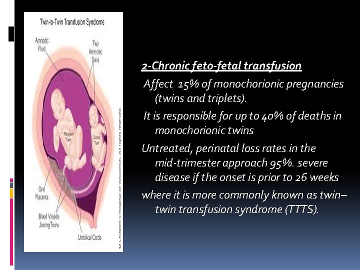
2 -Chronic feto-fetal transfusion Affect 15% of monochorionic pregnancies (twins and triplets). It is responsible for up to 40% of deaths in monochorionic twins Untreated, perinatal loss rates in the mid‐trimester approach 95%. severe disease if the onset is prior to 26 weeks where it is more commonly known as twin– twin transfusion syndrome (TTTS).
The underlying pathophysiology involves chronic shunting of blood from the donor twin to the recipient twin, leading to an inter‐twin haemodynamic imbalance. Such a vicious circle of events leads to the ‘donor’ twin becoming hypoperfused, growth restricted and with associated oliguria and the development of anhydramnios. The co‐twin, the ‘recipient’, becomes polyuric with often severe polyhydramnios and a hyperdynamic circulation that may cause both diastolic and systolic cardiac dysfunction, ending in the development of hydrops fetalis and death
Assessment of the a-wave in the DV at 11– 13 weeks’ gestation may help identify monochorionic pregnancies at risk of severe TTTS - as reversed a-wave is associated with an increased risk of developing severe TTTS, as well as other complications including aneuploidy, but due to high false positives, this is not widely advocated or must be interpreted with caution.
TTTS is diagnosed based on the following ultrasound criteria: 1 -Single placental mass (monochorionic ) 2 -Concordant gender. 3 -FFTS/TTTS is diagnosed when there is gross discordancy Oligohydramnios with maximum vertical pool (MVP) less than 2 cm in one sac and polyhydramnios in the other sac (MVP >8 cm). 4 -Discordant bladder appearances. 5 -Haemodynamic and cardiac compromise.
Quintero Staging system for FFTS/TTTS. Stage I Polyhydramnios /oligohydramnios with bladder of the donor still visible Stage II Bladder of the donor not visible Stage III Presence of absent end‐diastolic flow velocity in the umbilical artery, reverse flow in the ductus venosus or pulsatile umbilical venous flow in either twin Stage IV Hydrops in either twin Stage V Demise of one or both twin pregnancies may progress to the most adverse stages without warning
Monitoring for feto‐fetal transfusion syndrome Start diagnostic monitoring with ultrasound for feto‐ fetal transfusion syndrome (including to identify membrane folding) from 16 weeks Repeat monitoring fortnightly until 24 weeks. At every ultrasound examination, liquor volume in each of the amniotic sacs should be assessed and a deepest vertical pocket (DVP) depth measured and recorded, as well as the umbilical artery pulsatility index (UAPI). Fetal bladders should also be visualised
1‐. Although fi rst presentation of twin‐to‐twin transfusion syndrome (TTTS) is rare after 26+0 weeks of gestation, it can occur and therefore, scans should be performed at 2‐weekly intervals in uncomplicated monochorionic twins until delivery 2‐Carry out weekly monitoring of twin and triplet pregnancies with membrane folding or other possible early signs of feto‐fetal transfusion syndrome (specifically, pregnancies with intertwin membrane infolding and amniotic fluid discordance) to allow time to intervene if needed.
The principal clinical problem is severe polyhydramnios, which may be associated with premature rupture of membranes or preterm labour (or a combination of these) usually before 26 weeks’ gestation. Severe polycythaemia can occur, leading to fetal and placental thrombosis and hydrops fetalis in the anaemic twin. Rarely, mirror syndrome, the combination of fetal hydrops and maternal pre-eclampsia, has been reported. 2‐In addition, differences in inter‐twin haemodynamics may be associated with single or double twin demise (with antecedent CNS morbidity).
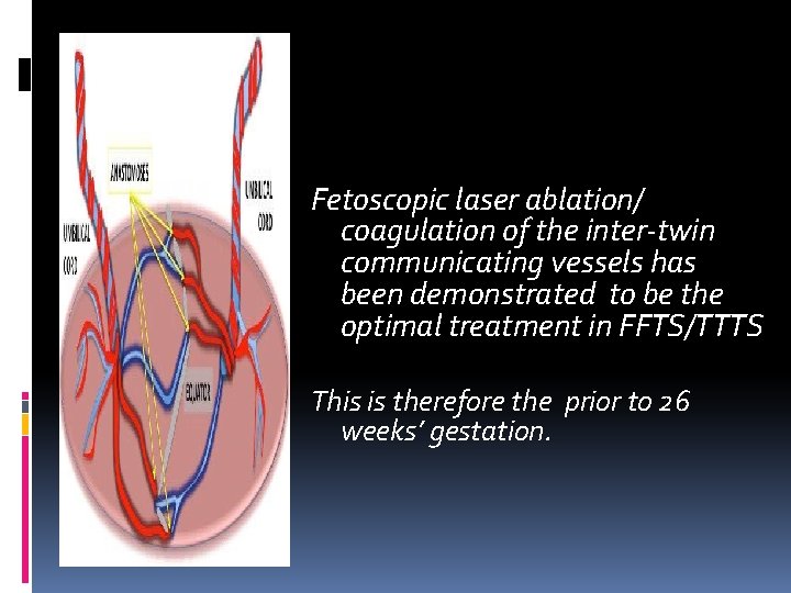
Fetoscopic laser ablation/ coagulation of the inter‐twin communicating vessels has been demonstrated to be the optimal treatment in FFTS/TTTS This is therefore the prior to 26 weeks’ gestation.
The procedure is performed either under local anaesthetic with intravenous sedation, with regional anaesthesia or occasionally under general anaesthesia. Under ultrasound guidance a 2– 3 mm diameter fetoscope is introduced into the amniotic cavity of the recipient twin. The location of the dividing twin membrane between the two amniotic cavities at the placental interface and the placental insertions of the umbilical cords are visualized. AV anastomoses are ablated using laser energy. Following the laser therapy, the fetoscope is removed an amnioreduction is performed until the amniotic fluid volume appears normal by ultrasound assessment.
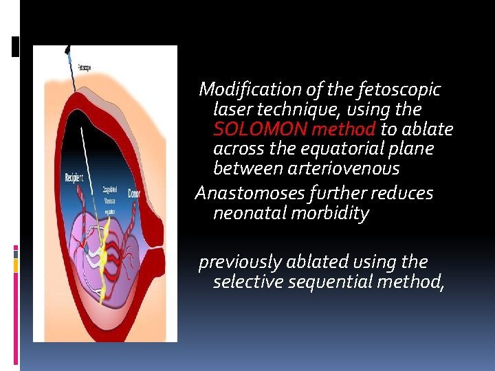
Modification of the fetoscopic laser technique, using the SOLOMON method to ablate across the equatorial plane between arteriovenous Anastomoses further reduces neonatal morbidity previously ablated using the selective sequential method,
3 -(TAPS) Twin anaemia polycythaemia sequence The incidence of TAPS occurring spontaneously in monochorionic diamniotic twins is up to 5%. it may complicate up to 13% of cases of TTTS following laser ablation. TAPS is believed to be due to the presence of Miniscule arteriovenous anastomoses (<1 mm) which allow slow transfusion of blood from the donor to the recipient, leading to highly discordant haemoglobin concentrations at birth
The postnatal diagnosis of TAPS is based on the finding of chronic anaemia (including reticulocytosis) in the donor and polycythaemia in the recipient. 1‐The criteria for diagnosis include a difference in haemoglobin concentration between the twins of more than 80 g/L 2‐ reticulocyte count ratio greater than 1. 7 or 3‐ small vascular anastomoses (<1 mm in diameter) in the placenta.
The prenatal diagnosis of TAPS is 1‐based on the finding of discordant middle cerebral artery (MCA) Doppler abnormalities, including an MCA peak systolic velocity (PSV) more than 1. 5 multiples of the median (Mo. M) in the donor suggesting fetal anaemia, and an MCA PSV less than 1. 0 Mo. M in the recipient, suggesting polycythaemia.
2‐ differences in placental echogenicity and thickness, with a bright thickened section for the donor twin and an echolucent thin recipient section. 3‐The polycythaemic twin might have a ‘starry sky’ appearance of the liver due to diminished echogenicity of the liver parenchyma and increased brightness of the portal venule walls.
The outcome of twin pregnancies complicated by TAPS is variable. 1‐Severe TAPS may result in intrauterine death of both twins. 2‐cases of severe cerebral damage have been reported in neonates with TAPS. Recent evidence suggests that in monochorionic twins at risk of neurodevelopmental delay is increased (20%) Therefore, brain imaging during the third trimester and neurodevelopmental assessment at the age of 2 years are recommended. 3‐mild TAPS may still allow the birth of two healthy neonates the main neonatal morbidity consists of anaemia (requiring transfusion)and polycythaemia partial exchange transfusion
The management of twin pregnancies complicated by TAPS should be individualized. 1‐ options depend on the gestational age at assessment, parental choice, the severity of the disease and technical feasibility of intrauterine therapy. The commonest options include 1‐conservative management, 2‐ early delivery, 3‐laser ablation 4‐intrauterine transfusion (IUT) for theanaemic twin, 5‐combined IUT for the anaemic twin and partial exchange transfusion to dilute the blood of the polycythaemic twin.
In order to screen for TAPS, The MCA PSV should be measured from 20 weeks onwards in both fetuses, and during the follow‐up of treated TTTS cases. Prevention of TAPS by modification of the fetoscopic laser ablation technique remains the best way to prevent morbidity.
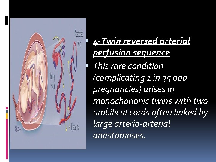
4 -Twin reversed arterial perfusion sequence This rare condition (complicating 1 in 35 000 pregnancies) arises in monochorionic twins with two umbilical cords often linked by large arterio‐arterial anastomoses.
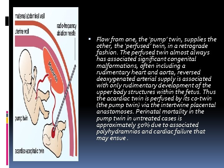
Flow from one, the ‘pump’ twin, supplies the other, the ‘perfused’ twin, in a retrograde fashion. The perfused twin almost always has associated significant congenital malformations, often including a rudimentary heart and aorta, reversed deoxygenated arterial supply is associated with only rudimentary development of the upper body structures within the fetus. Thus the acardiac twin is perfused by its co‐twin (the pump twin) via the intertwine placental anastomoses. Perinatal mortality in the pump twin in untreated cases is approximately 50% due to associated polyhydramnios and cardiac failure that may ensue.
1‐Although polyhydramnios may be elevated by amnioreduction, 2‐definitive treatments is occlusion of the perfused (acardiac) twin’s cord or rudimentary aorta may be achieved by a variety of fetoscopic or ultrasound‐guided techniques (intrafetal laser ablation, radiofrequency thermal ablation and fetoscopic cord occlusion).
There is still controversy whether to offer intervention electively, or only when signs of cardiac compromise occur, and at what gestational age intervention is best performed. Earlier intervention appears to be associated with a more advanced gestational age at birth. With careful case selection improved outcome for the pump twin is described in up to 85% complications include co‐twin demise or hypoperfusion (with cerebral morbidity) and/or preterm ruptured membranes.
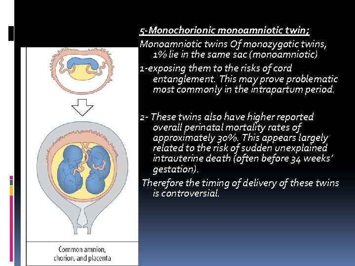
5 -Monochorionic monoamniotic twin; Monoamniotic twins Of monozygotic twins, 1% lie in the same sac (monoamniotic) 1‐exposing them to the risks of cord entanglement. This may prove problematic most commonly in the intrapartum period. 2‐ These twins also have higher reported overall perinatal mortality rates of approximately 30%. This appears largely related to the risk of sudden unexplained intrauterine death (often before 34 weeks’ gestation). Therefore the timing of delivery of these twins is controversial.
Treatment 1‐ suggested that use of prophylactic maternal sulindac (cyclooxygenase‐ 2 inhibitor) to reduce fetal urine output and thus amniotic fluid volume reduces the risk of cord entanglement 2‐ In combination with this, others have advocated hospitalization from 26– 28 weeks onwards and elective premature delivery of these twins between 32 and 34 weeks’ gestation. 3‐The most recent evidence indicates that the risk of these events are relatively low, with close outpatient surveillance being recommended rather than very early elective delivery 4‐most cases are delivered by elective caesarean section 5‐ suggests a course of prophylactic corticosteroids followed by elective caesarean delivery by 33 weeks in these rare twin pregnancies.
Labour and delivery of twins and multiple pregnancies it is best practice for intrapartum care to be discussed and a multidisplinary plan set out in the early third trimester of pregnancy. The indications for elective caesarean section are : . Congenital anomalies associated with significant risk of cephalopelvic disproportion (including conjoined twins) would be an obvious indication monoamniotic twins 3‐monochorionic pregnancies, complicated by placental anomalies associated with increased perinatal mortality (i. e. TTTS or TRAP), are generally delivered at 34– 36 weeks and usually by caesarean section
Relative indications Most current consensus based guidelines recommend delivery between 36 and 38 weeks’ gestation 1‐Perinatal mortality increases slightly after 38 weeks’ gestation in twins and there are therefore many obstetricians who elect for delivery then in dichorionic twin at 37 weeks and 0 days However, there are no data to indicate whether or not this rise in mortality applies to twins whose growth and well‐being are known to be normal on ultrasound. There is some evidence that the risk of perinatal loss is greater at the end of the third trimester in monochorionic twins compared with dichorionic twins so elective delivery 36 weeks and 0 days ,
Guid line triplet pregnancies elective birth from 35 weeks 0 days, after a course of antenatal corticosteroids has been offered. (all if continue will increases the risk of fetal death If elective birth is declined For women who decline elective birth, offer weekly appointments with the specialist obstetrician. At each appointment offer an ultrasound scan, and perform weekly biophysical profile assessments and fortnightly fetal growth scans 2‐Recent data have indicated that a planned elective caesarean section may achieve an up to 75% reduction in the risk of perinatal death compared with vaginal delivery by reducing risks of acidosis and anoxia (especially to the second twin)
3 -Those with previous caesarean section scars are probably best delivered by repeat caesarean section because of the greater risk of scar dehiscence/ rupture due to both uterine distension and intrauterine manipulation of the second twin. 4‐Caesarean section has been advised when the first twin is breech, which would obviate the rare risk of interlocking twins and entrapment of the head of a presenting breech above the second cephalic twin. The use of intrapartum ultrasound may allow detection of such problems with early recourse to emergency caesarean section. However, there is no evidence that vaginal delivery of a presenting breech that would otherwise satisfy criteria for vaginal delivery (EFW less than 3. 5– 4 kg, flexed head and not footling) is inappropriate in selected cases.
The presentation of the second twin is of no importance until after birth of the first. in twins where the first twin is cephalic presentation (at term), there may be the need for obstetric delivery of the second twin, with potentially an increased risk of perinatal morbidity. However, a Cochrane review (on delivery of the second twin not presenting cephalically) indicates that caesarean section increases maternal febrile morbidity without improving neonatal outcome This awaits critical appraisal. Caesarean section for second twin is occasionally indicated for disproportion, usually where the second twin is much bigger than the first 4%
Induction of labour is not contraindicated in twin pregnancies. Mode of delivery is decided on standard principles based on 1‐ presentation of the first twin (cephalic in 70%, breech in 30%) 2‐the documentation of optimal fetal growth 3‐well‐being.
Labour management; ‐continuous cardiotocography of both twins is best achieved by a combination of internal and external monitoring on a dual‐channel recorder. ‐An intravenous line is sited and maternal blood drawn for group and save, in view of the increased incidence of caesarean section and postpartum haemorrhage ‐Augmentation of labour with oxytocin may be used as in singletons. ‐An epidural anaesthetic is strongly advised in case of the unexpected need for internal manipulation of the second twin. If one is not sited, an anaesthetist will be required at delivery with early recourse to spinal or even general anaesthesia. ‐ The place of delivery is debatable but there is an increasing trend for twins to be delivered in operating theatres so that there is immediate redress to emergency caesarean section if necessary.
‐Delivery of the first twin proceeds as for a singleton. ‐ Its cord is clamped to prevent fetal haemorrhage (from the second twin along any placental anastomoses). ‐An experienced obstetrician discerns the presentation of the second twin, either by abdominal or vaginal examinationor, increasingly, by the use of transabdominal ultrasound. ‐Oblique or transverse lies are then converted to a longitudinal lie by external version and held in place by an assistant. ‐ Uterine contractions should be monitored and if necessary augmented using oxytocin. ‐The membranes should be left intact to facilitate version. ‐External cephalic version may be used to manipulate the fetal head over the pelvic inlet.
‐ Internal cephalic version is preferred as a primary procedure by many experienced obstetricians, as it seems to be associated with a higher success and lower complication rate than external cephalic version. ‐ One or preferably both feet are grasped and brought down into the vagina followed by an assisted breech delivery with contractions and maternal effort. Classically, intervals of greater than 30 min are acceptable providing the cardiotocograph is satisfactory and the presenting part is descending. After that the risk to the second twin is increased the greater the delay until delivery.
‐ Uterine inertia with a longitudinal lying second twin is corrected by oxytocininfusion. . ‐Fetal distress may be managed by ventouse delivery, even if the head is high ‐ breech extraction. The already stretched vaginal tissues after the birth of the first twin ‐ third stage of labour: An oxytocin infusion is given prophylactically to minimize the risks of postpartum haemorrhage.
Higher order multiples Perinatal and maternal risk increases exponentially with increasing fetal number. Most higher‐order multiple pregnancies are the result of assisted reproductive technologies and thus should be preventable with ‐closer monitoring of follicular response ‐ and single (or, at most, two) embryo transfers in IVF therapy. Indeed, there are proven arguments for restricting the number of embryos transferred to one in order to minimize twin and triplet risk, and this course of action appears to have limited adverse effect on live birth rates when more cycles are allowed
Every woman/couple with a higher‐order multiple pregnancy should have a discussion with a senior obstetrician relating to increased maternal and perinatal risks. ‐ This should involve the discussion and option of multiple fetal pregnancy reduction. ‐ In addition to perinatal mortality rates ‐ parents should be counselled as to the mean gestational age at delivery (33 weeks for triplets, 31 weeks for quadruplets). ‐ with severe neurological sequelae rates of 12% and 25% (respectively) in survivors
‐The chief perceived disadvantage of multiple fetal pregnancy reduction, usually accomplished by administration of a percutaneous fetal intrathoracic injection of abortifacient (commonly potassium chloride), is complete miscarriage. with rates for starting triplets and quadruplets of 7% and 15%, respectively There is now a consensus that multifetal pregnancy reduction between 10 and 12 weeks should be recommended for quadruplets and higher multiples so as to lower both maternal and fetal risks. The situation with triplets has been more controversial ‐with many considering this a social issue for parents.
‐Higher‐order multiple pregnancies should be managed in tertiary perinatal centres with a fetal medicine service. ‐Care is almost always individualized. Management is along standard lines for twins but with greater emphasis on preventing preterm delivery and on monitoring fetal growth and well‐being.
Although there have been successful reports of triplets and even quadruplets being delivered vaginally, most higher‐order pregnancies are now delivered by caesarean section. This alleviates difficulties with electronic fetal monitoring, avoids unrecognized hypoxaemia (especially given the high incidence of IUGR) and prevents birth trauma from manipulative delivery of non‐cephalic presenting fetuses. Given the higher incidence of preterm labour in the midtrimester, the option after delivery of the presenting fetus of conservative management with passive retention of residual fetus to prolong gestational age should be considered
The concept of a multiple pregnancy clinic Increasingly, there is consensus opinion that the management of multiple pregnancies should be concentrated in a designated ‘multiple pregnancy clinic’ with experienced midwifery and obstetric discussion and decision‐making and with access to immediate diagnostic ultrasound and multidisciplinary opinions (i. e. anaesthetic, neonatal paediatric and psychological services).
Thank you