1986 1987 Mini Pathria Michael Zlatkin Richard Rick
- Slides: 41
1986 -1987 • • • Mini Pathria Michael Zlatkin Richard (Rick) G Stiles
Mini Pathria • Middle-aged male • Hx of recent knee trauma
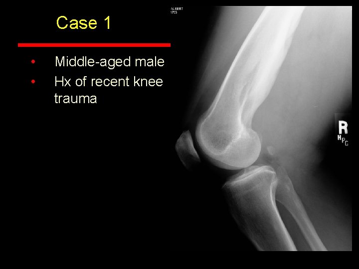
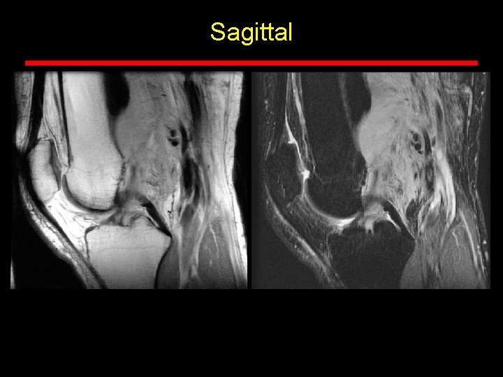
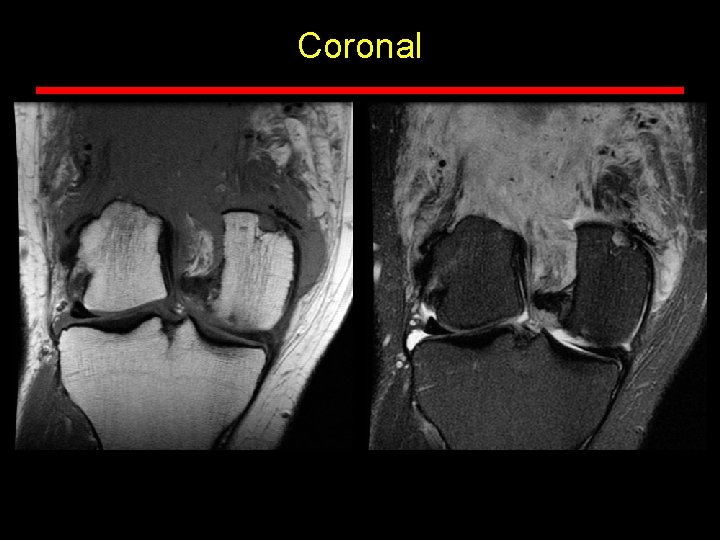
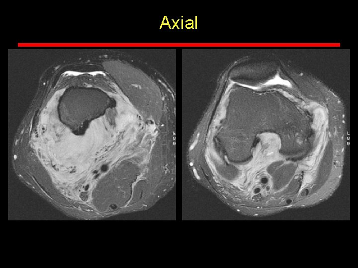
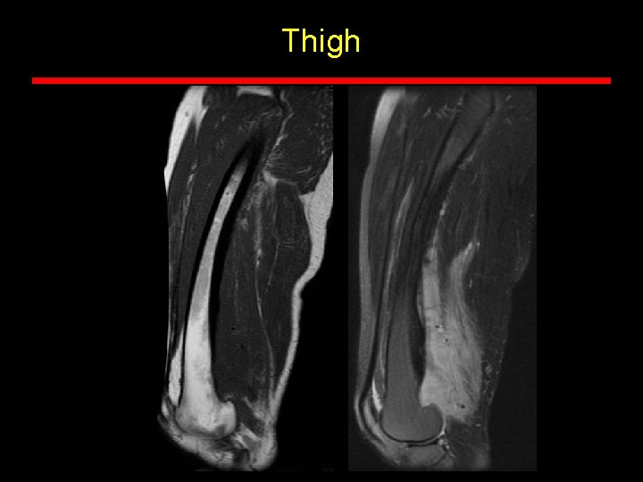
Case 1 • • Middle-aged male Hx of recent knee trauma
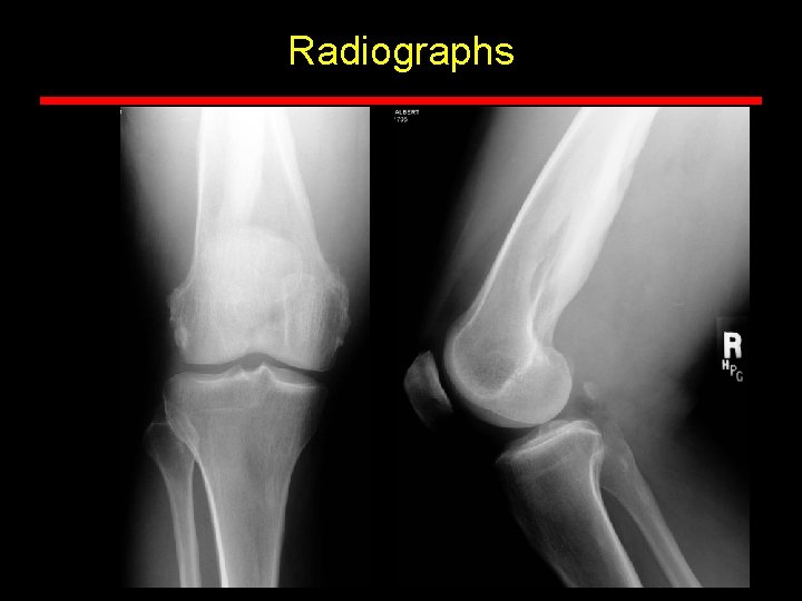
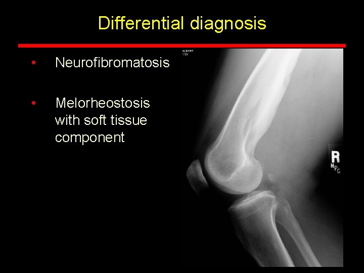
Radiographs
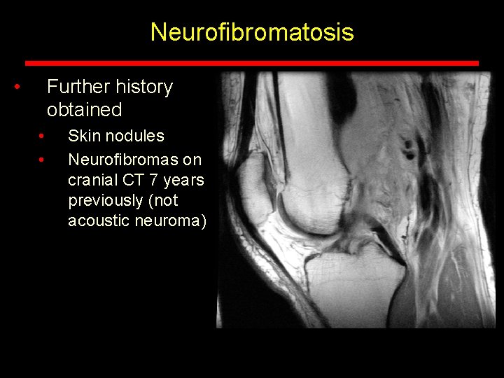
Sagittal
Coronal
Axial
Thigh
Differential diagnosis • Neurofibromatosis • Melorheostosis with soft tissue component
Neurofibromatosis • Further history obtained • • Skin nodules Neurofibromas on cranial CT 7 years previously (not acoustic neuroma)
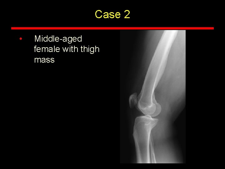
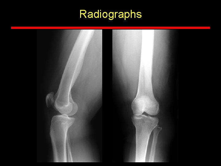
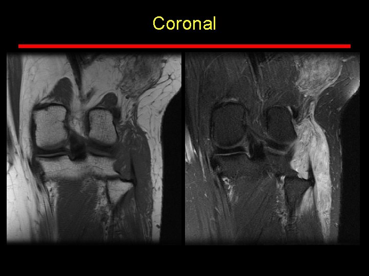
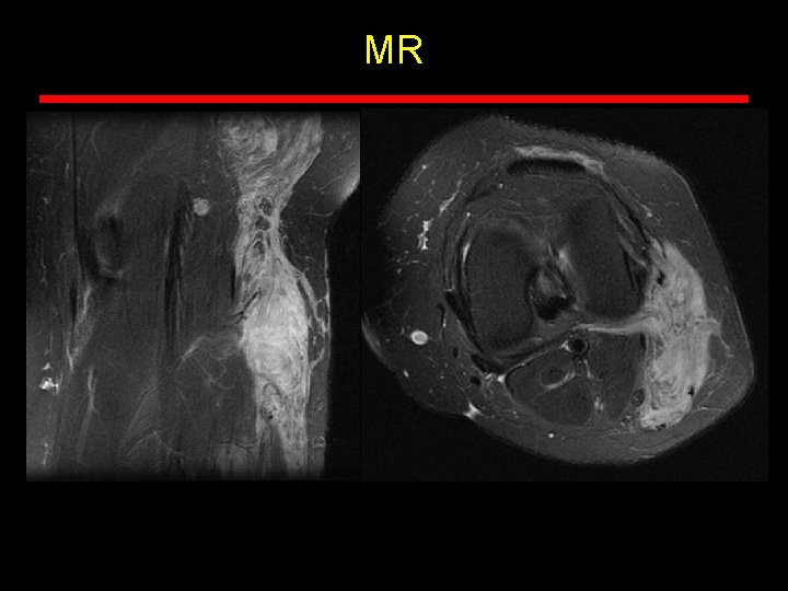
Case 2 • Middle-aged female with thigh mass
Radiographs
Coronal
MR
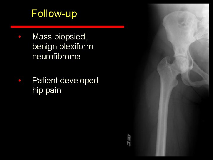
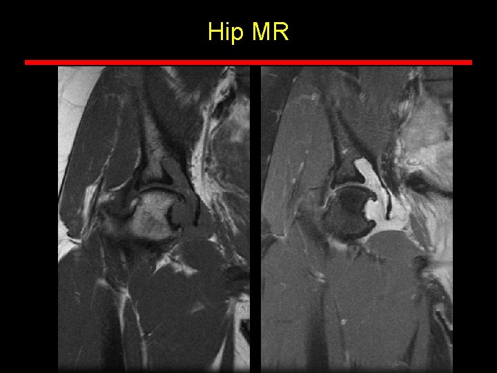
Follow-up • Mass biopsied, benign plexiform neurofibroma • Patient developed hip pain
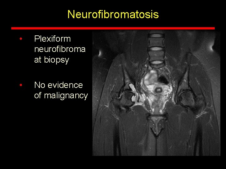
Hip MR
Neurofibromatosis • Plexiform neurofibroma at biopsy • No evidence of malignancy
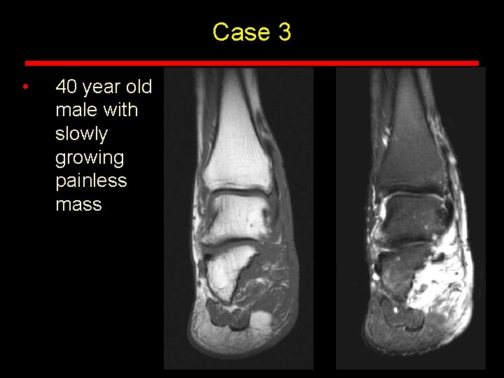
Case 3 • 40 year old male with slowly growing painless mass
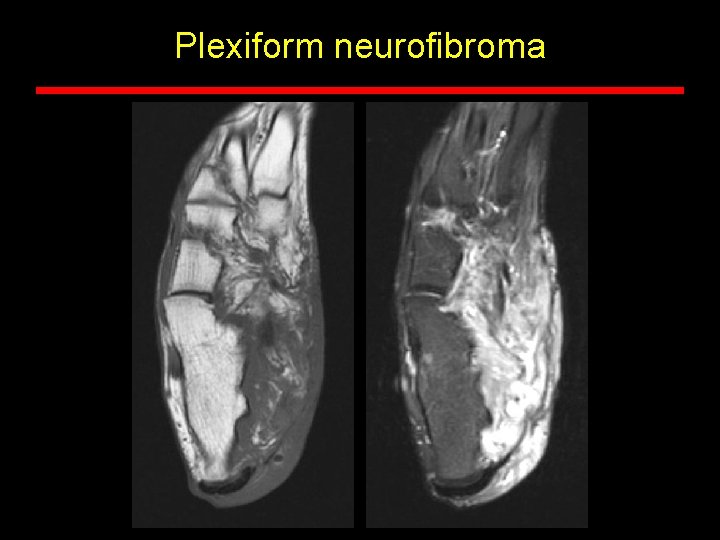
Plexiform neurofibroma
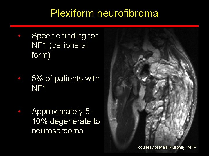
Plexiform neurofibroma • Specific finding for NF 1 (peripheral form) • 5% of patients with NF 1 • Approximately 510% degenerate to neurosarcoma courtesy of Mark Murphey, AFIP
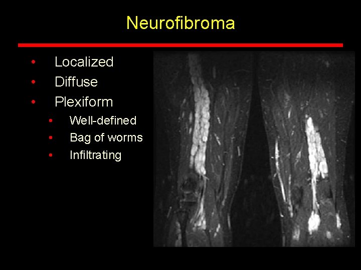
Neurofibroma • • • Localized Diffuse Plexiform • • • Well-defined Bag of worms Infiltrating
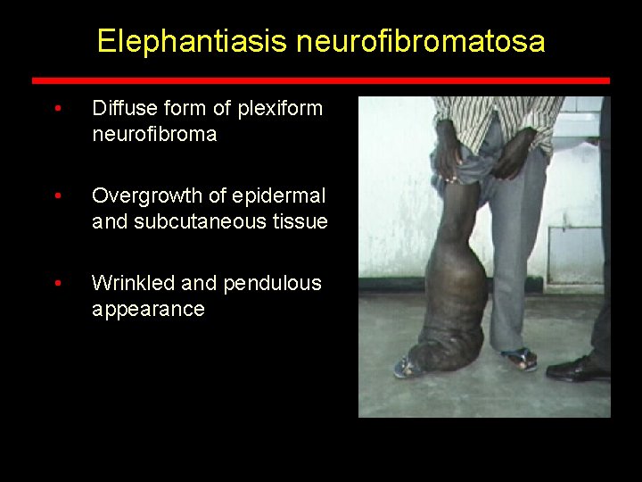
Elephantiasis neurofibromatosa • Diffuse form of plexiform neurofibroma • Overgrowth of epidermal and subcutaneous tissue • Wrinkled and pendulous appearance
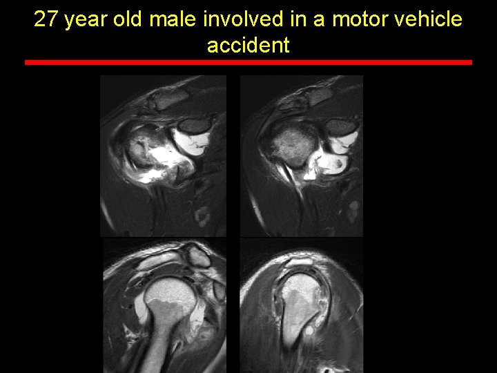
Michael B. Zlatkin and Alfredo Arraut NMSI • 27 year old male involved in a motor vehicle accident
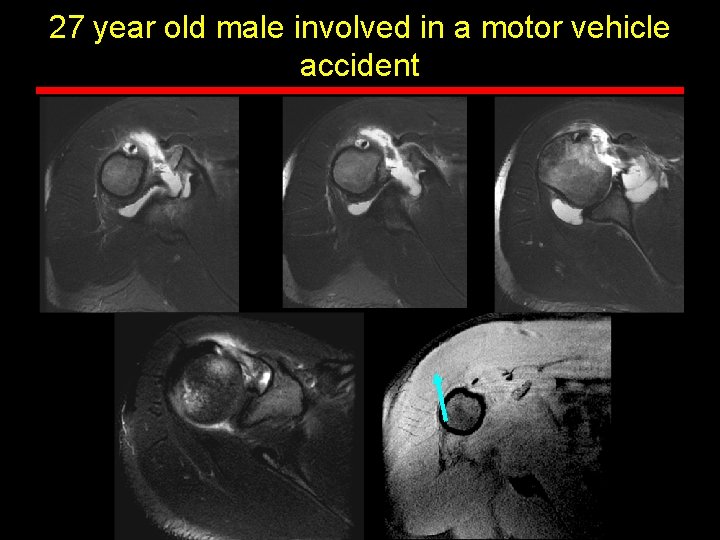
27 year old male involved in a motor vehicle accident
27 year old male involved in a motor vehicle accident
Findings • Subscapularis tendon is avulsed with bone from the lesser tuberosity • Humeral avulsion of the anterior band of the glenohumeral ligament (HAGL) • Middle glenohumeral ligament and possibly the superior glenohumral ligament may be avulsed as well • Possible reverse Hill-Sachs lesion
Subscap Rupture and HAGL (BHAGL) • Rupture of the subscapularis tendon uncommon but can be seen in younger patients injured by forced external rotation or extension of a partially abducted arm • Tears occur near insertion on the lesser tub. May occur at sup margin, where tendon may be weakened by degeneration • Avulsion fracture of the lesser tuberosity may occur, and is displaced medially and inferiorly • Subscapularis avulsions are also associated with injury to the anterior capsule and glenohumeral ligaments (HAGL) • When the AIGHL avulses a fragment of bone from the humerus, the lesion is known as a bony HAGL, or BHAGL • ? With MGHL and SGHL torn = Super BHAGL
Michael B. Zlatkin and Alfredo Arraut NMSI • 17 year old male with bony growth on the dorsum of hand
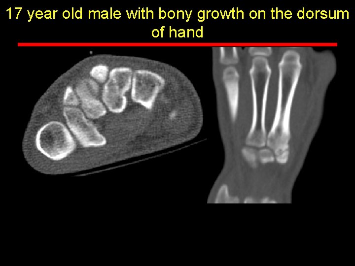
17 year old male with bony growth on the dorsum of hand
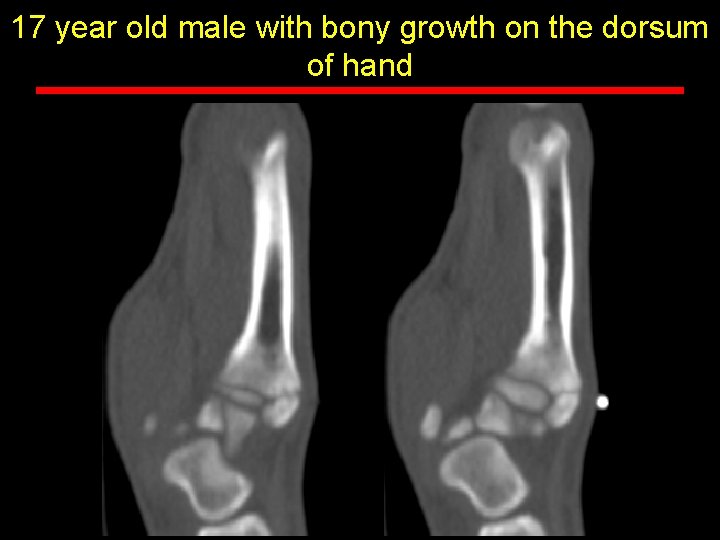
17 year old male with bony growth on the dorsum of hand
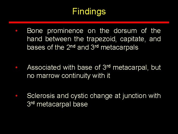
Findings • Bone prominence on the dorsum of the hand between the trapezoid, capitate, and bases of the 2 nd and 3 rd metacarpals • Associated with base of 3 rd metacarpal, but no marrow continuity with it • Sclerosis and cystic change at junction with 3 rd metacarpal base
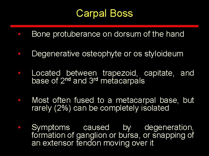
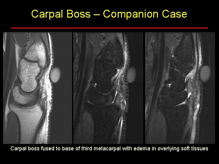
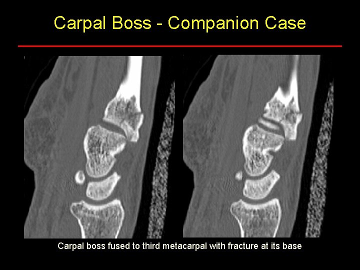
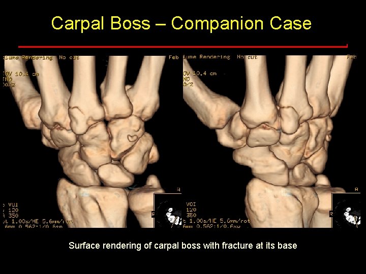
Carpal Boss • Bone protuberance on dorsum of the hand • Degenerative osteophyte or os styloideum • Located between trapezoid, capitate, and base of 2 nd and 3 rd metacarpals • Most often fused to a metacarpal base, but rarely (2%) can be completely isolated • Symptoms caused by degeneration, formation of ganglion or bursa, or snapping of an extensor tendon moving over it
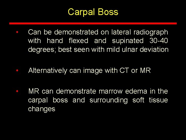
Carpal Boss • Can be demonstrated on lateral radiograph with hand flexed and supinated 30 -40 degrees; best seen with mild ulnar deviation • Alternatively can image with CT or MR • MR can demonstrate marrow edema in the carpal boss and surrounding soft tissue changes
Carpal Boss – Companion Case Carpal boss fused to base of third metacarpal with edema in overlying soft tissues
Carpal Boss - Companion Case Carpal boss fused to third metacarpal with fracture at its base
Carpal Boss – Companion Case Surface rendering of carpal boss with fracture at its base
Michael B. Zlatkin and Alfredo Arraut NMSI • 33 year old professional hockey player with pain in the flank after practice
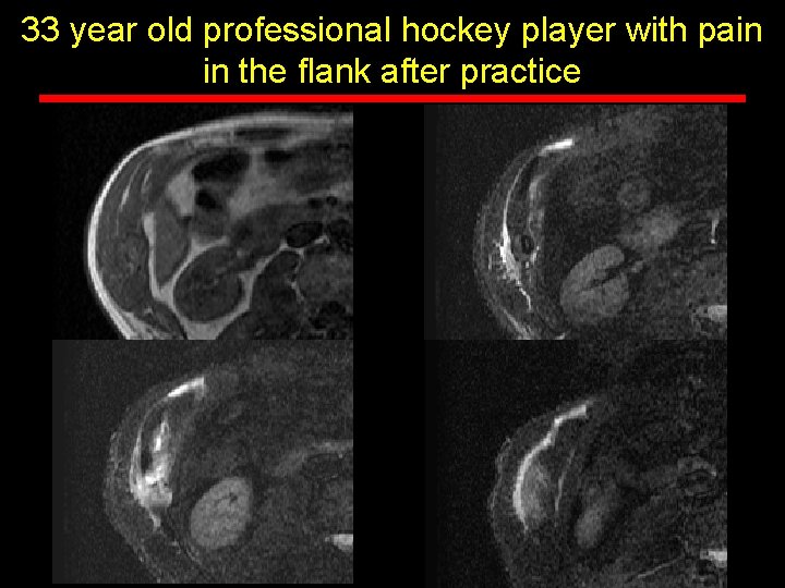
33 year old professional hockey player with pain in the flank after practice
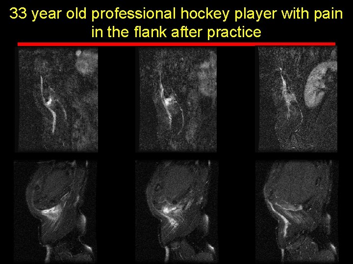
33 year old professional hockey player with pain in the flank after practice
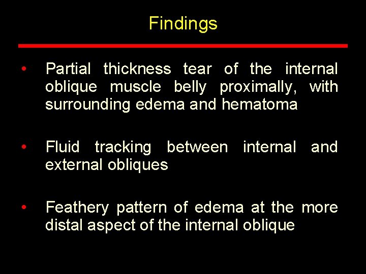
Findings • Partial thickness tear of the internal oblique muscle belly proximally, with surrounding edema and hematoma • Fluid tracking between internal and external obliques • Feathery pattern of edema at the more distal aspect of the internal oblique
Side Strain • Uncommon sporting injury presenting with pain and tenderness over anterolateral and posterolateral lower ribcage • Associated with cricket, golf, and ice hockey – eccentric contraction of trunk muscles • Partial or complete tear of lateral abdominal wall musculature (internal oblique > external oblique > transversus abdominis) • May also see avulsion at the muscular origins from the lower ribs • Recovery takes 6 -10 weeks. Full recovery is the norm