Trichinella spiralis T spiralis is the smallest human


. 1. 5 mm x")
 harboring")


")
 Pathogenic")


- Slides: 21
Trichinella spiralis T. spiralis is the smallest human nematode. The adults and larvae live in the same host, but they have to change a host to complete their life cycle. The disease that Trichinella spp. cause is referred to as trichinellosis or trichinosis, a zoonosis, which is spread by mammals that kill each other. Human infections result from eating raw or undercooked meat.
I. Morphology ♂1. 5 mm, ♀3 -4 mm, both have a single set of reproductive organs. 1. Adults: 2. Juvenile (larva): 124× 6 µm, one or more coil in a cyst in the skeletal muscle fibers. Newborn larva of T. spiralis
Adult male T. spiralis. Note claspers on tail (lower end). 1. 5 mm x 36 µm. Adult female T. spiralis. 3 mm x 36 µm. Note fully formed larvae in uterus.
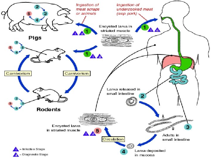
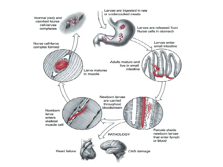
II. Life cycle Infection is initiated by ingesting raw or undercooked meats (pork) harboring the Nurse cell-larva complex. Larvae are released from muscle tissue by digestive enzymes in the stomach, and then locate to the upper two-thirds of the small intestine. The immature parasites penetrate the columnar epithelium at the base of the villus. They live within a row of these cells. Larvae molt four times, developing into adults. After mating, adult females produce newborn larvae which enter the lamina propria, to the bloodstream and become distributed throughout the body. Skeletal muscle cells are the only in which the parasites remain inside them after invasion, growth and development of the larva occurs. T. spiralis is infective by the 14 th day of infection.
Life cycle: 1. Infective stage: Encysted larva 2. Habitat: adults in small intestine (mainly in duodenum and jejunum), juveniles in skeletal muscles 3. Route of infection: by mouth 4. Life span of female: 1 -2 months 5. Final host and intermediate host: person 6. Reservoir host: pigs, cats, dogs, mice etc.
Newborn larva of T. spiralis entering muscle cell An adult female T. spiralis in situ
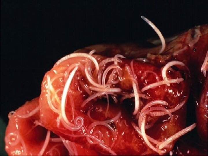
Infective larva of T. spiralis in muscle tissue
III. Clinical manifestation The process of the pathogenesis may be divided into 3 stages: 1. Invading stage(about 1 week): The damage is mainly found in the intestine. In this stage, abdominal pain, nausea, vomiting, diarrhea and fever may occur. 2. Migrating stage of the juveniles(2 -3 weeks): The damage is mainly in the skeletal muscles. In this stage, muscular pain with high fever is main symptoms, especially in active muscles. Wandering juveniles may also cause pneumonitis, pleurisy, encephalitis, nephritis and myocarditis etc. 3. Encysted stage (4 -16 weeks): In this stage, only muscular pain present without other symptoms.
IV. Diagnosis • Muscular biopsy • Examination of left food and xenodiagnosis; • Immunodiagnosis: ELISA & IHAT V. Treatment Albendazole and Mebendazole, cortisone, analgesics VII. Prevention • Quarantine of meat; • Avoid eating raw meat and • Avoid feeding of animals on raw meat
Trichuris trichiura ( Whipworm)
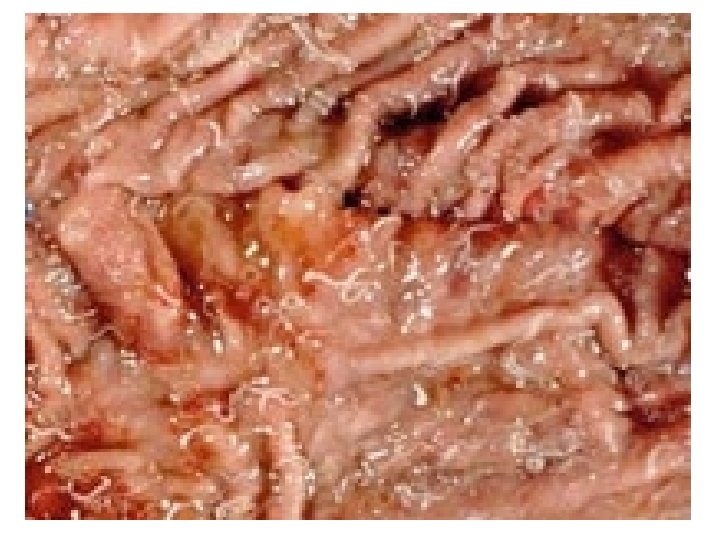
Disease: Trichuriasis Geographical Distribution: World wide Definitive host: man Habitat: large intestine (caecum) Pathogenic stage: adult. Adult embed their anterior thin portion into the mucosa and sub-mucosa. The life span of the adult is about 3 -5 years. No intermediate host and no reservoir host.
I. Morphology Adult: -looks like whip, cellular esophagus -The anterior 3/5 is slender - The posterior 2/5 is thick. -The female worm is 3 -5 cm. -The male is smaller and has a curved tail and one spicule. - Genitalia: one set
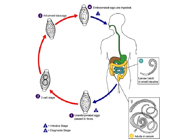
ü Egg: • it is barrel in shape • 50 x 20µm in size. • brownish • has a translucent polar plug at either ends. • The content is an undeveloped cell stage
II. Life Cycle 1. Infective stage: embryonated egg 2. Infective mode: orally by consumption of contaminated food & drink. 3. Diagnostic stage: immature egg Adults deposit Eggs Larvae hatch out develop 3 -10 days 3 weeks infective in small intestine ingested by man invade the intestinal wall return to the intestinal lumen Adults in caecum
III. Pathogenesis: ü Adult embed their anterior thin portion into the mucosa and submucosa extensive inflammation, traumatic & haemorrhagic effects at points of attachments secondary bacterial infection ulcers & abscess. This leads to the following clinical picture: ü Light infection: Asymptomatic ü Heavy infection: symptomatic abdominal pain, dysentry, tenesmus Complications: - Chronic dysentery - rectal prolapse - appendicitis
IV. Diagnosis: Detection of eggs in feces V. Treatment : Mebendazole 3 days VI. Prevention & Control: - Mass treatment - Sanitary disposal of feces - Avoid human fertilizers - Proper washing of vegetables - Pure water supply - Health education - Control of flies