Trichinella spiralis SALIENT FEATURES v COMMON NAME Trichina


- Slides: 34
Trichinella spiralis
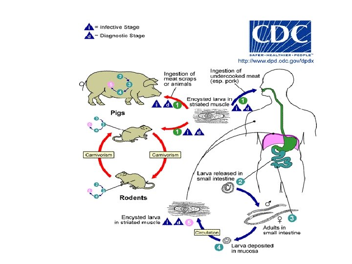
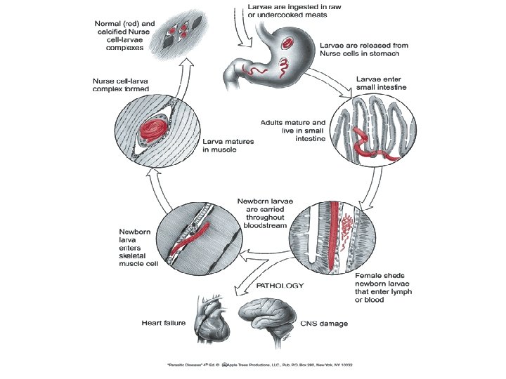
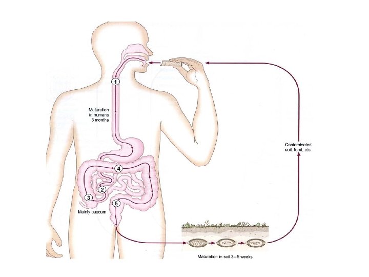
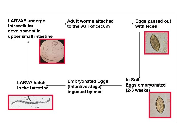
SALIENT FEATURES v COMMON NAME: Trichina worm- The Pork worm v Trichinella spiralis means spira, how this coils up in its host v DISEASE: Trichinosis v It is a zoonotic disease v HABITAT: Adult worms-Mucosa of small intestinal Encysted larvae- Striated muscles of hosts v The same animal acts as definitive and intermediated host v Is most common in Europe, North America, and Asia v INFECTIVE STAGE: Contaminated meat (muscle) containing encysted larvae (pig) v DIAGNOSTIC STAGE: Larvae encysted in muscle (human) v Can be fatal if large numbers of cysts form in the heart muscle.
MORPHOLOGY
LARVAE
VIVIPAROUS Expel active larvae
Epidemiology q The disease occurs among pigs, rats & humans q Rats and pigs feeding on garbage that includes infected offal q Dead or dying infected rats are themselves eaten by pigs q Raw or poorly cooked pork (sausage) harboring infective larvae then become the vehicle for human infections q Trichinellosis is a cosmopolitan disease that occurs most commonly in Europe and the US
PATHOGENESIS: Penetration of the adult females into mucosa The first symptoms appear between 1 - 2 days after ingestion The worms migrate in the intestinal epithelium Inflammation of duodenal and jejunal mucosa • • • This causes: INFLAMMATION NAUSEA VOMITING SWEATING DIARRHEA
The migrating larvae Ten days after infection the larvae will penetrate the muscle fibers & other parts of the body. • Muscular pain • Difficulty breathing • • Per orbital edema and conjunctivitis Heart (Myocarditis) Lungs (Pneumonitis) Brain (Encephalitis) – Can be fatal if large numbers of cysts form in the heart muscle. – Heart failure or respiratory or kidney malfunction
DIAGNOSIS : q Muscle biopsy at the encystment stage q Blood test for eosinophilia q Increased levels of creatine phosphokinase (CPK) q Serology test Immunoassays, such as ELISA q At the diarrheal stage, adult and larvae may be found in faeces
TREATMENT ü THIABENDAZOLE ü MEBENDAZOLE ü CORTICOSTEROIDS
Trichuris trichiura Whip worm
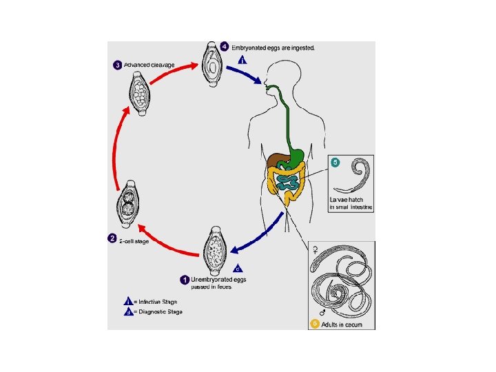
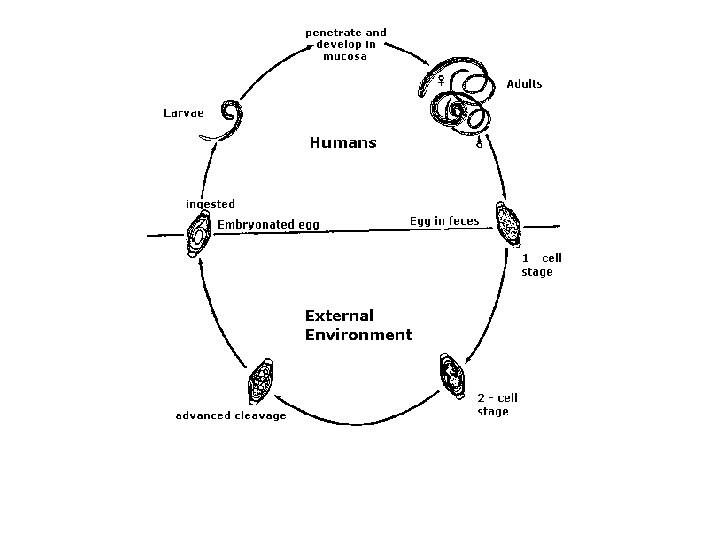
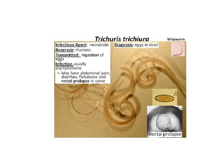
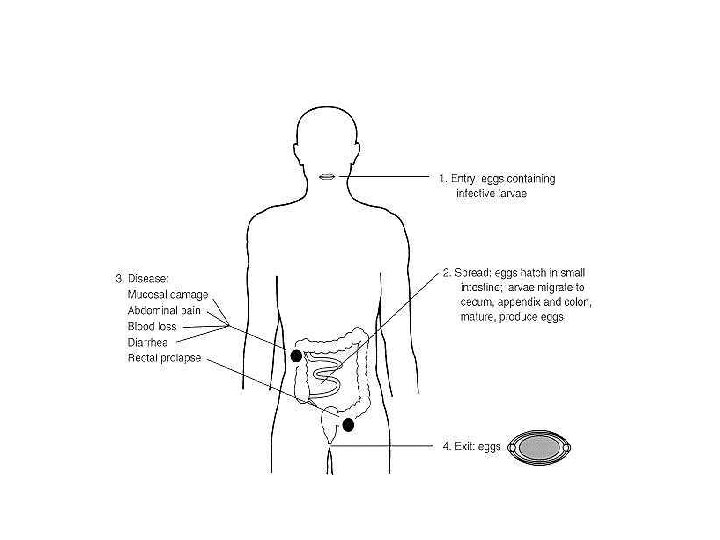
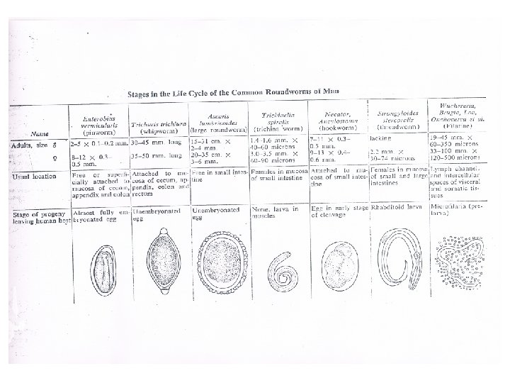
Trichuris trichiura Common name: whip worm Disease: trichuriasis, whip worm infection Final host: human, dogs, pig, monkey Habitat: large intestinal ( cecum, appendix, rectum) Geographical distribution: Cosmopolitan with poor sanitation. Children are more likely to be infected than adults because they are more likely to have close physical contact with contaminated soil q Infective stage: infective larva in egg q Transmission occurs through ingestion of eggs, usually on contaminated vegetables or soil. q Diagnostic stage: Egg barrel shape with polar plugs q q q
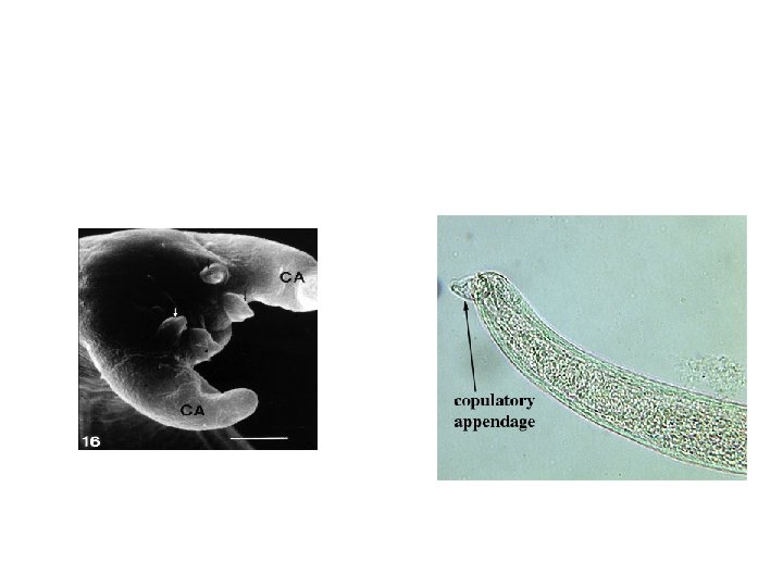
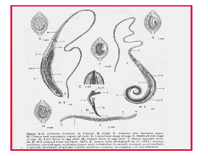
Morphology: Adult female worm: The anterior two-thirds of the body being very thin (looks like a whip) and the remaining posterior end is thick and linear. Size: 3. 5 -5 cm in length Adult Male worm: smaller than the female, 3. 0 -3. 5 cm. The posterior end is curved and has a single spicule enveloped with sheath.
q The anterior end two-thirds of the body being very thin (looks like a whip). q Adult worm penetrates into and embed its whip-like anterior portion in the intestinal mucosa, By small spear Adult female -Longer than the male. - posterior end is thick and linear. Adult male -Shorter than the female. posterior end curved and -has a single spicule enveloped with sheath. .
posterior end curved and has a single spicule enveloped with sheath
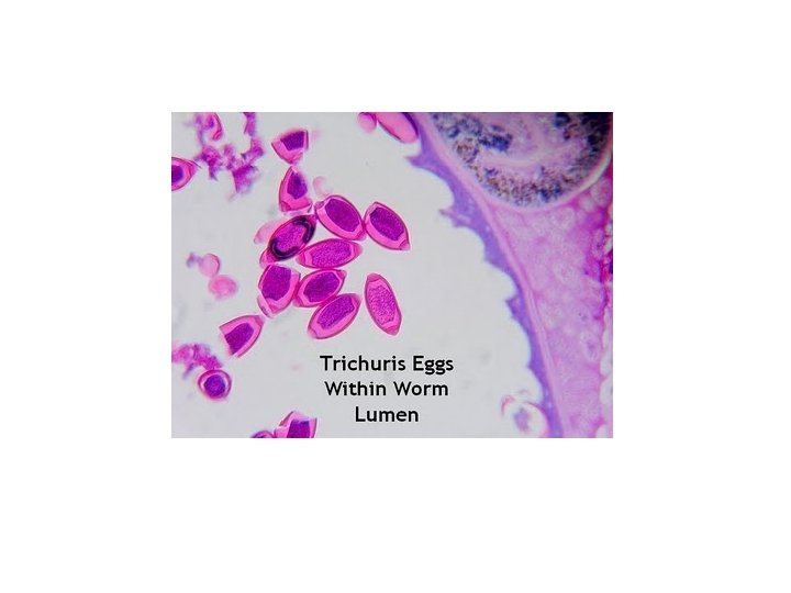
Eggs: Shape: barrel–shaped Size: 50 -55 x 25 -30μm Shell: thick egg shell with 2 polar plugs Color: Yellow-brown Content: immature egg cells 3000 -10000 eggs daily output
Life cycle: • Eggs pass out immature • Embryo develops inside the egg (that takes about 3 weeks at 25 C) • Mature eggs swallowed 1 st stage larvae hatch in small intestine and penetrate villi • Then migrate to large intestine and attach to mucosa with the thin anterior end • After 2 -4 month females mature and lay eggs.
Pathology: Light infection with Trichiuris are asymptomatic Heavier infections are characterized by 1 - diarrhea, 2 - anorexia, 3 - nausea 4 - abdominal pain 5 - anemia may be the result of hemorrhaging when the worms mucosal damage)(penetrate the intestinal wall Rectal prolapse. Children’s infection cause rectal prolapse, The reason is the cecum is damaged by the worm, the cecum can be pushed out from the anus.
Prolapsed Rectum
Laboratory diagnosis 1 - Eggs or worm in feces. Eggs are oval, barrel shaped, 2 - Eosinophilia may occur. 3 - In heavy infection proctoscopy or sigmoidoscopy, can show the worms attached to the mucosa. 4 - Visual detection of adult worms on prolapsed rectum.