The Heart and Circulatory System HEART CIRCULATION BLOOD

The Heart and Circulatory System HEART, CIRCULATION, BLOOD VESSELS BIOLOGY Mrs. S. Haughton

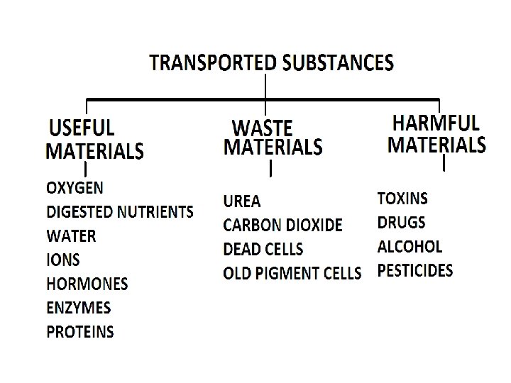
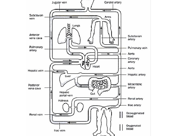
What is a transport system? • A transport system consists of: 1 a pump (e. g. the heart) 2 a series of channels (e. g blood vessels) 3 a fluid (e. g. blood) • Transport systems are necessary to carry useful, harmful and waste substances around an organism rapidly.

The Heart • The heart is a fist sized organ between the lungs closer to the left side of the chest.

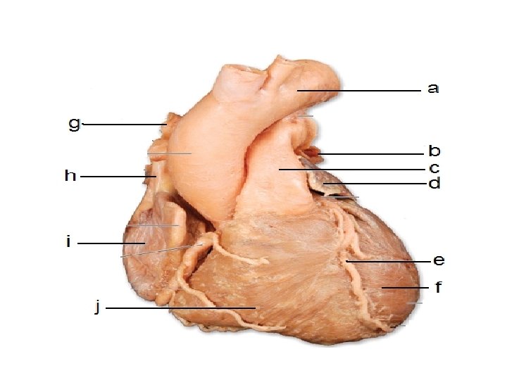
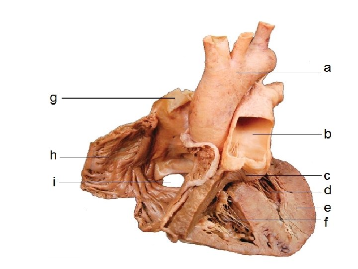
External Structure of the Heart

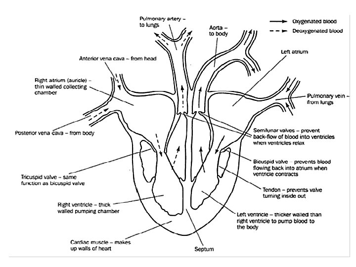
Internal Structure of the Heart • The heart has four chambers: the left and right atria (1 atrium) and the left and right ventricles. • The following diagram is the internal structure of the heart.
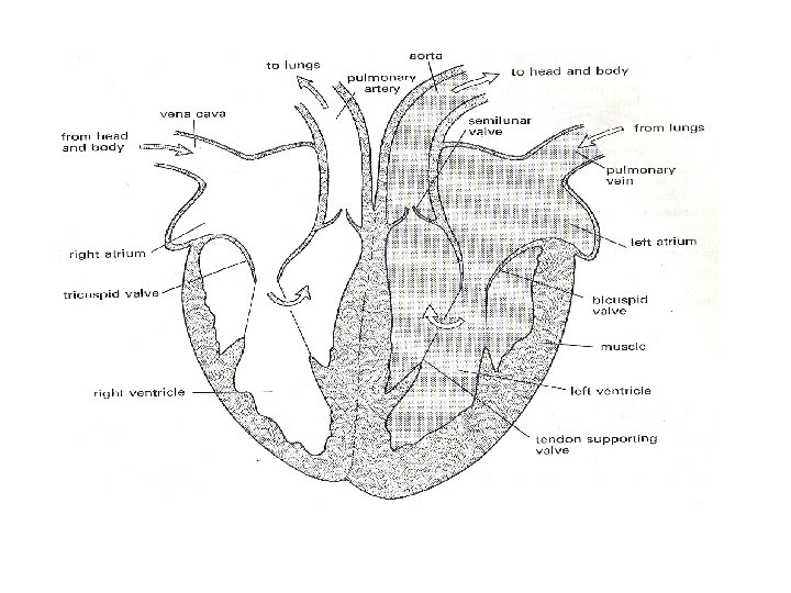
 accepts and pumps out oxygenated blood.")
• Remember that the left side (RED) accepts and pumps out oxygenated blood. • The right side of the heart (BLUE) accepts and pumps out deoxygenated blood.

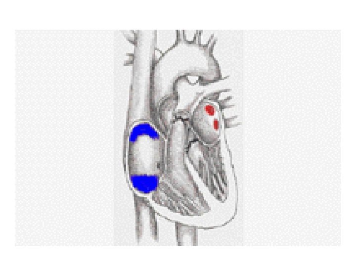
VALVES IN THE HEART • There are four valves in the heart: – Two semilunar valves, (one in each side of the heart). – Two atrioventricular valves (one each found between the atria and ventricles on both sides of the heart). – The function of the valves in the heart are to direct the flow of blood in only one direction.

The Heart Summary Fist sized; found on left side. Consists of cardiac muscle fed by coronary arteries Left side pumps O 2 -rich blood to head and body; Right side pumps O 2 -poor blood to lungs Two upper atria, two lower ventricles Septum separates left and right sides ensuring diffusion gradient • Left ventricle thicker to pump blood all over body • Valves prevent back-flow of blood • • •

OXYGENATED BLOOD • This is blood that is rich in oxygen because it has recently left the lungs where the process of gaseous exchange occurred in order for the oxygen to diffuse into the blood vessels. • The LEFT side of the heart pumps oxygenated blood which is very red.

DEOXYGENATED BLOOD • This is blood that is depleted of oxygen because it has travelled around the body supplying the body cells with the oxygen. • The RIGHT side of the heart pumps deoxygenated blood which has a slightly blue colour.
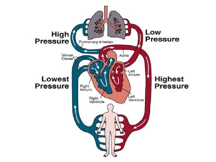

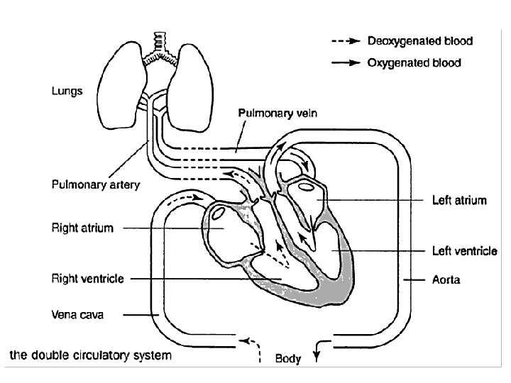
Circulation Summary • Consists of heart, blood and blood vessels • Double-circulation as blood travels twice through heart in one journey • Pulmonary circulation (heart lungs) • Systemic circulation (Heart body) • Oxygenated (O 2 rich) heart to body • Deoxygenated (O 2 poor) body to heart

Path of Oxygenated Blood • Pulmonary vein left atrium • Through mitral valve down into the left ventricle. • The ventricle then contracts forcing the mitral valve closed and opening the semilunar valve. • The blood then flows up into the aorta and around the body.

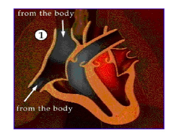
Path of Deoxygenated blood • Vena cava right atrium • Through tricuspid valve right ventricle. • Ventricle contracts, tricuspid valve closes, semilunar valve opens. • Blood flows to the pulmonary artery lungs.

THE HEARTBEAT • The heartbeat occurs in three stages and takes 0. 8 second to complete one cycle: • DIASTOLE: when the entire heart is at rest (0. 3 second) • ATRIAL SYSTOLE: after blood fills the atria and they contract to force blood down into the ventricles (0. 2 second). • VENTRICULAR SYSTOLE: after blood fills the ventricles and they contract to force blood out of the heart (0. 3 second).

Cardiac Cycle

SUMMARY • Heart muscle contract- systole • Heart muscle relax- diastole • Pacemaker or sinu-atrial node found at junction between vena cava and right atrium initiates heartbeat.

THREE MAJOR BLOOD VESSELS • The three major blood vessels are called the arteries, capillaries and veins.

PROPERT ARTERY Y Function Wall Lumen Pressure Blood direction Blood Valves CAPILLARY VEIN

ARTERIES vs. VEINS • All arteries carry blood AWAY from the heart. • Most arteries carry oxygenated blood. • Arteries have very thick external and muscular walls. • Arteries have small lumen with smooth interior walls. • All veins carry blood TO the heart. • Most veins carry deoxygenated blood. • have fairly thin external and muscular walls. • Veins have wide lumen with smooth interior walls and VALVES.

HEPATIC VEIN AND HEPATIC PORTAL VEIN • There is no mesenteric artery, because it would be a waste of time and nutrients if digested food were to go back directly to the heart without being filtered by the liver. • Therefore, deoxygenated blood from the gut goes to the kidney by a special vein called the hepatic portal vein. • Afterwards, the hepatic vein carries the filtered blood back the heart.

BLOOD COMPONENTS
- Slides: 32