Structure and Function of Skeletal Muscle Skeletal Muscle


- Slides: 43
Structure and Function of Skeletal Muscle
Skeletal Muscle § Human body contains over 400 skeletal muscles – 40 -50% of total body weight § Functions of skeletal muscle – Force production for locomotion and breathing – Force production for postural support – Heat production during cold stress
Movement and breathing § How do our muscles help us breath? § https: //www. youtube. com/watch? v=O 3 n. LJg RO-d 8 § Internal pressure is changed when the ribs and cavity expand.
Postural Support § Postural muscles are constantly contracting and relaxing different fibers so that you can stay upright.
Heat production § Muscles are the main site of energy use. § When we become too cold we shiver – This is because muscle action uses energy and that energy creates heat. – That is why our body naturally reacts to shiver when we are cold.
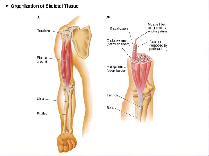
Structure of Skeletal Muscle: Connective Tissue Covering § Facia – Outer layer that hold muscles down § Epimysium – Surrounds entire muscle § Perimysium – Surrounds bundles of muscle fibers § Fascicles § Endomysium – Surrounds individual muscle fibers
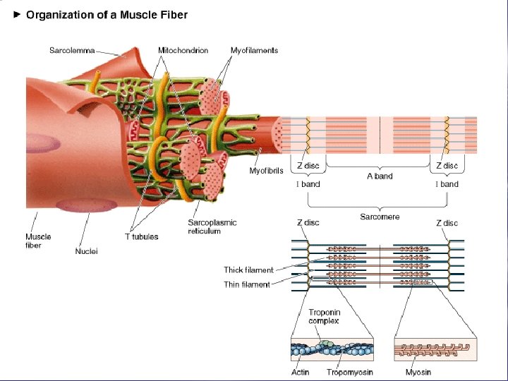
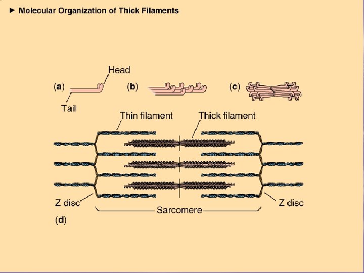
Structure of Skeletal Muscle: Microstructure § Sarcolemma – Muscle cell membrane § Myofibrils – Threadlike strands within muscle fibers – Actin (thin filament) – Myosin (thick filament)
Structure of Skeletal Muscle: The Sarcomere § Further divisions of myofibrils – Z-line § (Divider for each Actin/Myosin Group) – A-band § (Active area where actin and Myosin reside) – I-band § (In-between area which gets smaller as muscles contract and larger as they relax)
See Video on Breakdown of muscle anatomy § Chap 8 b § Now we will look at the Sarcomere Subunit and how muscle contractions happen.
Muscles Packet 1. Insertion: the moveable bone § Bicep insertion is the radius 2. Origin: the stationary bone § bicep originates in two different places in scapula
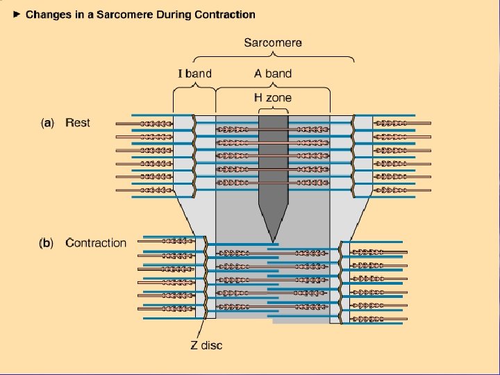
Muscular Contraction § The sliding filament model – Muscle shortening occurs due to the movement of the actin filament over the myosin filament – Formation of cross-bridges between actin and myosin filaments – Reduction in the distance between Z-lines of the sarcomere
The Sliding Filament Model of Muscle Contraction
Cross-Bridge Formation in Muscle Contraction
Sliding Filament Theory § Rest – uncharged ATP cross-bridge complex § Excitation-coupling – charged ATP cross-bridge complex, “turned on” § Contraction – actomyosin – ATP > ADP & Pi + energy § Recharging – reload cross-bridge with ATP § Relaxation – cross-bridges “turned off”
See Video on Sliding Filament theory in motion § Chap 8 a
Muscle Function § All or none law – fiber contracts completely or not at all § Muscle strength gradation – Multiple motor unit summation – more motor units per unit of time – Wave summation – vary frequency of contraction of individual motor units
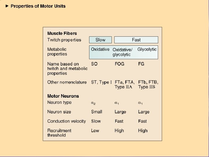
Motor Unit § Single motor neuron & muscle fibers it innervates § Eye muscles – 1: 1 muscle/nerve ratio § Hamstrings – 300: 1 muscle/nerve ratio
Illustration of the Neuromuscular Junction
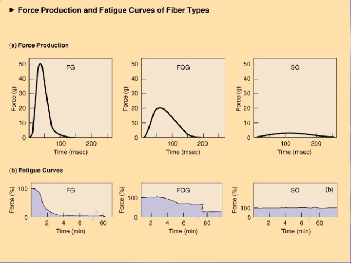
Individual Fiber Types Fast fibers § Type IIb fibers – Fast-twitch fibers – Fast-glycolytic fibers § Type IIa fibers – Intermediate fibers – Fast-oxidative glycolytic fibers Slow fibers § Type I fibers – Slow-twitch fibers – Slow-oxidative fibers
Staining of Fiber Type
Comparison of Maximal Shortening Velocities Between Fiber Types
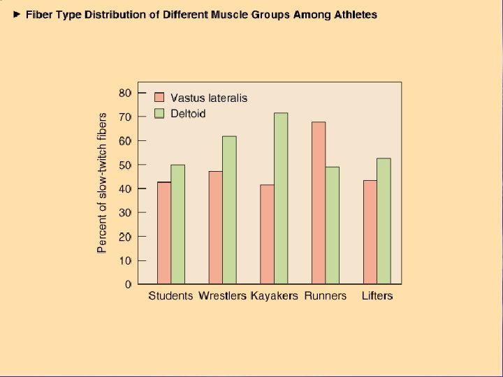
Fiber Types and Performance § Power athletes – Sprinters – Possess high percentage of fast fibers § Endurance athletes – Distance runners – Have high percentage of slow fibers § Others – Weight lifters and nonathletes – Have about 50% slow and 50% fast fibers
Alteration of Fiber Type by Training § Endurance and resistance training – Cannot change fast fibers to slow fibers – Can result in shift from Type IIb to IIa fibers § Toward more oxidative properties
Training-Induced Changes in Muscle Fiber Type
Hypertrophy and Hyperplasia § Increase in size § Increase in number
Age-Related Changes in Skeletal Muscle § Aging is associated with a loss of muscle mass – Rate increases after 50 years of age § Regular exercise training can improve strength and endurance – Cannot completely eliminate the age-related loss in muscle mass
Age-Related Changes in Skeletal Muscle § Aging is associated with a loss of muscle mass – Rate increases after 50 years of age § Regular exercise training can improve strength and endurance – Cannot completely eliminate the age-related loss in muscle mass
Types of Muscle Contraction § Isometric – – – Muscle exerts force without changing length Pulling against immovable object Postural muscles § Isotonic (dynamic) – Concentric § Muscle shortens during force production – Eccentric § Muscle produces force but length increases
Isotonic and Isometric Contractions
Illustration of a Simple Twitch
Force Regulation in Muscle § Types and number of motor units recruited – More motor units = greater force – Fast motor units = greater force § Initial muscle length – “Ideal” length force generation § Nature of the motor units neural stimulation – Frequency of stimulation § Simple twitch, summation, and tetanus
Relationship Between Stimulus Frequency and Force Generation
Simple Twitch, Summation, and Tetanus
Receptors in Muscle § Muscle spindle – Detect dynamic and static changes in muscle length – Stretch reflex § Stretch on muscle causes reflex contraction § Golgi tendon organ (GTO) – Monitor tension developed in muscle – Prevents damage during excessive force generation § Stimulation results in reflex relaxation of muscle
Muscle Spindle
Golgi Tendon Organ