Physiology of Skeletal muscle Structure Skeletal muscle fiber

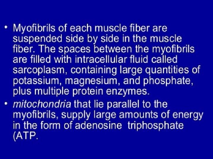
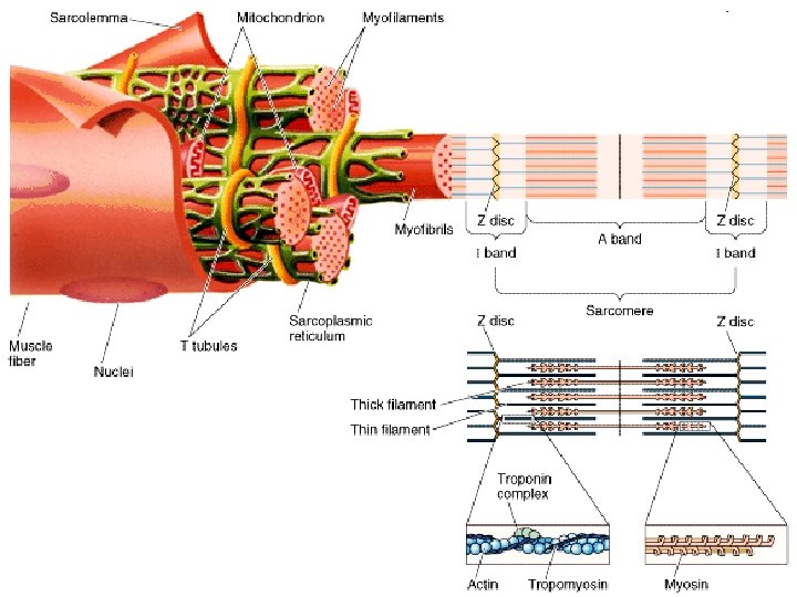
Physiology of Skeletal muscle • Structure: Skeletal muscle fiber represents a single cell of a muscle, each fiber is a thin, elongated cylinder with rounded ends. Beneath it is cell membrane or sarcolemma, the cytoplasm or sarcoplasm of the fiber contains many small, oval nuclei and mitochondria. • The sarcoplasm contains numerous, threadlike myofibrils that lie parallel to one another. They contain two kinds of protein filaments, thick filaments composed of the protein myosin, and thin filaments composed of protein actin. The arrangement of these filaments produces alterating light and dark striations of skeletal muscle fiber.

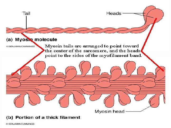
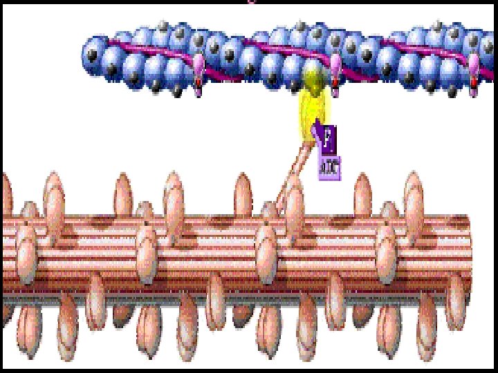
• A myosin molecule is composed of two twisted protein strands with globular parts called cross- bridges projecting outward along their lengths, many of these molecules comprise a myosin filament. • An actin molecule is a globular structure with a binding site to which the cross-bridges of the myosin molecules can attach. Many of these actin molecules, arranged together in adouble twisted strand( helix), form an actin filament.
 Light area at center of")
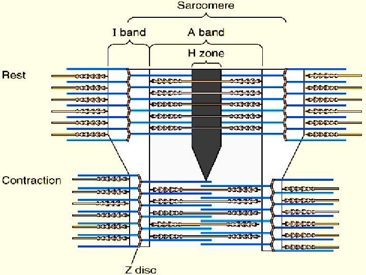
A band is dark, contains thick filaments (mostly myosin) Light area at center of A band is H band = area where actin and myosin don’t overlap I band is light, contains thin filaments (only actin) At center of I band is Z line/disc where actins attach

Sarcomeres: Are contractile units of skeletal muscle consisting of components between 2 Z discs M lines are structural proteins that anchor myosin during contraction Titin is elastic protein attaching myosin to Z disc that contributes to elastic recoil of muscle
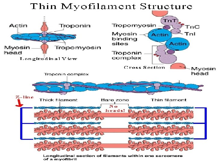

• Two types of proteins, tropomyosin and troponin, associate with actin filaments. Tropomyosin molecules are rod- shaped, and occupy the longitudinal grooves of the actin helix. • Troponin protein which is a complex of three globular protein molecules: 1 -Troponin I/ has strong affinity for actin. 2 -Troponin. T/has strong affinity for tropomyocin. 3 -Troponin. C/has strong affinity for Ca+ ion.

• Troponin I&T complex is believed to attach the tropomyocin to the actin. • Troponin C is believed to initiate the contraction process. • Each tropomyosin has a troponin molecule attached to it is surface, forming a tropomyosintroponin complex. In the resting state, the tropomyosin strands physically cover the active sites of the actin strands, so that interaction cannot occur between the actin and myosin to cause contraction.

• Within the cytoplasm is a network of membranous channels that surrounds each myofibril and runs parallel to it sarcoplasmic reticulum. • Set of membranous channels called transverse tubules (T-tubules), extends inward as invaginations from the fibers membrane, and passes all the way through the fiber. • The site where the nerve fiber and muscle fiber meet is called neuromuscular junction (myoneural junction). There, the muscle fiber membrane is specialized to form a motor end plate, where nuclei and mitochondria are abundant, and the sarcolemma is extensively folded.

• The membrane of nerve fiber and the membrane of the muscle fiber are separated by a small gap called the synaptic cleft. • The cytoplasm at distal ends of the nerve fibers is rich in mitochondria and contains many tiny vesicles ( synaptic vesicles) that store chemicals called neurotransmitters. • Each muscle fiber receive a single axon terminal from a somatic motor neuron. The motor neuron stimulates the muscle fiber to contract by liberating acetyl choline at the neuromuscular junction. A motor neuron, together with all of the muscle fibers that it innervates is known as a motor unit.

Electrical properties of skeletal muscle Skeletal muscle is an excitable tissue, when it is stimulated it will cause action potential as muscle impulse. The differences of action potential of skeletal muscle from that of nerve action potential: 1. RMP(- 90 mv). 2. Firing level is (- 60 mv). 3. Duration of muscle impulse is about ( 2 -4 ms). 4. It will not pass in after hyper polarization period, even in repeated stimuli. . The potential of muscle can be recorded during contraction and this record known as Electro myograph (EMG), which record the algebraic summation of action potential of all muscle fibers.

• Contractile property of skeletal muscle v. When muscle is stimulated, it contracts and after the contraction, and when there is no stimulus, it relax. The contraction is done by shortening in length, but return to it is original length in relaxation. v. The muscle cannot contract without stimulus, this is called: Excitation- contraction coupling, this mean that there is no contraction without excitation of the muscle.

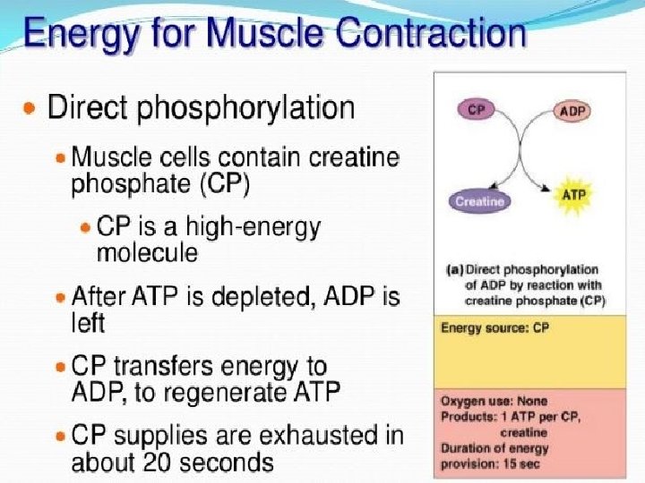
Mechanism of skeletal muscle contraction Sliding filament theory of muscle contraction: Suggests that the head of a myosin cross-bridge can attach to an actin binding site and bend slightly, pulling the actin filament with it. Then the head can release and combine with another binding site farther down the actin filament. The cross-bridges of myosin filaments contain the enzyme ATPase, which catalyzes the breakdown of ATP to ADP and phosphate, and release energy that provides the force with which a cross-bridges pulls. This cycle can repeated over and over.

Major events of muscle contraction and relaxation: Muscle fiber contraction: 1. The distal end of a motor neuron release acetylcholine. 2. Acetylcholine diffuses across the gap at the neuromuscular junction. 3. The sarcolemma is stimulated, and a muscle impulse travels over the surface of the muscle fiber and deep into the fiber through the transverse tubules and reaches the sarcoplasmic reticulum.

4. Calicum ions diffuse from the sarcoplasmic reticulum into the sarcoplasm and bind to troponin molecules. 5. Tropomyosin molecules move and expose specific sites on actin filaments. 6. Actin and myosin filaments form linkages. 7. Actin filaments are pulled inward by myosin crossbridges. 8. Muscle fiber shortens as a contraction occurs.

Muscle fiber relaxation: 1. Acetylcholinesterase enzyme which present at the neuromuscular junction on the membrane of the motor end plate, decomposes acetylcholine, and the muscle fiber membrane is no longer stimulated. 2. Calcium ions are actively transported into sarcoplasmic reticulum. 3. Linkages between actin and myosin filaments break.

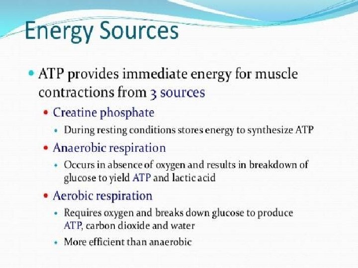
4. Troponin and tropomyosin molecules inhibit the interaction between myosin and actin filaments. 5. Actin and myosin filaments slide apart. 6. Muscle fiber relaxes and it is resting state is reestablished. 7. When a muscle fiber is at rest, tropomyosin-troponin complexes block the binding sites on the actin molecules and thus prevent the formation of linkages. v. Both contraction and relaxation are an active process and need energy. v. Fatigue of the skeletal muscle is due to the ATP source is lost in the muscle with continues stimulus.

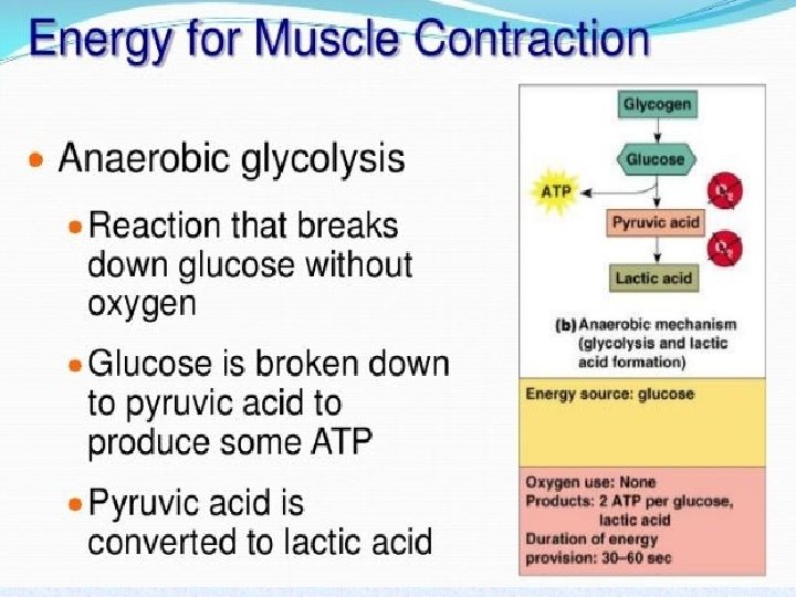
• There is depletion of muscle glycogen due to the interruption of blood flow through muscle contraction, and loss of nutrient supply especially loss of O 2. Therefore, muscle stop contractions. • Muscle hypertrophy: forceful muscular activity cause the muscle size to increase. Most hypertrophy result from increase in the diameter of muscle fiber and partly due to hyperplasia (increase the number of muscle fiber). • Muscle atrophy: results from non-using muscle for prolonged period of time, also result from muscle denervation.

General characters of skeletal muscle contraction Types of contractions: The muscle contains an elastic elements, and can do both types of contractions: • Isotonic contraction ( constant tension)= means the tension developed during contraction is constant, while the length of muscle is changed e. g. walking. • Isometric contraction ( constant length) = tension is changed while the length is constant e. g. standing, pushing an un movable thing. So the muscle contract isometrically by shortening the contractile elements, and elongation of elastic component, so the length of entire muscle is constant. In isometric contraction, the muscle does not work, while in isotonic it works, so that isotonic contraction need more energy than isometric contraction, it does work, so isotonic contraction is stronger than isometric contraction(need more energy).

Relationship between the length of muscle fiber and it is tension during contraction Each muscle have got a constant length and this called resting length, depend on age, sex , growth, and it is constant for each muscle. If the length of the muscle is decrease or increase than resting length, the tension will decrease. So the relationship between the tension of muscular fiber is inversely proportional with muscle length. The reason of this relationship is due to:

1. If the muscle length increase, the cross bridges( which are responsible for muscle tension), will become away from the actin filaments, and cannot pull actin, and the tension produced by few number of cross bridges sharing in the sliding of actin on myosin. 2. In shortening of muscle fiber, the actin filament will overlap on each other, and some cross bridges fail to reach the active sites on the actin filaments, and decrease the tension. In conclusion: Increasing or decreasing the muscle fiber length, will decrease the tension, due to decrease the number of cross bridges sharing in the tension formation.

Summation of contraction The muscle convert the electrical energy to mechanical energy, so summation means adding together of individual contractions. In the muscle, a group of muscle fibers supplied by a single axon, called motor unit. If we apply a stimulus to skeletal muscle, we gain simple muscle twitch. (muscle contraction followed by relaxation). If we increase the strength of continuous stimulus, increasing the number of motor units contracting by increase the magnitude of stimulus. ( 2 impulses/ ms < 3 impulses/ms < 4 stimulus/ ms). This is called Multiple- motor units summation( spatial summation). There will be increase in the number of motor units contractions.

If the duration of simple muscle twitch is 1 ms, so if we apply 2 stimulus/ms (same strength of stimulus), the second contraction stronger than the first one, and so on, because the Ca+ remain in the sarcoplasm, and the muscle not relax ( no refractory period), the curve will continue as a wave until we stop stimulus. Wave summation(temporal summation). There will be increase in the number of contraction for the same motor units in a constant period, the tension will increase because there will be accumulation of Ca+ in the sarcoplasm, not return to sarcoplasmic reticulum. When there is continues contractions without relaxations, the condition called complete tetanus.

Physiology of smooth muscle: They are called Unstriated, involuntary muscle, they are of two types: 1. Visceral smooth muscle. 2. Multi units smooth muscle. In visceral smooth muscle: the muscle fibers are spindleshaped cells that are held in close contact by gap junctions, and attached to each other to present in a sheet, so that if we give stimulus to one muscle fiber, the stimulus will spread to other muscle fibers, so it has syncytiam character, so that the whole sheet will contract as one unit, therefore, the visceral smooth muscle also called single unit smooth muscle, which form about 99% of the smooth muscles in the body, the contraction is called syncytial contraction.

In multi unit smooth muscles: The muscle fibers each one contracts individually, and have a single axon e. g. the muscle surround the iris of the eye to control the opening of the pupil. IN SMOOTH MUSCLES: 1. There is no well developed sarcoplasmic reticulum. 2. Actin and myosin filaments are distributed randomly. 3. Has no troponin, but contain calmodulin, which got the same function of troponin C in skeletal muscle. 4. The myosin is the same but it got an enzyme called myosin light chain kinase, (convert ATP→ ADP +energy), and it also got cross bridges.

Electrical properties of smooth muscle Resting membrane potential: The resting membrane potential of smooth muscle is un stable and fluctuated and vary from(-55 mv to -50 mv), there is unstable Na+-K+ pump and it can be hyper active or hypo active i. e. waxing (activation) or wanning (inhibition) of Na+- K+ pump lead to slow wave rhythm, that occur at regular intervals and considered as electro tonic potential. Smooth muscle under normal condition can produce: • Action potential by nerve stimulus. • Spontaneous action potential without stimulus because of prolonged inhibition of Na+- K+ pump that may cause the potential to reach(-45 mv) which is the firing level, and cause action potential spontaneously. • The myoneural junction of smooth muscle, the smooth muscle receive nerve impulses from:
")
Sympathetic and parasympathetic nerve supply, so the neurotransmitters either: 1}. excitatory ( acetyl choline) in parasympathetic stimulation. Acetyl choline will attach to receptors and lead to opening of Na+ channels, Na+ will enter the cell and initiate action potential. 2}. inhibitory ( nor adrenaline or nor epinephrine)in sympathetic stimulation. There will be opening to Cl- channels, and Cl- will enter to the cell or opening of K+ channels and K+ get outside the cell, both cause hyper polarization, and inhibition to the muscle.

• Smooth muscle contraction resembles skeletal muscle contraction in a number of ways, both mechanisms reflect reactions of actin and myosin; both are triggered by membrane impulses and release of calcium ions; both use energy from ATP molecules. • There are significant differences: smooth muscle fibers lack troponin , instead, it use a protein called calmodulin, which bind to calcium ions released when it is fibers are stimulated, thus actinmyosin contraction mechanism activating. The calcium necessary for smooth muscle contraction diffuses into the cell from the extra cellular fluid.

• Smooth muscle is slower to contract and slower to relax than skeletal muscle, and can maintain a forceful contraction for a longer time with the same amount of ATP. • Smooth muscle fibers can change length without changing tautness; stretching of smooth muscle fibers can also trigger contractions. The ability of smooth muscle to make it is tension constant despite the change in smooth muscle fibers length called plasticity.
: a pattern of")
• Types of smooth muscle contraction: 1. Rhythmic contraction (rhythmicity): a pattern of repeated contractions, this caused by self- exciting fibers that deliver spontaneous impulses that transmitted from cell to cell and rhythmicity responsible for the wavelike motion called peristalsis(alterate contraction and relaxation) e. g. in GIT. 2. Tonic contraction: continuous contraction of smooth muscle, this type is found in the sphincters and wall of blood vessels.


- Slides: 42