Skin Color Determinants Normal Skin Color Determinants Melanin


- Slides: 17

Skin Color Determinants

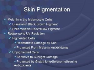
Normal Skin Color Determinants • Melanin • Yellow, brown or black pigments • Carotene • Orange-yellow pigment from some vegetables • Hemoglobin • Red coloring from blood cells in dermis capillaries • Oxygen • Content determines the extent of red coloring • The more oxygen, the more red the skin

Skin colors resulting from conditions • Cyanosis • Hemoglobin is poorly oxygenated, and the skin appears blue • Skin becomes cyanotic when a person has difficulty breathing or heart attack • Skin color can indicate emotions or disease • Redness or erythema • Red skin may indicate embarrassment, fever, hypertension, inflammation, or allergy

Skin colors cont’d • Pallor or blanching – looking pale – may be anger, fear, anemia, low blood pressure, or impaired blood flow • Jaundice – yellow cast – shows as a yellow skin tone – indicates liver** disorder in which excess bile pigments are absorbed into the blood • Black and blue marks – bruises – blood has escaped from the circulation and clotted in tissue spaces – also called hematomas**

SKIN APPENDAGES

Appendages of the Skin • Cutaneous glands are all exocrine glands that release their secretion to the skin surface via ducts • 2 groups • Sebaceous (oil) glands • Sweat glands

Sebaceous glands (oil glands) • Found everywhere except palms of hands and soles of the feet • Produce oil or sebum • Lubricant for skin • Kills bacteria • Most with ducts that empty into hair follicles

Sebaceous Glands cont’d • Glands are activated at puberty • Blocked sebum gland’s duct forms a whitehead, a blackhead forms if it oxidizes • Acne is an active infection of the sebaceous glands

Sweat glands • More than 2. 5 million person • Widely distributed in skin • Produce sweat • Two types • Eccrine • Apocrine

Eccrine (Merocrine) glands • Open via duct to pore on skin surface • More numerous and are found all over the body • Help regulate body temperature • If body temperature or external temperature is high, they secrete sweat

Apocrine glands • Ducts empty into hair follicles • Found mainly in the axillary (arm pit) and genital regions • Begin to function in puberty • Function minimally in thermoregulation • Responsible for odor in humans

Sweat and Its Function • Composition • Mostly water • Some metabolic waste • Fatty acids and proteins (apocrine only) • Function • Helps dissipate excess heat • Excretes waste products • Acidic nature inhibits bacteria growth • Odor is from associated bacteria

• Produced by hair bulb • Consists of hard keratinized epithelial cells • Melanocytes provide pigment for hair color Hair Figure 4. 7 c

Hair Anatomy • Central medulla • Cortex surrounds medulla • Cuticle on outside of cortex • Most heavily keratinized Figure 4. 7 b

Associated Hair Structures • Hair follicle • Dermal and epidermal sheath surround hair root • Arrector pilli • Smooth muscle • Sebaceous gland • Sweat gland Figure 4. 7 a

Appendages of the Skin Nails • Scale-like modifications of the epidermis • Heavily keratinized • Stratum basale extends beneath the nail bed • Responsible for growth • Lack of pigment makes them colorless

Nail Structures • • Free edge Body Root of nail Eponychium – proximal nail fold that projects onto the nail body Figure 4. 9
 Melanin sentezi nedir
 Lizin ve hidroksilizin metabolizmasi bozukluklari
 Chapter 26 pedicuring test answers
 Skin information
 Thin skin vs thick skin
 Milady facial steps
 4 quadrants of abdomen
 Normal percussion of abdomen
 Normal skin
 Hymen
Hymen Carotenemia
 Skin color map of the world
 Flesh coloured ppt
 Inspect uniformity of skin color rationale
 Biology of skin color video questions
 Regardless of skin color
 Human skin color wheel
 Color blindness punnett square


