Sir Isaac Newton 1672 viewed light through a

 – Hypothesized the Electromagnetic Spectrum: light waves 1 micron Hair")

 Copyright © 2002 Wadsworth Group. Wadsworth is an imprint of the")
 (close your right eye,")


 of the lens • The closer an object is to the eye")


 of communication within the eye • Light passes through the")

 -Within Outer Segment of photoreceptors are stacks")
 -photon of light (smallest particle of light)")


 are the cones")


: example of closely")


 • Looked at")

")
- Slides: 52
Sir Isaac Newton: 1672 viewed light through a prism – hypothesizing light waves Sir Isaac Newton (1643 - 1727) R O Y G B I V
James Clerk Maxwell (1760’s) – Hypothesized the Electromagnetic Spectrum: light waves 1 micron Hair 1 mm 1 meter 1 km Wavelength in nanometers (billionth of a meter)
Light - The Visual Stimulus • Light can be described as both a particle and a wave Wave: through air Particle: striking receptor • Wavelength of a light is the distance of one complete cycle of the wavelength The distance from the peak of a wavecrest to the peak of the subsequent wavecrest, or from one trough to the next trough. Expressed in units of distance (e. g. km, m, cm, micron, nm) • Visible light has wavelengths from about 400 nm (violet) to 700 nm (red) • Wavelength of light is related to its perceived color
Page 154 (40) Copyright © 2002 Wadsworth Group. Wadsworth is an imprint of the Wadsworth Group, a division of Thomson Learning Cross-section of the vertebrate eye The eye works like a camera, using a lens to focus light onto a photosensitive surface at the back of a sealed structure.
Filling-in the blind spot up plus with your left eye) (close your right eye, line
Focusing light on the Retina Changing the shape of the lens: accommodation (“bending” the light) retina cornea lens
Feeling and seeing accommodation • Near and far always seem to be in focus, but this is not true – we just accommodate unconsciously – Hold pencil point at arms length and star at point 20 feet away…. move pencil toward you while keeping distant focus…see how pencil blurs – When the pencil is 12” away, switch your focus to the pencil and notice how the background blurs. – The shifting blur is the shifting accommodation
Accommodation (bending) of the lens • The closer an object is to the eye the more light must bend to have the image fall (focus) on the retina • Focusing “experiment” to “see” accommodation • “Near point” is the closest to your eye in which an object remains in focus • Where is your near point?
Line of Sight & the Fovea • Accommodation of the lens results in light converging on (not in front or behind) the retina • Eye, head, body movements (“accommodations”) are made to place the line of sight (straight line of the light) on the Fovea • The Fovea is the place of detailed vision – The size is about 2 -degrees “visual angle” • “Experiment” – stare at a single point and read the words either left or right of it.
Organization of Retina Photochemical is located here Back of the eye Light To optic Ganglion Amacrine Bipolar Horizontal Cone nerve cell Rod
The Eye & Retina • The human eye • http: //www. youtube. com/watch? v=Xk. Lnp. Pi 3 Jp. U • Cross section of the retina
The pathway (“Neural Circuits”) of communication within the eye • Light passes through the entire maze of cells to the back of the retina to the Outer Segment of the receptors • Activation of the receptors activates the Bipolar cells • Bipolar cells communicate with the Ganglion cells • Ganglion cell axons combine to form the Optic Nerve • Chain of interconnecting neurons is called a “neural circuit”
Transduction at the Retina -sensory transduction occurs in the photoreceptors rods and cones
Transduction in the photoreceptors (rods and cones) -Within Outer Segment of photoreceptors are stacks of internal membranes called “disks” -Disks are made up of lipids (fat) and proteins. These proteins are photo “pigment” molecules. -Photoreceptor pigment is called rhodopsin
Transduction in the photoreceptors (rods and cones) -photon of light (smallest particle of light) strikes receptors causing the separation of rhodopin into opsin and retinal -opsin is an enzyme that effects sodium channels of neurons (causing channels to close) resulting in hyperpolarization (remember, this is an “inhibitory response”? )
How can a hyperpolarization cause to “see” something? • Hyperpolarization causes release of LESS neurotransmitters to postsynaptic neurons in the retina (inhibitory response) • “Coding” in nervous system only needs systematic changes to detect a stimulus that could be either inhibitory or excitatory • Neural coding of light is analogous to coding a photographic “negative”
Seeing in hyperpolarization and inhibition
Structure of the Eye: Where are the Rods and Cones? Lens Retina Fovea (point of central focu Iris Cornea Light rays Pupil Blind spot Optic nerve The eye works like a camera, using a lens to focus light onto a photosensitive surface at the back of a sealed structure.
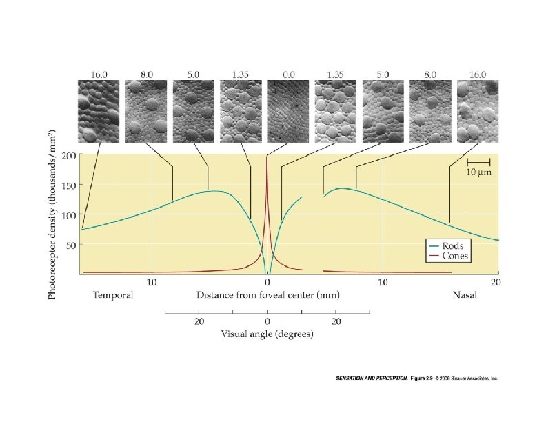
per human retina. . there are 120 million rods there are 6 million cones
The Rods and Cones are Differential Distributed on the Retina Thousands of receptors per square millimeter 180 140 Fovea Rods 100 60 Cones Blind spot 20 0 60 40 20 40 Distance on retina from fovea (degrees) 60
Figure 2. 12, page 44 Copyright © 2002 Wadsworth Group. Wadsworth is an imprint of the Wadsworth Group, a division of Thomson Learning Distribution of rods and cones Adapted from Human Information Processing by P. Lindsay and D. A. Norman , 1977, 2 nd ed. , p. 126. Copyright © 1977 Academic Press, Inc. Adapted with permission.
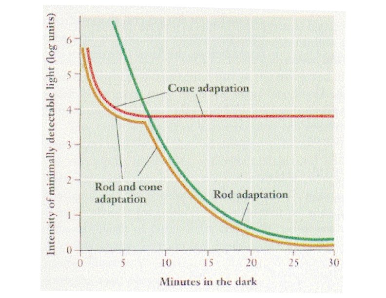
Copyright © 2002 Wadsworth Group. Wadsworth is an imprint of the Wadsworth Group, a division of Thomson Learning Figure 2. 21, page 51 Dark adaptation: how long does it take to see in the dark (and why)?
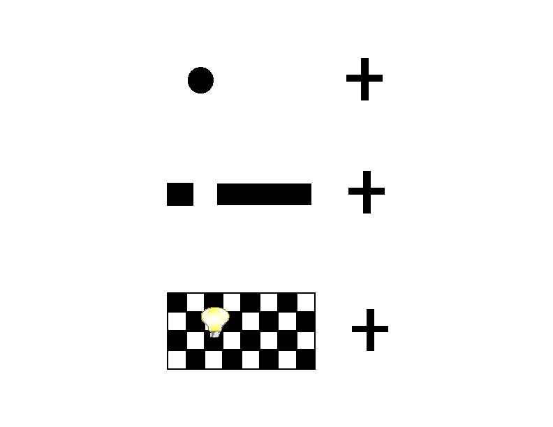
Dark Adaptation -the increase in sensitivity that occurs when illumination is reduced from light to dark -Exp’t 1 – Fixate “X” while adjusting the “test light” so that you can just barely see it (method of adjustment) Exp't 1 findings – Two Stage Dark Adaptation Curve test light Fixatio (peripher n y) -sensitivity(fovea increases for 3 -4 mins (you can see ) improvements levels off better) then X -sensitivity increases again after 7 -10 mins
Seeing in the dark • 1 st adaptation (3 -to-4 -minutes) are the cones adapting • 2 nd adaptation (after 7 -min) are the rods adapting • Best “dark vision” is mediated by rods – This is due to the “convergence” of many rods (explained below)
Actually, in human retina there are three types of cone pigments Relative responsiveness of cones short wavelength pigment “S-Cones” 419 nm medium wavelength pigment - “M-Cones” - 531 nm long wavelength pigment – “L-Cones” 558 nm “Blue” cones “Green” cones “Red” cones Wavelength in nanometers (billionths of a meter)
Short-, Medium- & Long-Cone densities
The difference between Rods & Cones? Cones • Found in the fovea • Population of about 6 million • Low sensitivity (not good in the dark) • High acuity (very good detail in the light) • Processes color Rods • Found in the periphery • Population of about 120 million • Higher sensitivity (better in the dark) • Good for “global information” (big objects), poor acuity (not good for details) • Processes black, white and shades of gray
per human retina. . there are 120 million rods there are 6 million cones there are 1 million ganglion cells on average receive input from 120 rods, but only about 6 cones per ganglion cell. When more than one neuron synapses on a postsynaptic neuron, there must be a lot of neural “convergence” –
Copyright © 2002 Wadsworth Group. Wadsworth is an imprint of the Wadsworth Group, a division of Thomson Learning Figure 2. 6, page 40 Eye cross-section Notice the convergence
There is more Convergence from Rods than from Cones Spots of light can be much smaller, but needs to be quite bright in the fovea needs to be bigger, but can be dark in peripheral retina Receptive fields Light (a) Fovea Ganglion cells Bipolar cells Photoreceptors (cones) Photoreceptors (rods) Pigmented epithelium
The amount of convergence that Rods and Cones have on Ganglion cells explains why… Cones are more precise and specific (high acuity): • Only 6 cones per ganglion (on average) • Some “private lines” between single cones and ganglion cells Rods are more sensitive in the dark • Takes less light to trigger a response • Can make out more detail in the dark
Why are Rod Systems more Sensitive than Cone Systems in the dark 2 units each per receptor 2 2 2 1 0 ganglion cells 2 2 2 10 10 10 (threshold needed for Ganglion Cell to fire = 10
Rod Systems are more Sensitive than Cone Systems in the dark because of Spatial Summation (and implicit Temporal Summation) 2 units 2 2 2 ganglion cells (threshold = 10 units) 2
Rod Systems are less Acute than Cone Systems (regulating “Visual Acuity”): example of closely spaced stimuli 2 light sources 3 3 2 3 0 2 2 ganglion 1 cells = 10 (threshold 0 units) 2 sources of light seen as 1 source 10 10 10 10 5 sources of light seen as 5 different sources
How cone systems offer greatest Visual Acuity: example of closely spaced stimuli 2 0 2 0 2 2 0 0 0 2 2 0 0 1 ganglion cell activates for Rods; 4 ganglion cells activate to the cones AND there is a “silent” (separating) (threshold = 10
Tools for visual specificity • Convergence in the neural code varies sensitivity between rods and cones • “Lateral Inhibition” from the Horizontal and Amacrine cells – First seen in the retina – But, also directly or indirectly most neurons throughout the nervous system
Lateral Inhibition - + -
Organization of Retina Photochemical is located here Back of the eye Light To optic Ganglion Amacrine Bipolar Horizontal Cone nerve cell Rod
Herman Grid
Figure 2. 42, page 64 Hermann grid and lateral inhibition Firing at neuron “B” is relatively stronger (perceived as “brighter” in relative contrast to the surroundings), than is the firing of “A, ” hence “A” looks darker (and you see the “shadow. ”
Tools for visual specificity • Convergence of rods and cones on to the ganglion cells create differential sensitivity • “Lateral Inhibition” from the Horizontal and Amacrine cells help differentiate stimuli • Ganglion cells have different “receptive fields”
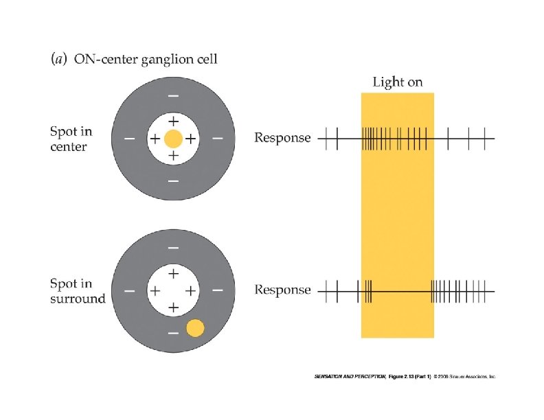
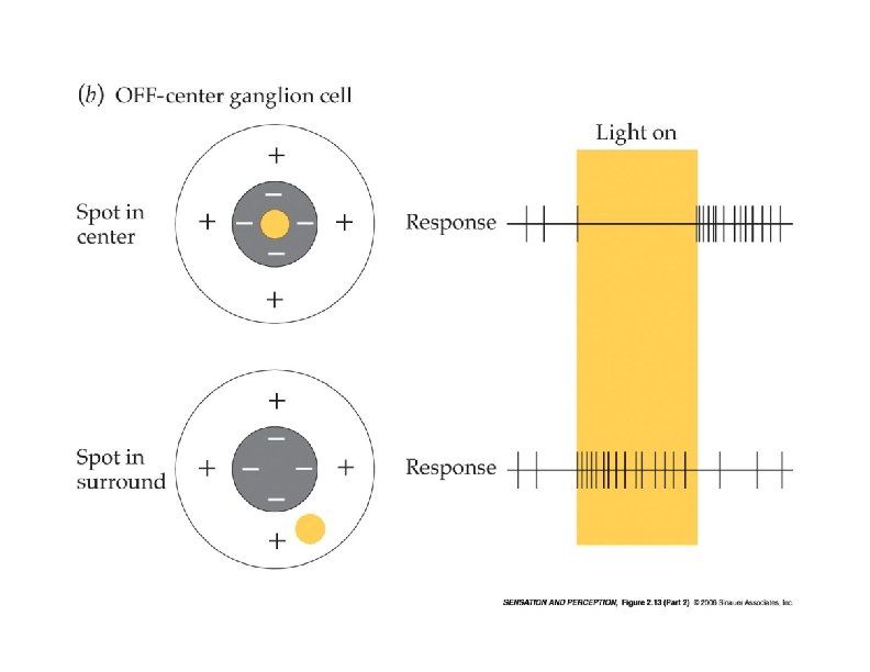
Centre-Surround Receptive Fields -retinal ganglion cells inhibition excitation
Finding the “receptive field” of Ganglion Cells: Hubel & Wiesel (1961) • Looked at cat’s eyes and ganglion cells (remember, Ganglion axons are the optic nerve) • Cat is immobilized while looking at a screen of “spots” of light • Activity of Ganglion cells measured as light spots are moved around
Mapping Receptive Fields • Go to Virtual Lab – Mapping Receptive Fields – Stimulus size and Receptive Fields • Both excitatory and inhibitory
Tools for visual specificity • Differential specificity of rods (i. e. , dark sensitivity) and cones (i. e. , precise responses (“private lines” to Ganglion cells); color processing) • Convergence in the neural code varies sensitivity • Lateral inhibition: activity of neighboring cells influence one another (effect of Horizontal and Amacrine cells on bipolar cells) • Organization of “receptive fields” and Center-Surround antagonism