Serous Fluid Pleural Lung cavity Pericardial heart Peritoneal


 are not normally seen in body fluids.")


- Slides: 18
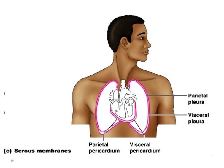
Serous Fluid Pleural: Lung cavity Pericardial: heart Peritoneal: abdominal cavity
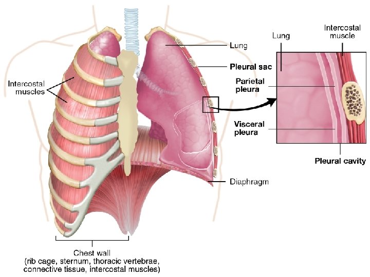
All body cavities are lined by a thin membrane which has 2 parts: 1. Parietal membrane: lines the body cavity 2. Visceral membrane: outer lining of the organ The serous fluid is in the space between the 2 membranes.
Serous fluid: Is an ultra filtrate of plasma derived from the capillary network of the membrane. - The accumulation of serous fluid is called an effusion.
Categorization of effusions: Effusions are the accumulation of fluids in the tissue spaces. Transudate effusions: Occur during various systemic disorders that disrupt fluid filtration, fluid reabsorption, or both. -Example of systemic disorders that may result in the formation of transudates include congestive heart failure.
Exudate effusions: Occur during inflammatory processes that result in damage to blood vessel walls, body cavity membrane damage, or decreased reabsorption by the lymphatic system. Examples include infections, inflammations, hemorrhages and malignancies.
Transudate is fluid pushed through the capillary due to high pressure within the capillary. Exudate is fluid that leaks around the cells of the capillaries caused by inflammation.
Types of serous fluids: Serous body fluids are found in the cavities surrounding the vital organs. - This fluid is normally clear and slightly yellow, resembling serum. - Serous cavities include the pericardium, pleural, and peritoneum.
Pericardial fluid: normally, the pericardium contains less than 50 ml of fluid. - Pericardial effusions are an accumulation of fluid around the heart. - The procedure for removing excess pericardial fluid is peri-cardiocentesis. - Pericardial effusions are usually always exudates. -
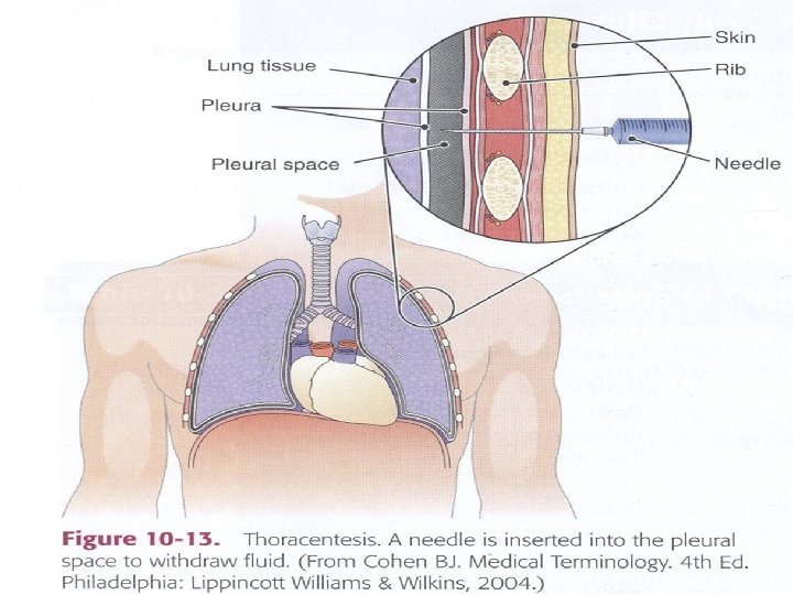
Pleural fluid: The pleural cavity normally contains less than 30 ml of fluid. - Pleural effusions occur when fluid accumulates around the lungs. - A thoracentesis is performed to remove this excess fluid. - Pleural effusions can be transudate or exudate. -
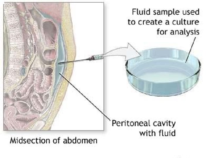
Peritoneal fluid: - A peritoneal effusion is the accumulation of peritoneal fluid, also called ascites, in the abdominal cavity.
Laboratory testing of serous fluid: Macroscopic examination: - Serous body fluids normally resemble serum, clear and pale yellow. - Turbid (cloudy) fluid indicates a high white blood cells which correlates with bacterial infection.
Microscopic examination: - - Red blood cells(RBC’s) are not normally seen in body fluids. When present in RBCs may indicate hemorrhage. WBCs are normally present in low numbers. The presence of increased numbers of WBCs correlate with various pathologies.
Thoracentesis It is the procedure in which the fluid is obtained by a needle and syringe
Pleural Effusion