Proteomics Proteome proteome is a mix of protein


- Slides: 19
Proteomics
Proteome : proteome" is a mix of "protein" and "genome While genome is the entire heredity information of an organism , proteome is the organism's complete complement of proteins. Proteomic : Is the study of an organism's complete complement of proteins especially their function and structure. While genomic is the study of the whole genome.
Why not m. RNA : • Proteomic consider the amount and physiology of the protein in the cell m. RNA is not always translated to protein. • A lot of proteins are modified after the RNA been translated.
Protein Biochemistry Westerns pull-down assays Enzyme Assays Sub-mg quantities Protein Structure NMR: Nuclear magnetic resonance spectroscopy X-ray crystallography Structural Biologists 10’s of mg quantities
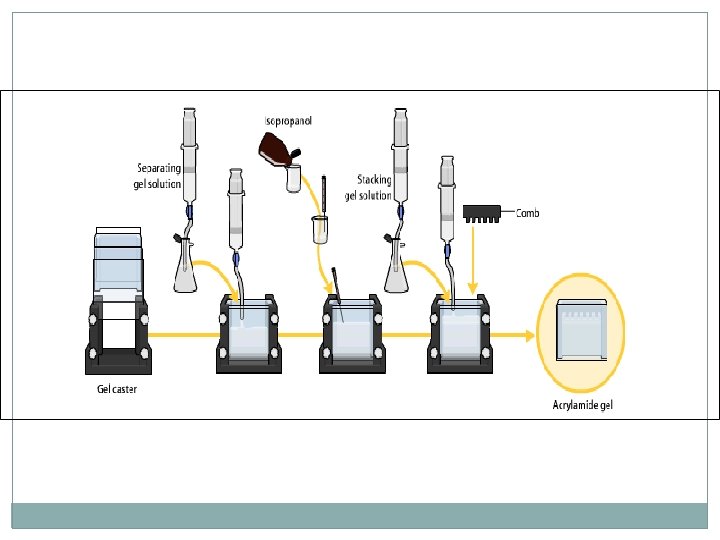
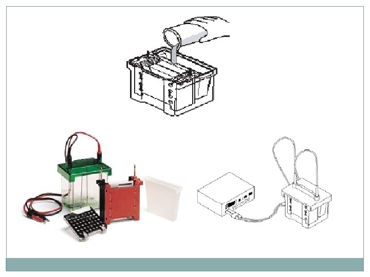
Western Blot : 1 - Making the SDS gel
SDS-PAGE Gel Component Table
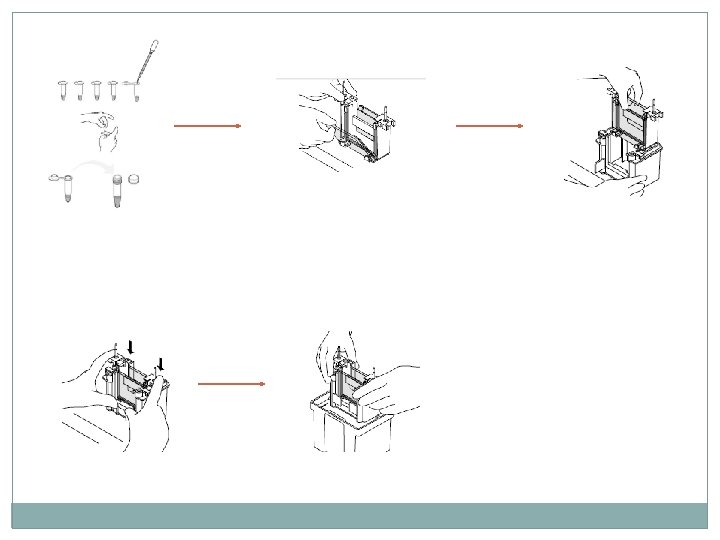
Finally the cascade system is ready
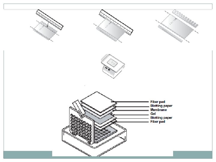
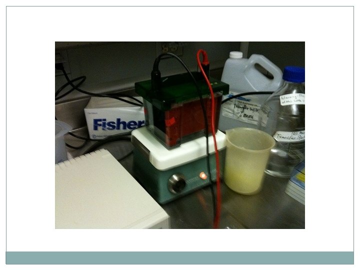
2 - transfer the proteins to nitrocellulose membrane : By Trans-Blot that is specifically designed to pass electric current horizontally through the gel forcing the negatively charged proteins to migrate out of the gel onto the nitrocellulose membrane.
3 - Primary antibody is added to the membrane and incubated to allow the antibody to bind to the myosin protein on the membrane. The unbound antibody is then washed away.
4 - Secondary antibody is added to the membrane and incubated to allow the secondary antibody to bind to the primary antibody. The unbound secondary antibody is then washed away.
5 -Colorimetric enzyme substrate is added to the membrane and incubated to allow color to develop. Purple/gray bands will develop on the membrane exactly where the myosin protein bands are located.