Protein Kinases Second Messenger Hypothesis Protein kinase A
- Slides: 40
Protein Kinases
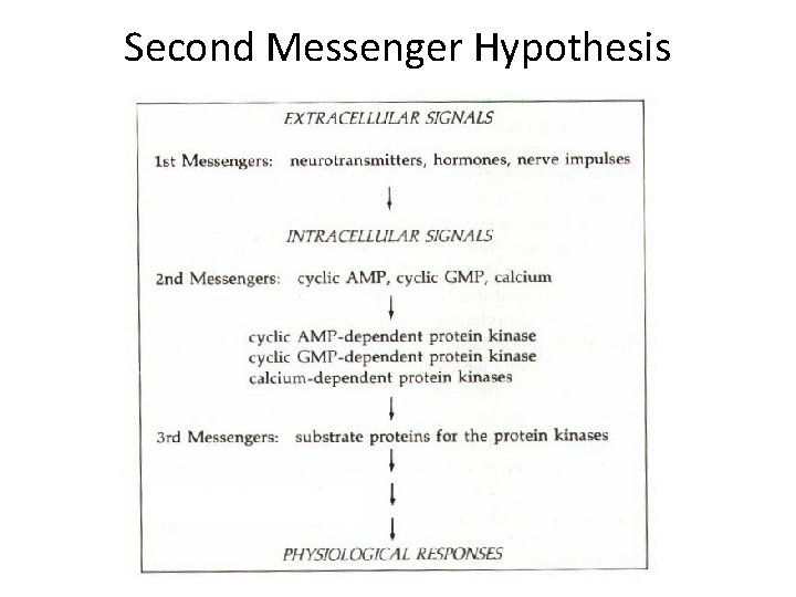
Second Messenger Hypothesis
Protein kinase • A protein kinase is a kinase enzyme that modifies other proteins by chemically adding phosphate groups to them (phosphorylation). • Phosphorylation usually results in a functional change of the target protein by changing enzyme activity, cellular location, or association with other proteins. • The human genome contains about 518 protein kinase genes and they constitute about 2% of all human genes. • Kinases are known to regulate the majority of cellular pathways, especially those involved in signal transduction. • Protein kinases are also found in bacteria and plants, where they take an active participation in mechanistic cellular signaling.
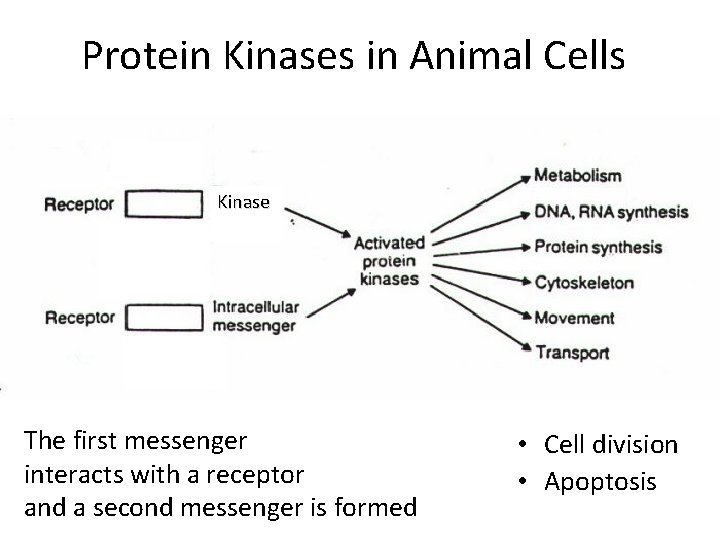
Protein Kinases in Animal Cells Kinase s The first messenger interacts with a receptor and a second messenger is formed • Cell division • Apoptosis
History and Importance • About 518 genes in humans encode protein kinases; • Protein kinases are the fourth largest gene family in humans – C 2 H 2 zinc finger proteins (3%) – G-protein coupled receptors (2. 8%) – Major histocompatibility (MHC) complex protein family (2. 8%)
How Many Protein Kinases Are There? • The kinome refers to all protein kinases in the genome • There are 478 conventional eukaryotic protein kinases (e. PKs) plus 106 pseudogenes – 388 Protein-serine/threonine kinases – 90 Protein-tyrosine kinases • 58 receptor PTKs • 32 Non-receptor PTKs • There are 40 atypical protein kinases (e. g. EF 2 K/alpha kinases) • 478 + 40 = 518
Protein Kinase Classifications • Protein-Serine/threonine • Protein-Tyrosine – Receptor: ligand binding domain and catalytic site on the same polypeptide – Non-receptor: catalytic domain separate from the receptor • Dual Specificity (both serine/threonine and tyrosine) also occur • Broad Specificity: have several substrates, e. g. , PKA • Narrow Specificity: have one or a few substrates, e. g. , pyruvate dehydrogenase kinase with one substrate • Classified by activator: PKA, PKG, PKC
General Classes I • ACG Group • Protein kinase A; cyclic AMP-dependent protein kinase • Protein kinase C • Protein kinase G • Basic amino acid-directed enzymes that phosphorylate serine/threonine.
General Classes II • Ca. MK • Calcium-calmodulin-dependent protein kinases – III – IV • Type II is a broad specificity kinase • The others are dedicated kinases with a limited substrate specificity
General Classes III • CMGC • Cyclin-dependent protein kinases – These are important regulators of the cell cycle • MAP (Mitogen activated protein/microtubule associated protein) kinases – Many of these promote cell division • GSK 3 (glycogen synthase kinase-3) • Clk (Cyclin-dependent like kinase)
General Classes IV • PTK (Protein-tyrosine kinases) – Receptor, e. g. , epidermal growth factor receptor, insulin receptor – Non-receptor, e. g. , Src, Abl protein kinases • Specifically phosphorylate protein-tyrosine (note they are not tyrosine kinases but protein-tyrosine kinases)
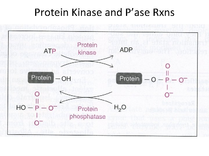
Protein Kinase and P’ase Rxns
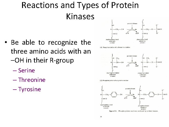
Reactions and Types of Protein Kinases • Be able to recognize three amino acids with an –OH in their R-group – Serine – Threonine – Tyrosine
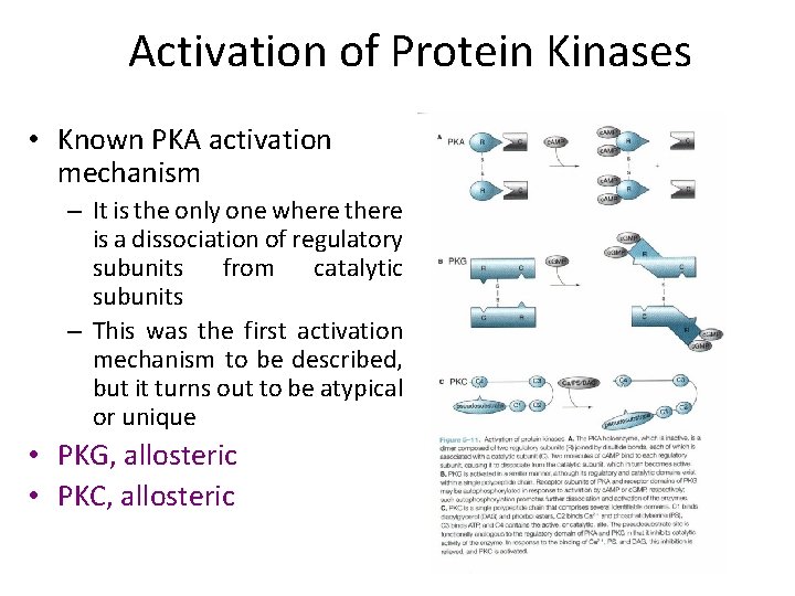
Activation of Protein Kinases • Known PKA activation mechanism – It is the only one where there is a dissociation of regulatory subunits from catalytic subunits – This was the first activation mechanism to be described, but it turns out to be atypical or unique • PKG, allosteric • PKC, allosteric
Protein Kinase A • Activated by c. AMP • Regulation of glycogen, sugar and Lipid metabolism. • Secondary Messenger • The protein enzyme contains two catalytic subunits and two regulatory subunits. • When there is no c. AMP, the complex is inactive. • When c. AMP binds to the regulatory subunits, their conformation is altered, causing the dissociation of the regulatory subunits, which activates protein kinase A and allows further biological effects.
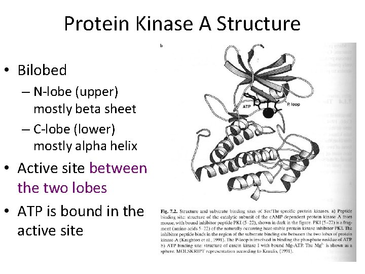
Protein Kinase A Structure • Bilobed – N-lobe (upper) mostly beta sheet – C-lobe (lower) mostly alpha helix • Active site between the two lobes • ATP is bound in the active site
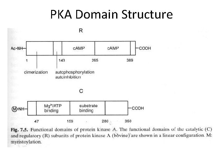
PKA Domain Structure
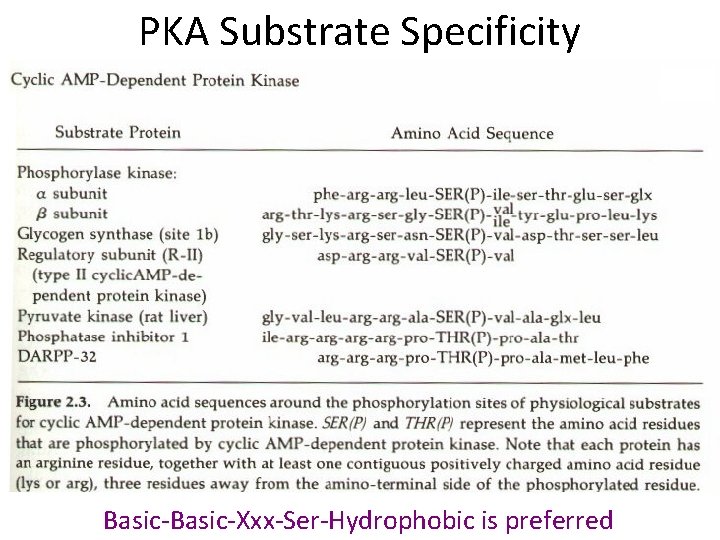
PKA Substrate Specificity Basic-Xxx-Ser-Hydrophobic is preferred
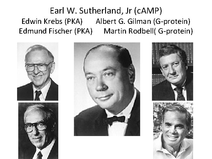
Earl W. Sutherland, Jr (c. AMP) Edwin Krebs (PKA) Albert G. Gilman (G-protein) Edmund Fischer (PKA) Martin Rodbell( G-protein)
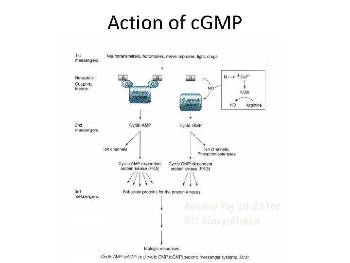
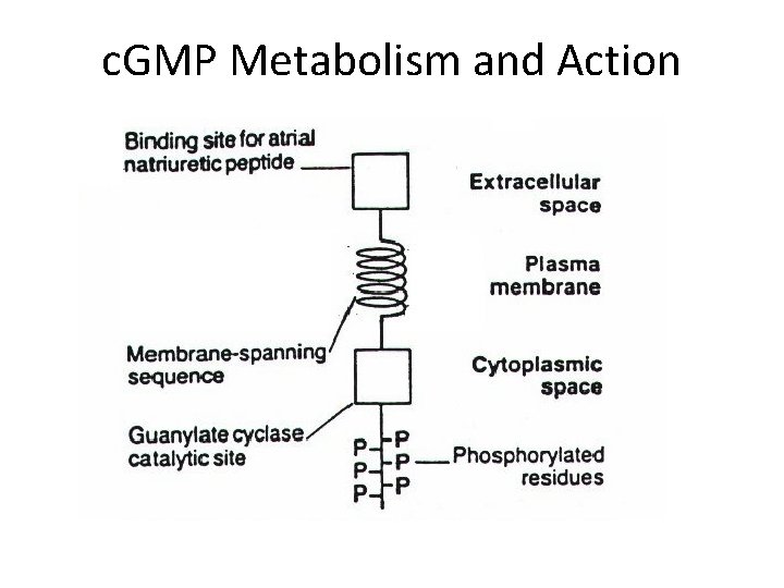
Protein Kinase G • Activated by c. GMP • Second second messenger protein kinase (after c. AMP) • Little known about physiological protein substrates despite needs extensive investigation • Two types of guanylyl cyclase – Second messenger generated by atrial naturetic factor as an integral membrane guanylyl cyclase. – Second messenger generated by NO action on the soluble guanylyl cyclase reaction.
Action of c. GMP Review Fig 19 -23 for NO biosynthesis
c. GMP Metabolism and Action
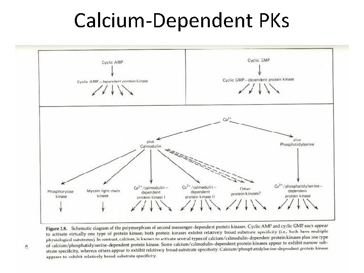
Calcium-Dependent Protein Kinases • Protein Kinase C – Requires calcium, diacylglycerol, and phospholipid for activity – Diacylglycerol is generated by the action of phospholipase C, C refers to calcium – DAG and phospholipid were also described as necessary for the activation of this enzyme – It is paradoxical to have PKCs that are independent of Ca 2+ and DAG • Calcium-Calmodulin-Dependent Protein Kinases – CAM Kinases I, III, IV – Several other protein kinases activated by calmodulin including myosin-light chain kinase, phosphorylase kinase, some isoforms of adenylyl cyclase and some isoforms of phosphodiesterase
Calcium-Dependent PKs
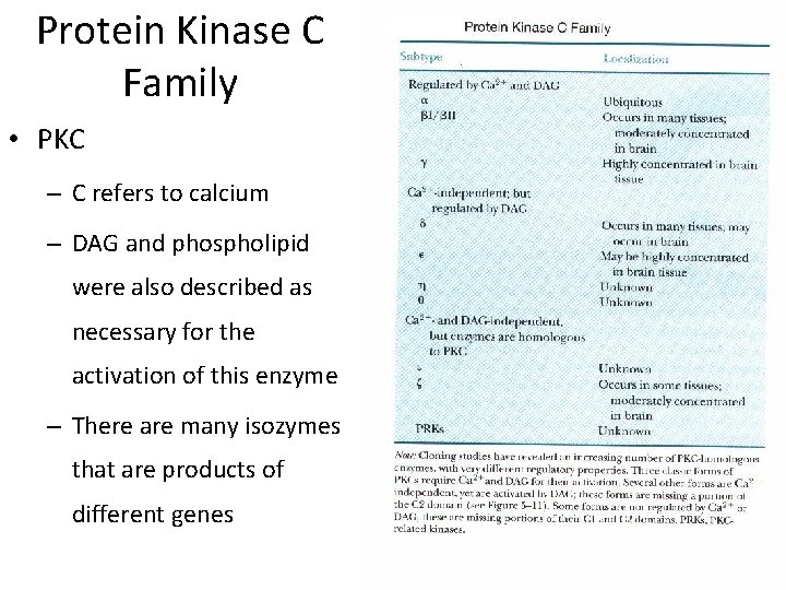
Protein Kinase C Family • PKC – C refers to calcium – DAG and phospholipid were also described as necessary for the activation of this enzyme – There are many isozymes that are products of different genes
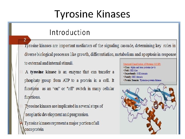
Tyrosine Kinases
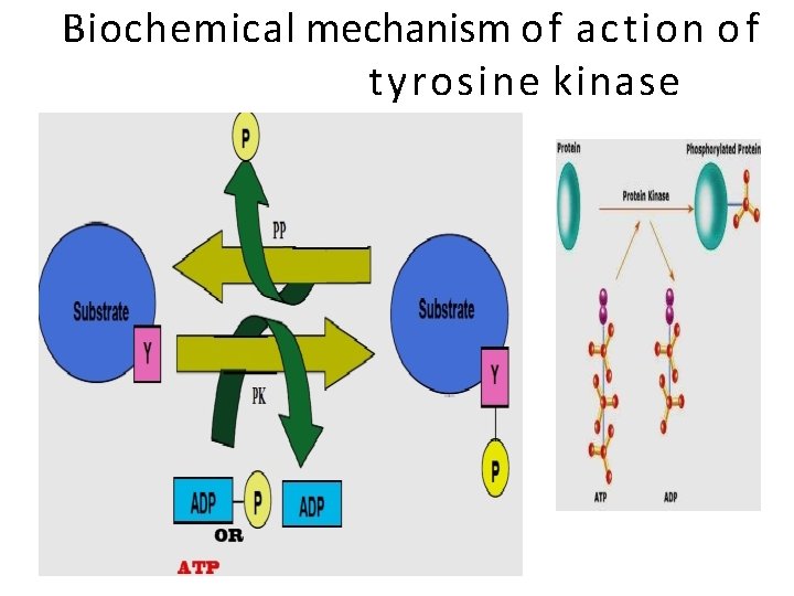
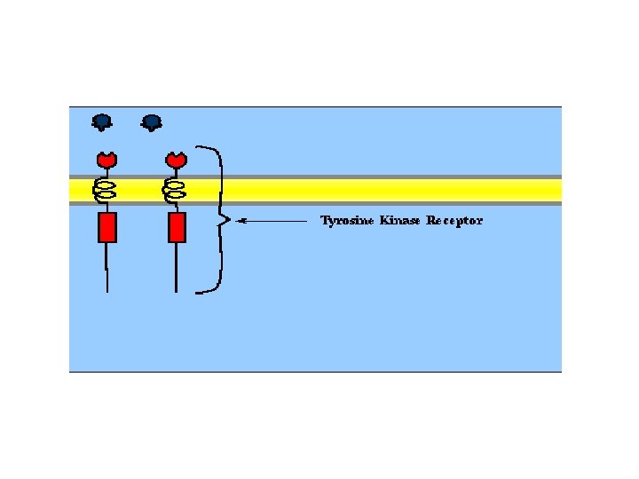
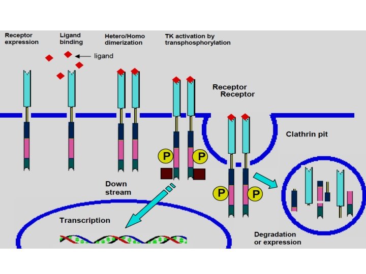
Tyrosine kinase mechanism • Tyrosine kinase receptors are a family of receptors with a similar structure. • They each have a tyrosine kinase domain (which phosphorylates proteins on tyrosine residues), a hormone binding domain, and a carboxyl terminal segment with multiple tyrosines for autophosphorylation. • When hormone binds to the extracellular domain the receptors aggregate.
Biochemical mechanism o f action o f tyrosine kinase
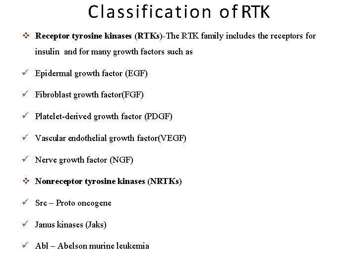
Classification o f RTK Receptor tyrosine kinases (RTKs)-The RTK family includes the receptors for insulin and for many growth factors such as Epidermal growth factor (EGF) Fibroblast growth factor(FGF) Platelet-derived growth factor (PDGF) Vascular endothelial growth factor(VEGF) Nerve growth factor (NGF) Nonreceptor tyrosine kinases (NRTKs) Src – Proto oncogene Janus kinases (Jaks) Abl – Abelson murine leukemia
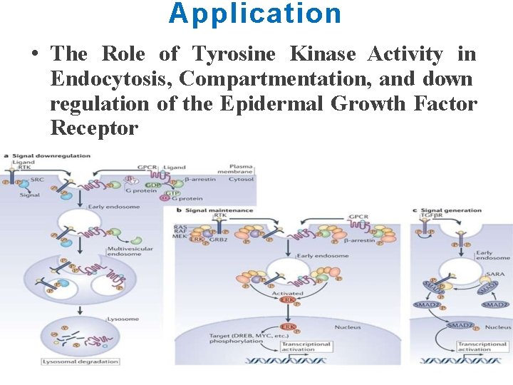
Application • The Role of Tyrosine Kinase Activity in Endocytosis, Compartmentation, and down regulation of the Epidermal Growth Factor Receptor
Ribosomal s 6 kinase • Ribosomal s 6 kinase (rsk) is a family of protein kinases involved in signal transduction. • There are two subfamilies of rsk, p 90 rsk, also known as MAPK-activated protein kinase-1 (MAPKAP-K 1), and p 70 rsk, also known as S 6 -H 1 Kinase or simply S 6 Kinase. • There are three variants of p 90 rsk in humans, rsk 1 -3. • Rsks are serine/threonine kinases and are activated by the MAPK/ERK pathway. • There are two known mammalian homologues of S 6 Kinase: S 6 K 1 and S 6 K 2.
Cross talk b/w signaling pathways • Crosstalk refers to instances in which one or more components of one signal transduction pathway affects another. • Most common form being crosstalk between proteins of signaling cascades. • In these signal transduction pathways, there are often shared components that can interact with either pathway. • A more complex instance of crosstalk can be observed with transmembrane crosstalk between the extracellular matrix (ECM) and the cytoskeleton.
c. AMP pathway • One example of crosstalk between proteins in a signalling pathway can be seen with cyclic adenosine monophosphate's (c. AMP) role in regulating cell proliferation by interacting with the mitogen-activated protein (MAP) kinase pathway. • c. AMP is a compound synthesized in cells by adenylate cyclase in response to a variety of extracellular signals. . • c. AMP primarily acts as an intracellular second messenger whose major intracellular receptor is the c. AMP-dependent protein kinase (PKA) that acts through the phosphorylation of target proteins. • The signal transduction pathway begins with ligand-receptor interactions extracellularly. • This signal is then transduced through the membrane, stimulating adenylyl cyclase on the inner membrane surface to catalyze the conversion of ATP to c. AMP.
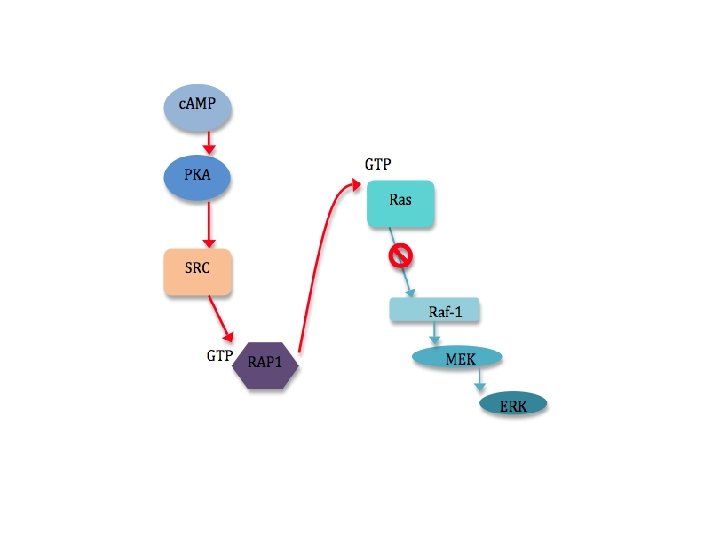
• ERK, (Extra cellular signal regulated kinases) a participating protein in the MAPK signaling pathway, can be activated or inhibited by c. AMP. • c. AMP can inhibit ERKs in a variety of ways, most of which involve the c. AMPdependent protein kinase (PKA) and the inhibition of Ras-dependent signals to Raf 1. • c. AMP can also stimulate cell proliferation by stimulating ERKs. • This occurs through the induction of specific genes via phosphorylation of the transcription factor CREB by PKA. • Though ERKs do not appear to be a requirement for this phosphorylation of CREB, the MAPK pathway.
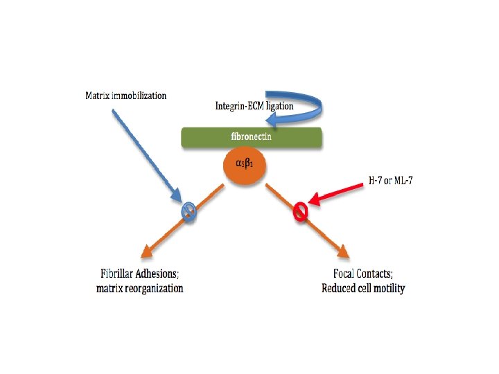
ECM • Crosstalk can even be observed across membranes. • Membrane interactions with the extracellular matrix (ECM) and with neighboring cells can trigger a variety of responses within the cell. • However, the topography and mechanical properties of the ECM also come to play an important role in powerful, complex crosstalk with the cells growing on or inside the matrix.
• For example, integrin-mediated cytoskeleton assembly and even cell motility are affected by the physical state of the ECM. • Binding of the α 5β 1 integrin to its ligand (fibronectin) activates the formation of fibrillar adhesions and actin filaments. • In turn, binding of the same integrin (α 5β 1) to an immobilized fibronectin ligand is seen to form highly phosphorylated focal contacts/focal adhesion (cells involved in matrix adhesion) within the membrane and reduces cell migration rates and matrix immobilization. • Matrix immobilization inhibits the formation of fibrillar adhesions and matrix reorganization. • Likewise, players of other signaling pathways inside the cell can affect the structure of the cytoskeleton and thereby the cell’s interaction with the ECM.