Pregnancy Growth and Development Chapter 23 Conception A


 • Stage one – the period from the onset of true labor")


- Slides: 44
Pregnancy, Growth and Development Chapter 23
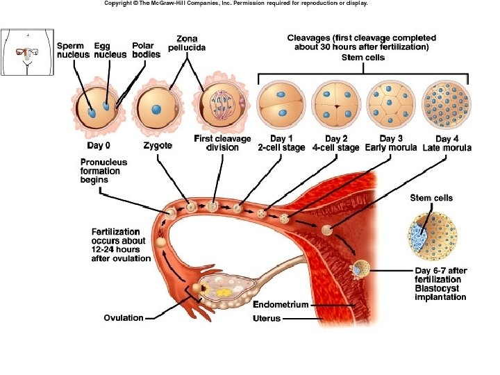
Conception • A secondary oocyte can be fertilized for about 24 hours after ovulation • Sperm remain viable for up to 48 hours within the female reproductive tract • This gives a three day “window” for intercourse to result in fertilization: two days before to one day after ovulation
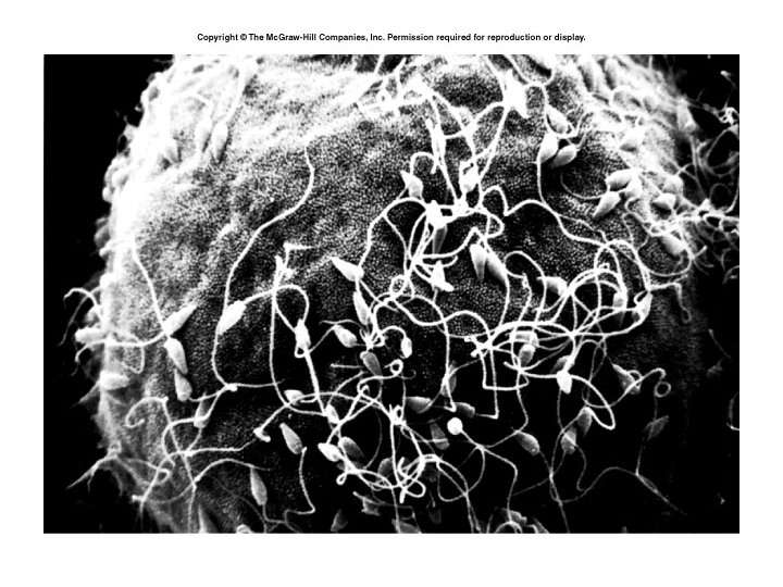
• Fertilization usually takes place in the outer one-third of the uterine tube, but can take place in the abdominal cavity • Sperm swim up the female reproductive tract, aided by muscular contractions of the uterus stimulated by prostaglandins in the semen. • The oocyte may also secrete a chemical that attracts sperm
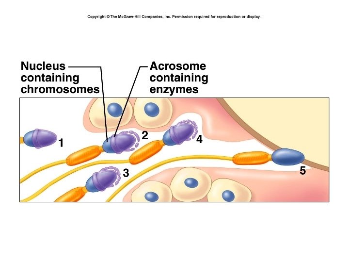
• Sperm undergo a functional change in the female tract – called capacitation • During this process the membrane around the acrosome becomes fragile, and its enzymes are released. • It requires the combined action of many sperm to allow one sperm to penetrate the oocyte.
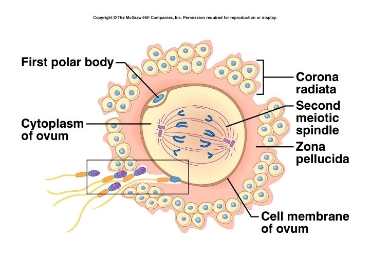
• When the first sperm enters the egg, the cell depolarizes causing the release of calcium ions inside the cell. • This stimulates the release of granules that cause changes in the zona pellucida to prevent entry of other sperm. • Secondary oocyte completes division, and nuclei of ovum and sperm unite to form a zygote.
Twins • Dizygotic or fraternal twins occur when two separate eggs are ovulated. May be of different sexes. • Monozygotic or identical twins occur when a single egg is fertilized but dividing cells break into two groups and develop into two individuals. Genetically identical (clones)
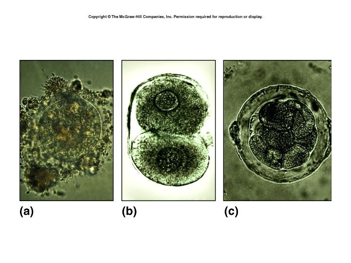
• Zygote undergoes rapid mitotic cell division, but these do not increase the size of the zygote – called cleavage divisions • Cleavage produces a solid sphere of cells, still surrounded by zona pellucida – now called a morula. • At 4. 5 to 5 days, cells have developed into a hollow ball of cells – blastocyst. • It is at this stage that it enters the uterus.
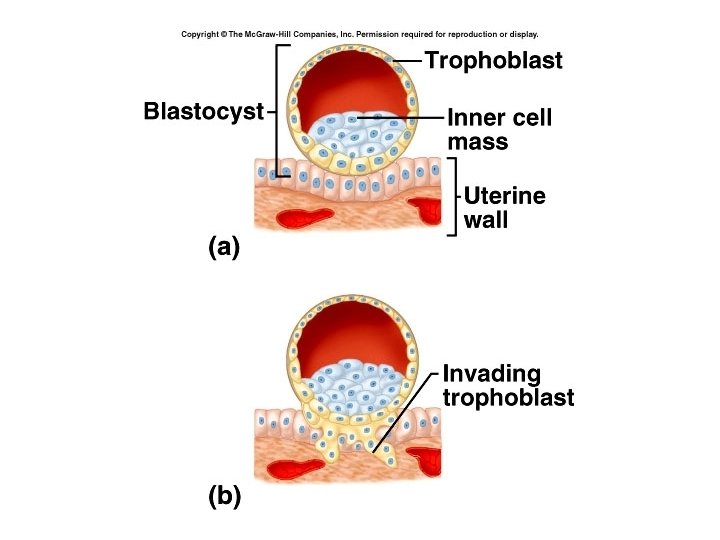
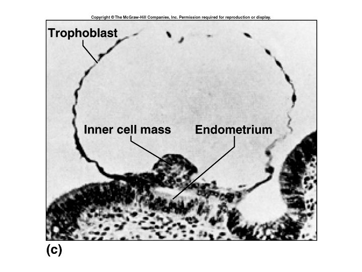
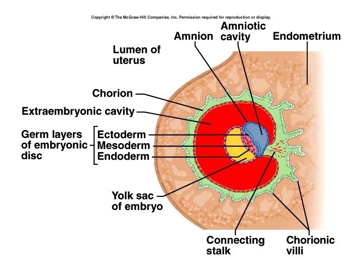
• Blastocyst has an outer layer of cells called the trophoblast, an inner cell mass, and a fluid filled cavity called the blastocele. • The trophoblast and part of the inner cell mass will form the membranes of the fetal portion of the placenta, the rest of the inner mass forms the embryo.
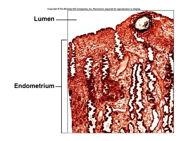
Implantation • The blastocyst remains free in the uterus a short time, during which the zona pellucida disintegrates. • Blastocyst nourished by glycogen from glands of the endometrium. • At about 6 days after ovulation blastocyst implants – orients cell mass toward endometrium, and secretes enzymes which allow it to penetrate (digest) the endometrial wall. This nourishes the blastocyst for about a week after implantation.
• Implantation can also occur in uterine tube, cervix, or the abdominal cavity. • Implantation anywhere outside the uterus is called an ectopic pregnancy. • It is possible for fetus to grow in the abdominal cavity, but growth inside the uterine tube causes the tube to rupture, resulting in severe bleeding.
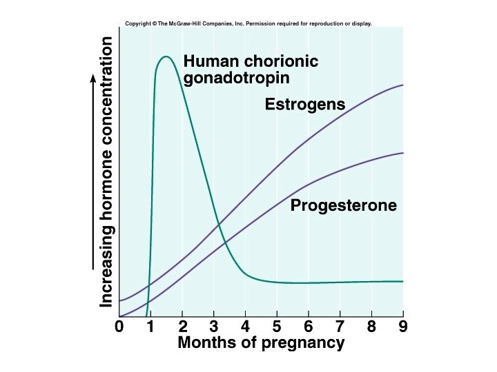
• As early as 8 -12 days after fertilization, the blastocyst begins to secrete human chorionic gonadotropin or h. CG. • h. CG keeps the corpus luteum active until the placenta can produce estrogens and progesterone. • The presence of h. CG is the basis for pregnancy tests.
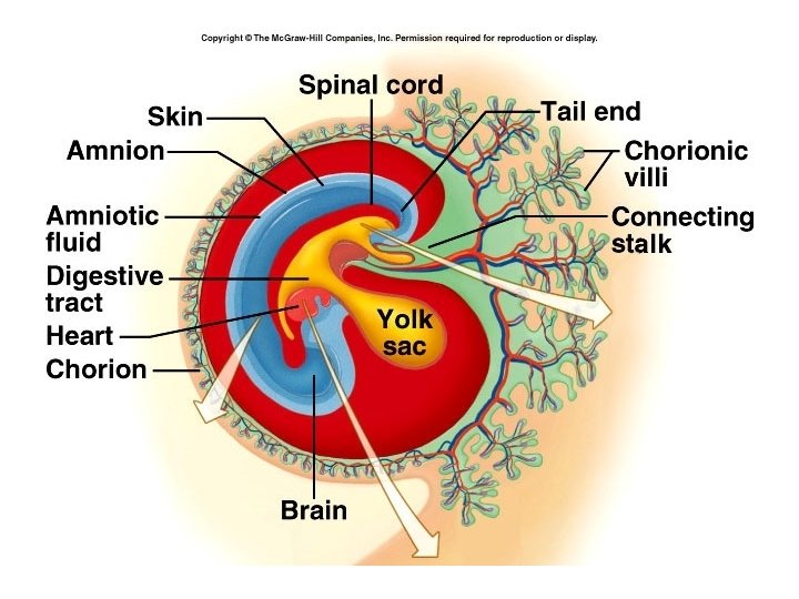
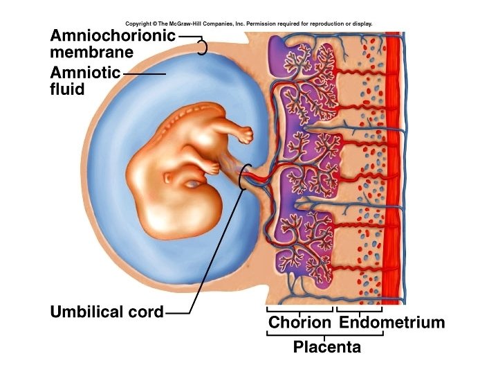
• Inner cell mass forms two cavities: – The yolk sac – Amniotic cavity • In humans the yolk sac produces blood cells and future sex cells • The amniotic cavity becomes the cavity in which the embryo floats. Fluid is produced from fetal urine, and secretions from the skin, respiratory tract, and amniotic membranes.
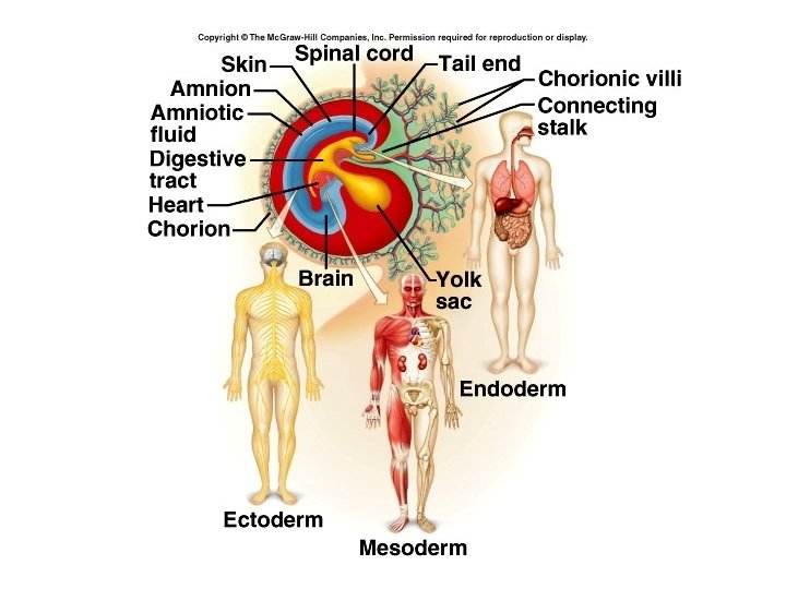
Primary germ layers • In between the yolk sac and the amniotic cavity is the embryonic disc, which gives rise to the primary germ layers: – Endoderm – Mesoderm – Ectoderm
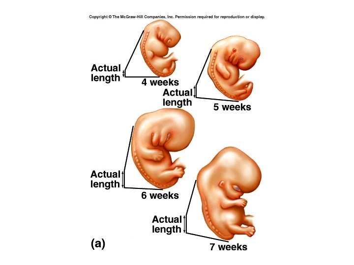
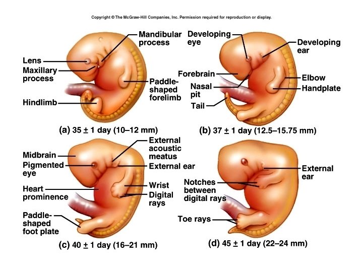
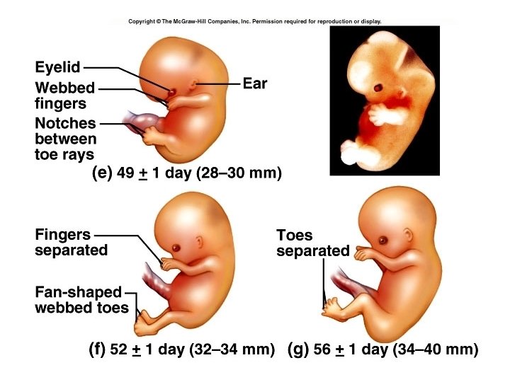
Gestation period • Divided into three trimesters. • During first trimester individual starts out as a zygote, then morula, blastocyst, and after implantation, is called an embryo. • Embryonic phase of development lasts from fertilization until the 8 th week of gestation, when it becomes a fetus. • By day 35 the heart is beating, and eye and limb buds are present.
• By month four, the rudiments of all organ systems are formed and functioning, and from then on, fetal development is primarily a matter of growth. • By the end of the third month the placenta is functioning.
The placenta • The chorion develops into the fetal part of the placenta. • The chorionic villi connect the fetal circulation to the placenta • Composed of both fetal and maternal tissues
Functions of the placenta: 1 Transfer gasses 2 Transport nutrients 3 Excretion of wastes 4 Hormone production – temporary endocrine organ – estrogen and progesterone 5 Formation of a barrier – incomplete, nonselective – alcohol, steroids, narcotics, anesthetics, some antibiotics and some organisms can cross
Quickening • The first movement of the fetus felt by the mother, usually occurring during the fourth or fifth month of pregnancy • By month seven the fetus is quite active • During the last month the fetus becomes less active (usually due to space considerations. )
• At the end of pregnancy both the mother and the uterus become “irritable” • The uterus undergoes Braxton-Hicks contractions: intermittent, painless contractions which can come 10 to 20 minutes apart. • Become more frequent as gestation progresses, and can be mistaken for onset of labor • Cervix begins to thin and dilate
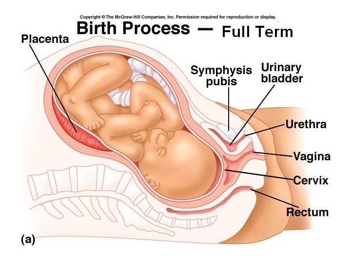
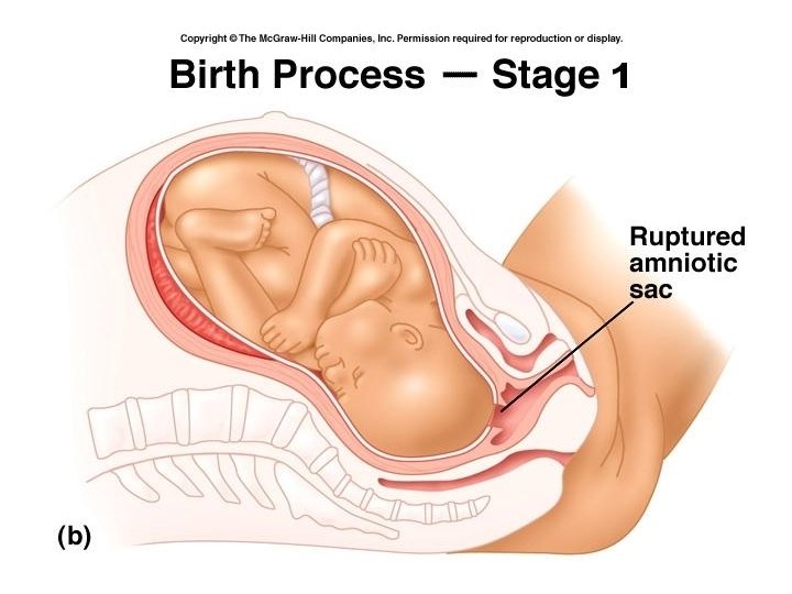
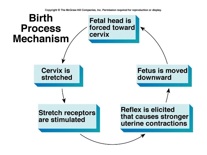
Labor (parturition) • Stage one – the period from the onset of true labor contractions until the cervix is completely dilated at 10 cm. • The uterine contractions cause the cervix to dilate, and the amniotic sac may rupture. • Usually lasts 6 – 24 hours depending on the number of previous deliveries.
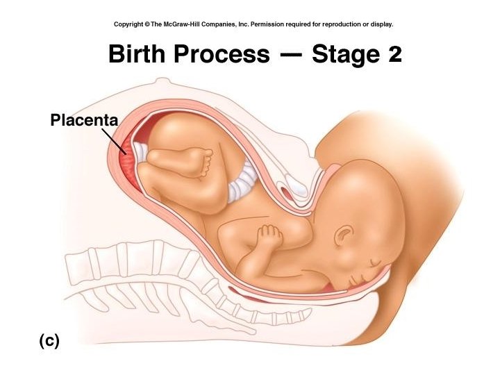
Stage 2 • Period from maximal cervical dilation until the birth of the baby • Lasts minutes to an hour • Contractions become more intense and frequent.
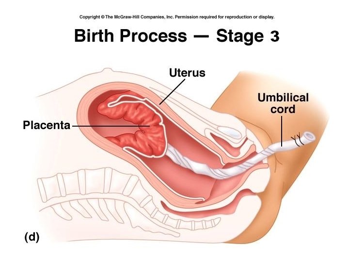
Stage 3 • The expulsion of the placenta • Usually occurs within 15 minutes after the birth of the baby, but can range from 5 to 60 minutes.
The End !! • • That’s it ! You’ve made it ! Study well ! Good luck !