PERIMETRY Riffat Sultana GOLDMAN PERIMETER IS THE MOST


 Has the following parts • 1. Stand: A heavy stand")


")


- Slides: 26
PERIMETRY Riffat Sultana
• GOLDMAN PERIMETER IS THE MOST WIDELY USED INSTRUMENT FOR MANUAL PERIMETRY
• The part of the external world visible to one eye when a person fixes his gaze on one point is called the field of vision for that eye. • Traquair defined visual field as island of vision in the sea of darkness. Sea represents the area of no light perception.
• The process of charting the monocular field of vision is called perimetry. • It is employed for the diagnosis of various lesions of the visual pathways.
Perimetry(also called Goldmans apparatus) Has the following parts • 1. Stand: A heavy stand on which a metal arc is fixed on a pivot, provides stability to the apparatus. • 2. Metal arc: A broad metal arc shaped like a half circle is mounted on the stand can be rotated in any meridian around its central pivot. one half of the arc, the concavity of which is directed towards the subject, has a scale of 0 to 90 degrees marked on its convex surface.
• Meridian locating dial: it gives the location of semi-circular dome when its position is being changed. it is present on the back of perimeter. • Chin rest: An adjustable chin rest is provided to keep the head steady. The chin of the subject rests on the right cupwhen the left eye is to be tested and the left cup is used when the right eye is tested.
• • Wooden base or stage. Fixation point. Eye piece (with a white tip) Chart:
Perimetry(charting the field of vision)
Chart • The perimeter chart on which the field of vision is to be plotted is divided by circles from 0 to 90 degrees and by meridians at 15 degree interval. • Both the angles and the meridians are printed on the chart. • The limits for the normal peripheral field of vision for the two eyes, and the blind spots are printed on the chart for comparison with the plotted field of vision. • The term peripheral field refers to the peripheral or outer limits of the field
Principle of student perimeter • In this model the inclination of the arc is read from a plastic dial fitted behind. • So when an object is moved along the inside of the arc, becomes visible, the angle it subtends at the fixation point in a given meridian can be read from the scale engraved on the outside of the arc. • The reading the meridian and the angle are then transferred to the corresponding points on the chart.
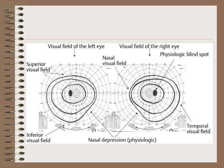
Divisions of visual field • The visual field of human eye has an angle of 160 degree in horizontal meridian and 135 degree in vertical meridian. The visual field is divided into four parts. 1. Temporal field. 2. Nasal field. 3. Upper field. 4. Lower field.
Temporal and Nasal fields • The visual field of each eye can be divided into two unequal parts namely outer or temporal field and the inner or nasal field by a vertical line passing through the fixation point. • The fixation point is the meeting point of visual axis with the object. • The temporal part of visual field extends up to about 100 degree but the nasal part extends only up to 60 degree because of restriction of nose.
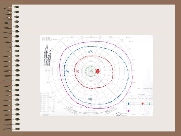
Isopter • All the locations where the stimulus is first seen have equal sensitivity these locations can be connected to form a ring shaped locus of points
Upper and lower fields • The visual field of each eye is also divided into an upper field and a lower field by horizontal line passing through the fixation point. • The extent of the upper field is about 60 degree and it is restricted by the upper eye lid and orbital margin. • The extent of the lower field is about 75 degree. This is restricted by check. Thus the visual field is restricted in all the sides except in the temporal part.
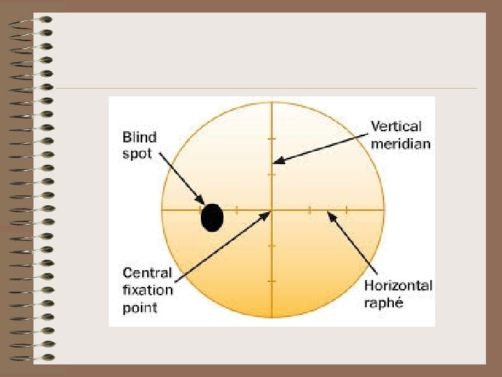
Blind spot • The optic disc in the retina does not have any visual receptors and if the image of any object falls on the optic disc the object cannot be seen. This part of the retina is blind. Hence it is known as blind spot.
Procedure 1. Apply eye pad on one eye. 2. Put chin on chin rest keeping the eye level at eye piece, ask the subject to fix his gaze at the fixation point(0 degree semi circular). 3. Slowly move white object from the periphery towards the centre and ask the subject to observe the object without moving the eyes.
procedure 4. Ask the subject identifies the object, stop the movement of object and take the reading(mark dot). 5. Rotate the arc at 15 degree interval for each eye till 360 degree. 6. After completing the whole test for 360 degree for both eyes join the dots.
Precautions 1. Room should be well illuminated. 2. The subject should use glasses only if there is a major refractive error. 3. In case of white object the background should be black. 4. In case of coloured object the back ground should be white. 5. One eye should be covered. 6. The subject should keep his eye fixed at fixation point.
SCOTOMA • Focal region of abnormally decreased sensitivity surrounded by the area of normal sensitivity. • Blind spot other than physiological blind spot is called scotoma. If multiple blind spots are present then they are called Scotomata.
CAUSES OF ABNORMAL BLIND SPOT. • Toxicity to eyes particularly to retina due to lead poisoning which may cause blind spot formation. • Allergic reactions involving rods and cones may causeblind spot. • Retinitus pigmentosa is degeneration pof retina and causes blind spot. • Excessive tobacco intake.
CAUSES OF ABNORMAL BLIND SPOT. • Glucoma • Retrobulbar neuritis • Hemianopia(loss of half of field of vision)
Colour vision • Red line appear in the centre of graph followed by green blue and in the periphery is white line. • Why the red line appear in the centre. • Cones are meant for colour • The concentration of cones is also more in the centre. An orange monochromatic light with a wavelength of 580 nanometers stimulates the red cones to a value of about 99 percent.
• Thank you for your attention.