PELVIS SACRUM Dr Jamila ElMedany Dr Essam Eldin


- Slides: 30
PELVIS & SACRUM Dr. Jamila El-Medany Dr. Essam Eldin Salama
Learning Objectives v. At the end of the lecture, the students should be able to : § Describe the bony structures of the pelvis. § Describe in detail the hip bone, the sacrum, and the coccyx. § Describe the boundaries of the pelvic inlet and outlet. § Identify the articulations of the bony pelvis. § List the major differences between the male and female pelvis. § List the different types of female pelvis.
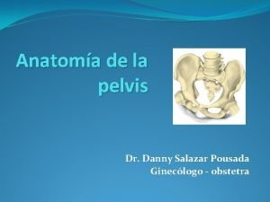
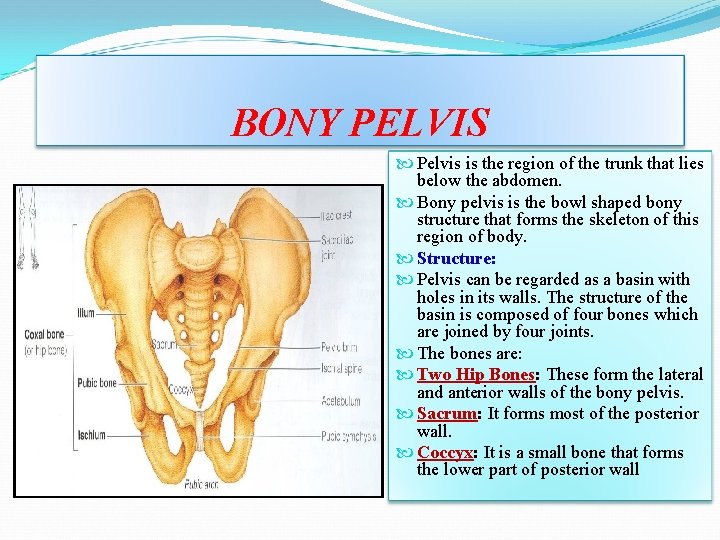
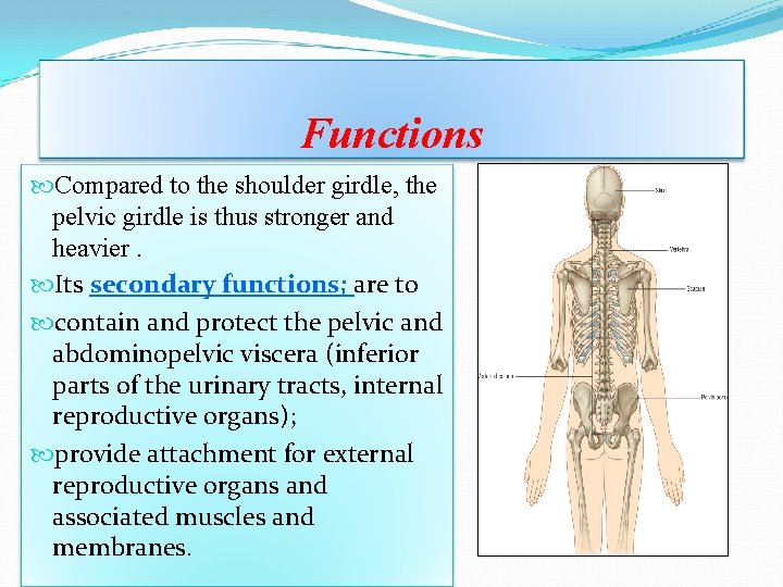
BONY PELVIS Pelvis is the region of the trunk that lies below the abdomen. Bony pelvis is the bowl shaped bony structure that forms the skeleton of this region of body. Structure: Pelvis can be regarded as a basin with holes in its walls. The structure of the basin is composed of four bones which are joined by four joints. The bones are: Two Hip Bones: These form the lateral and anterior walls of the bony pelvis. Sacrum: It forms most of the posterior wall. Coccyx: It is a small bone that forms the lower part of posterior wall
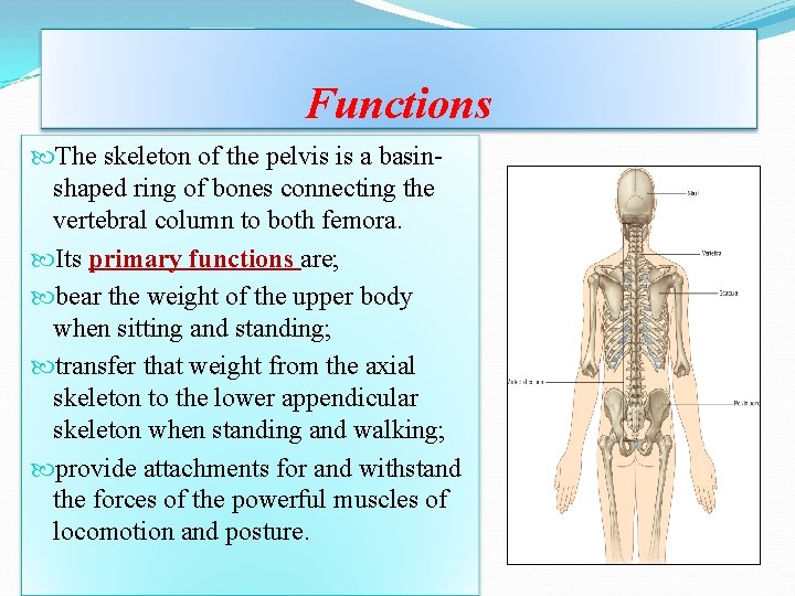
Functions The skeleton of the pelvis is a basinshaped ring of bones connecting the vertebral column to both femora. Its primary functions are; bear the weight of the upper body when sitting and standing; transfer that weight from the axial skeleton to the lower appendicular skeleton when standing and walking; provide attachments for and withstand the forces of the powerful muscles of locomotion and posture.
Functions Compared to the shoulder girdle, the pelvic girdle is thus stronger and heavier. Its secondary functions; are to contain and protect the pelvic and abdominopelvic viscera (inferior parts of the urinary tracts, internal reproductive organs); provide attachment for external reproductive organs and associated muscles and membranes.
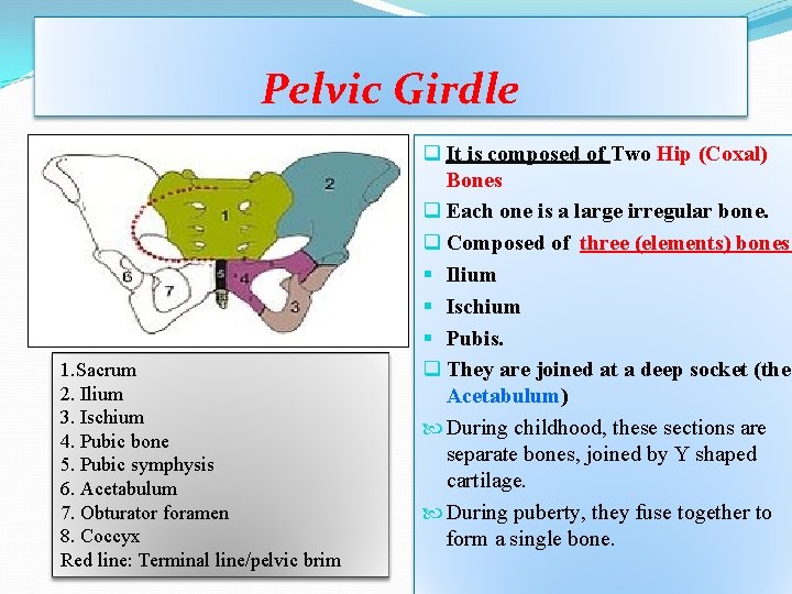
Pelvic Girdle 1. Sacrum 2. Ilium 3. Ischium 4. Pubic bone 5. Pubic symphysis 6. Acetabulum 7. Obturator foramen 8. Coccyx Red line: Terminal line/pelvic brim q It is composed of Two Hip (Coxal) Bones q Each one is a large irregular bone. q Composed of three (elements) bones: § Ilium § Ischium § Pubis. q They are joined at a deep socket (the Acetabulum) During childhood, these sections are separate bones, joined by Y shaped cartilage. During puberty, they fuse together to form a single bone.
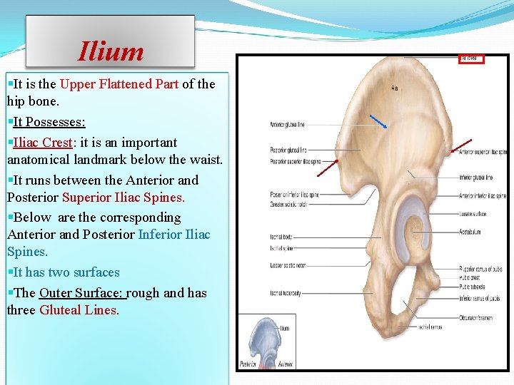
Ilium §It is the Upper Flattened Part of the hip bone. §It Possesses: §Iliac Crest: it is an important anatomical landmark below the waist. §It runs between the Anterior and Posterior Superior Iliac Spines. §Below are the corresponding Anterior and Posterior Inferior Iliac Spines. §It has two surfaces §The Outer Surface: rough and has three Gluteal Lines.
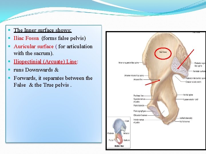
§ The Inner surface shows: § Iliac Fossa (forms false pelvis) § Auricular surface ( for articulation with the sacrum). § Iliopectinial (Arcuate) Line: § runs Downwards & § Forwards, it separates between the False & the True pelvis.
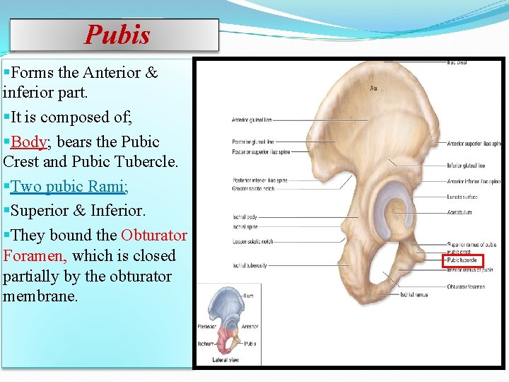
Pubis §Forms the Anterior & inferior part. §It is composed of; §Body; bears the Pubic Crest and Pubic Tubercle. §Two pubic Rami; §Superior & Inferior. §They bound the Obturator Foramen, which is closed partially by the obturator membrane.
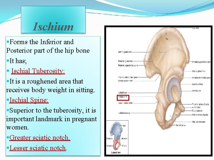
Ischium §Forms the Inferior and Posterior part of the hip bone §It has; § Ischial Tuberosity: §It is a roughened area that receives body weight in sitting. §Ischial Spine: §Superior to the tuberosity, it is important landmark in pregnant women. §Greater sciatic notch. §Lesser sciatic notch.
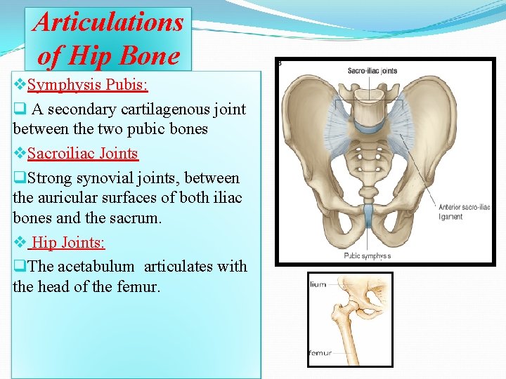
Articulations of Hip Bone v. Symphysis Pubis: q A secondary cartilagenous joint between the two pubic bones v. Sacroiliac Joints q. Strong synovial joints, between the auricular surfaces of both iliac bones and the sacrum. v Hip Joints: q. The acetabulum articulates with the head of the femur.
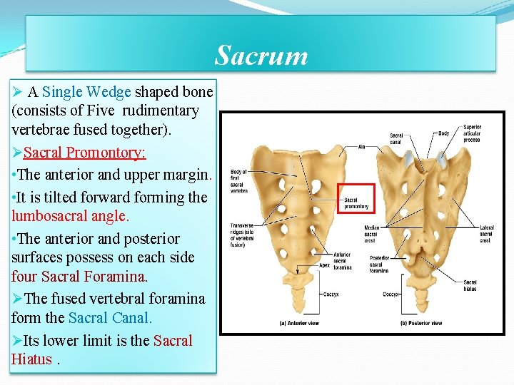
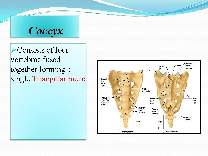
Sacrum Ø A Single Wedge shaped bone (consists of Five rudimentary vertebrae fused together). ØSacral Promontory: • The anterior and upper margin. • It is tilted forward forming the lumbosacral angle. • The anterior and posterior surfaces possess on each side four Sacral Foramina. ØThe fused vertebral foramina form the Sacral Canal. ØIts lower limit is the Sacral Hiatus.
Coccyx ØConsists of four vertebrae fused together forming a single Triangular piece
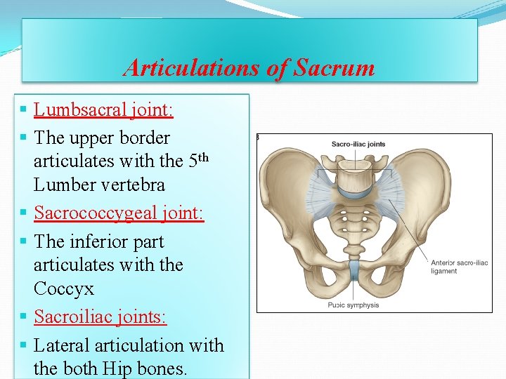
Articulations of Sacrum § Lumbsacral joint: § The upper border articulates with the 5 th Lumber vertebra § Sacrococcygeal joint: § The inferior part articulates with the Coccyx § Sacroiliac joints: § Lateral articulation with the both Hip bones.
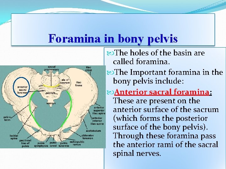
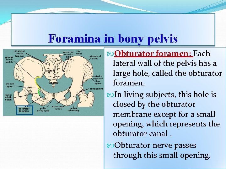
Foramina in bony pelvis The holes of the basin are called foramina. The Important foramina in the bony pelvis include: Anterior sacral foramina: These are present on the anterior surface of the sacrum (which forms the posterior surface of the bony pelvis). Through these foramina pass the anterior rami of the sacral spinal nerves.
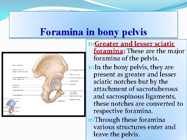
Foramina in bony pelvis Greater and lesser sciatic foramina: These are the major foramina of the pelvis. In the bony pelvis, they are present as greater and lesser sciatic notches but by the attachment of sacrotuberous and sacrospinous ligaments, these notches are converted to respective foramina. Through these foramina various structures enter and leave the pelvis.
Foramina in bony pelvis Obturator foramen: Each lateral wall of the pelvis has a large hole, called the obturator foramen. In living subjects, this hole is closed by the obturator membrane except for a small opening, which represents the obturator canal. Obturator nerve passes through this small opening.
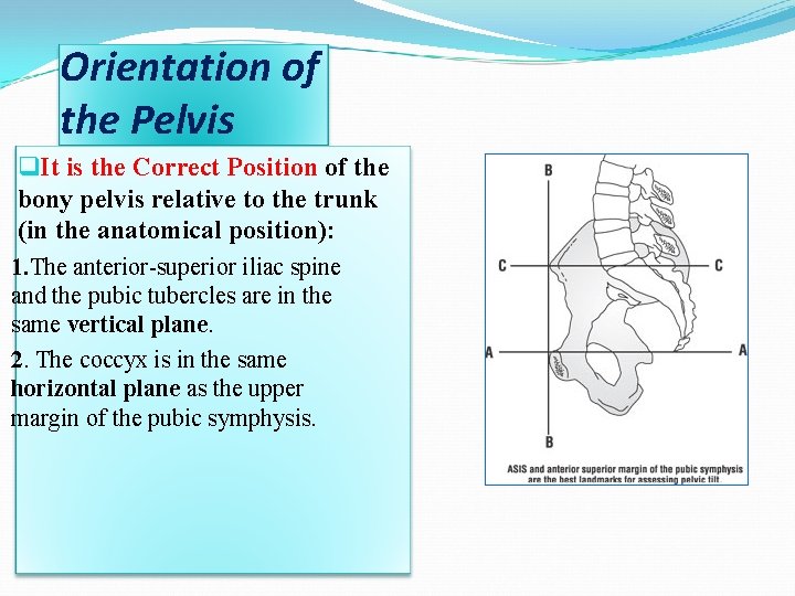
Orientation of the Pelvis q. It is the Correct Position of the bony pelvis relative to the trunk (in the anatomical position): 1. The anterior-superior iliac spine and the pubic tubercles are in the same vertical plane. 2. The coccyx is in the same horizontal plane as the upper margin of the pubic symphysis.
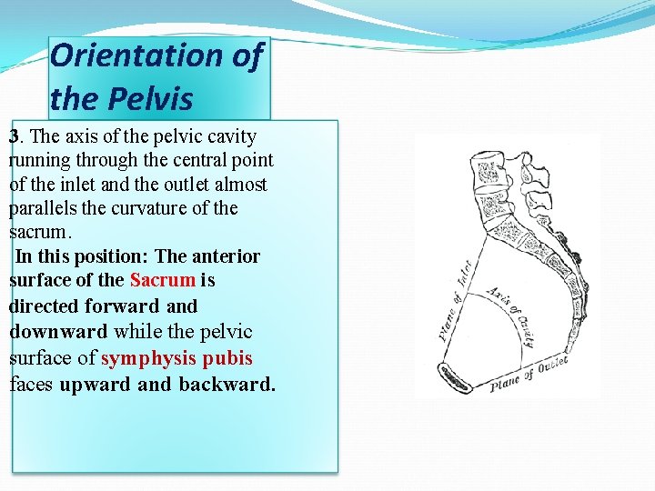
Orientation of the Pelvis 3. The axis of the pelvic cavity running through the central point of the inlet and the outlet almost parallels the curvature of the sacrum. In this position: The anterior surface of the Sacrum is directed forward and downward while the pelvic surface of symphysis pubis faces upward and backward.
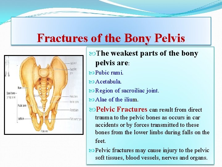
Fractures of the Bony Pelvis The weakest parts of the bony pelvis are: Pubic rami. Acetabula. Region of sacroiliac joint. Alae of the ilium. Pelvic Fractures can result from direct trauma to the pelvic bones as occurs in car accidents or by forces transmitted to these bones from the lower limbs during falls on the feet. Pelvic fractures may cause injury to the pelvic soft tissues, blood vessels, nerves and organs.
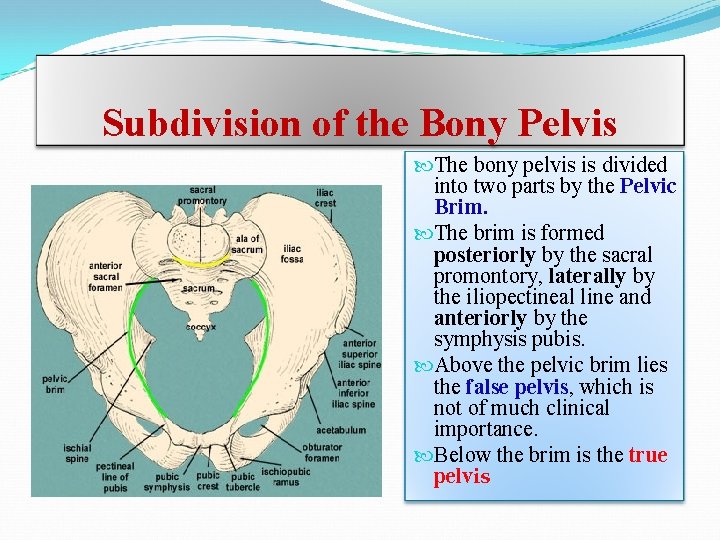
Subdivision of the Bony Pelvis The bony pelvis is divided into two parts by the Pelvic Brim. The brim is formed posteriorly by the sacral promontory, laterally by the iliopectineal line and anteriorly by the symphysis pubis. Above the pelvic brim lies the false pelvis, which is not of much clinical importance. Below the brim is the true pelvis
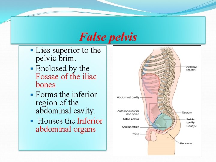
False pelvis § Lies superior to the pelvic brim. § Enclosed by the Fossae of the iliac bones § Forms the inferior region of the abdominal cavity. § Houses the Inferior abdominal organs
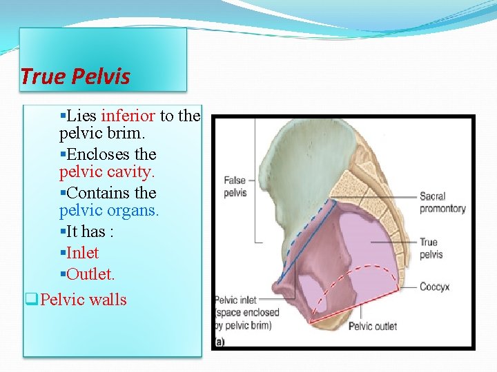
True Pelvis §Lies inferior to the pelvic brim. §Encloses the pelvic cavity. §Contains the pelvic organs. §It has : §Inlet §Outlet. q. Pelvic walls
Pelvic Inlet (pelvic Brim) q. Bounded by: §Sacral promontory §Iliopectineal lines. §Symphysis pubis.
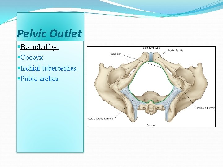
Pelvic Outlet §Bounded by: §Coccyx §Ischial tuberosities. §Pubic arches.
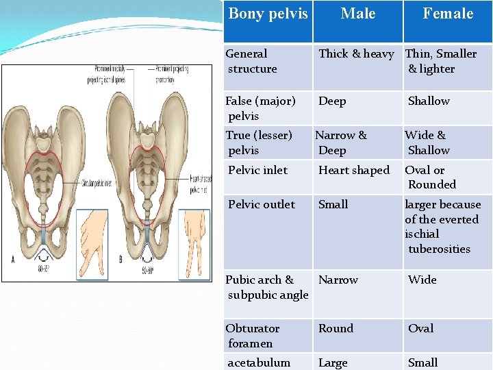
Bony pelvis Male Female General structure Thick & heavy Thin, Smaller & lighter False (major) pelvis Deep Shallow True (lesser) pelvis Narrow & Deep Wide & Shallow Pelvic inlet Heart shaped Oval or Rounded Pelvic outlet Small larger because of the everted ischial tuberosities Pubic arch & Narrow subpubic angle Wide Obturator foramen Round Oval acetabulum Large Small
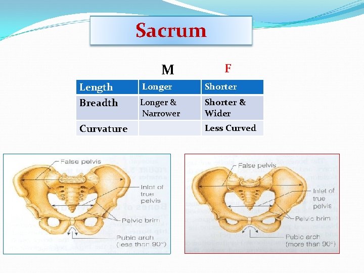
Sacrum M F Length Longer Shorter Breadth Longer & Narrower Shorter & Wider Curvature Less Curved
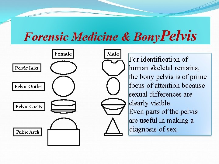
Forensic Medicine & Bony. Pelvis Female Pelvic Inlet Pelvic Outlet Pelvic Cavity Pubic Arch Male For identification of human skeletal remains, the bony pelvis is of prime focus of attention because sexual differences are clearly visible. Even parts of the pelvis are useful in making a diagnosis of sex.
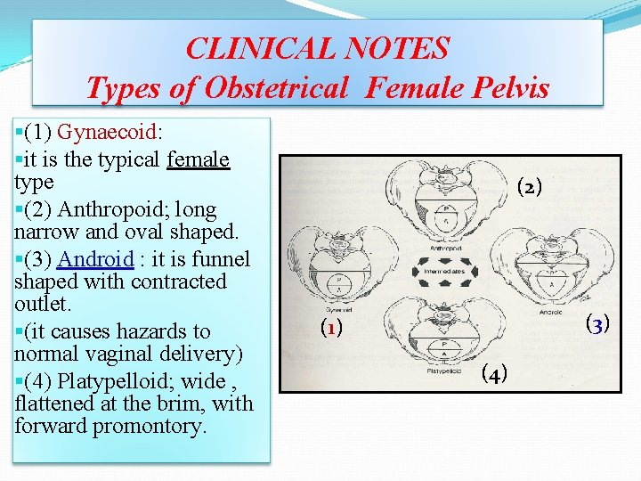
CLINICAL NOTES Types of Obstetrical Female Pelvis §(1) Gynaecoid: §it is the typical female type §(2) Anthropoid; long narrow and oval shaped. §(3) Android : it is funnel shaped with contracted outlet. §(it causes hazards to normal vaginal delivery) §(4) Platypelloid; wide , flattened at the brim, with forward promontory. (2) ( 3) ( 1) (4)
Thank you