
This file reviews materials in Lab 4 and
This file reviews materials in Lab 4 and it gets you ready for Lab 6. Lab#4 -- Ex. 7 (Overview of the skeleton) and Ex. 8 (Axial skeleton) Lab#5 -- Ex. 9 (Appendicular skeleton), Ex. 10 (Fetal skeleton), and Ex. 11 (Articulations and body movements) Lab#6 -- the second lab practical (200 points) by Dr. Shaw, Zoology 251 Lab Coordinator, x 7176; donalds@utm. edu 1
Lab#4– Ex. 7: Overview of the skeleton 2
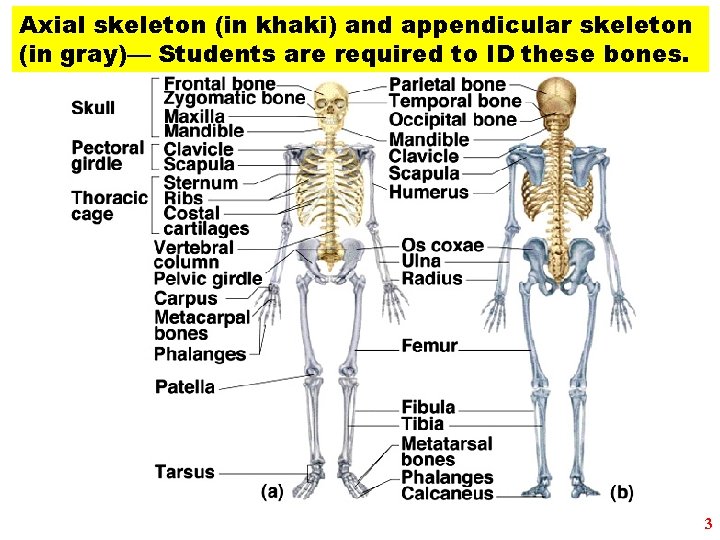
Axial skeleton (in khaki) and appendicular skeleton (in gray)— Students are required to ID these bones. 3
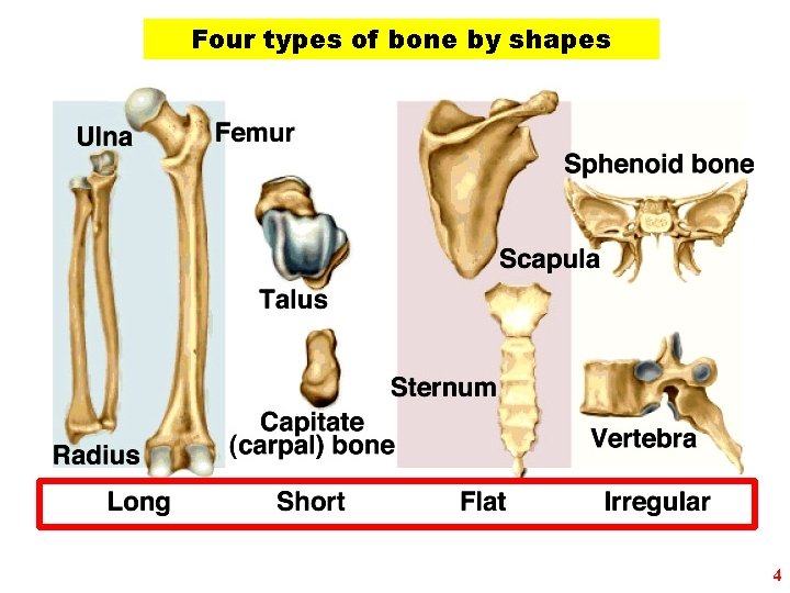
Four types of bone by shapes 4
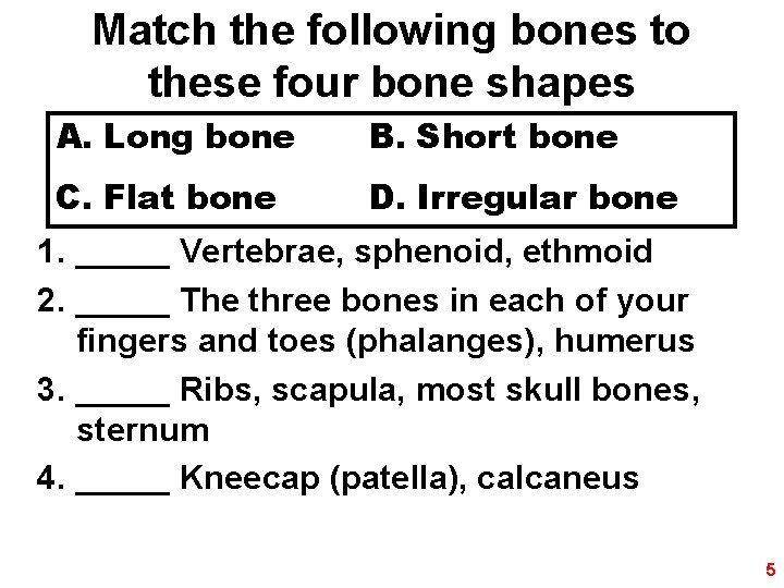
Match the following bones to these four bone shapes A. Long bone B. Short bone C. Flat bone D. Irregular bone 1. _____ Vertebrae, sphenoid, ethmoid 2. _____ The three bones in each of your fingers and toes (phalanges), humerus 3. _____ Ribs, scapula, most skull bones, sternum 4. _____ Kneecap (patella), calcaneus 5
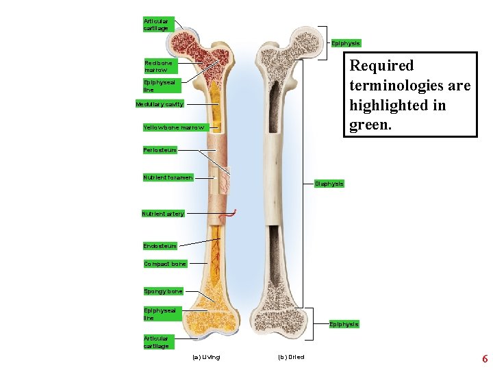
Articular cartilage Epiphysis Required terminologies are highlighted in green. Red bone marrow Epiphyseal line Medullary cavity Yellow bone marrow Periosteum Nutrient foramen Diaphysis Nutrient artery Endosteum Compact bone Spongy bone Epiphyseal line Epiphysis Articular cartilage (a) Living (b) Dried 6
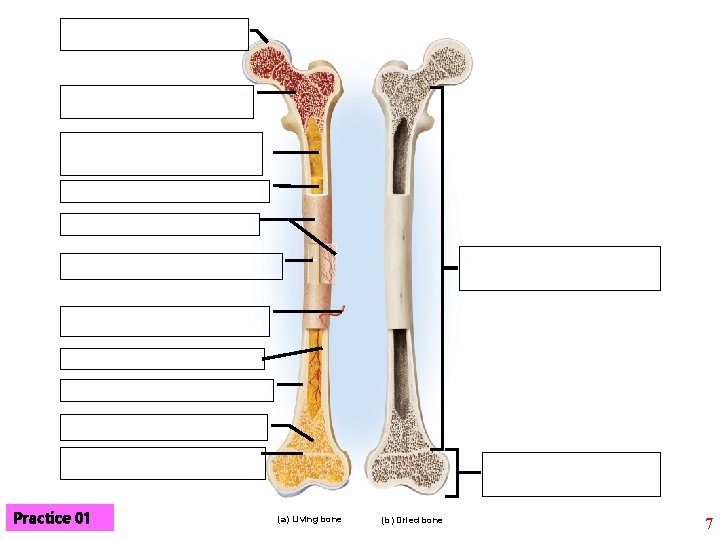
Practice 01 (a) Living bone (b) Dried bone 7
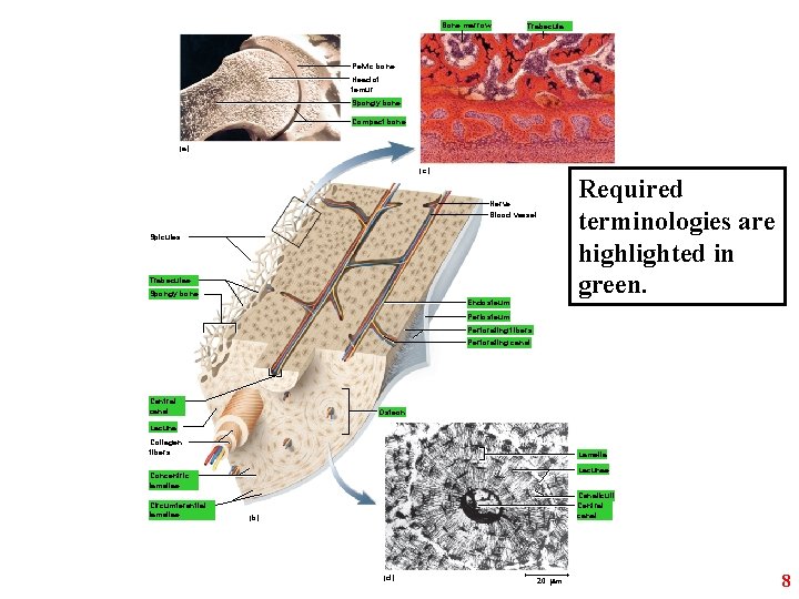
Bone marrow Trabecula Pelvic bone Head of femur Spongy bone Compact bone (a) (c) Nerve Blood vessel Spicules Trabeculae Spongy bone Endosteum Required terminologies are highlighted in green. Periosteum Perforating fibers Perforating canal Central canal Osteon Lacuna Collagen fibers Lamella Lacunae Concentric lamellae Circumferential lamellae Canaliculi Central canal (b) (d) 20 m 8
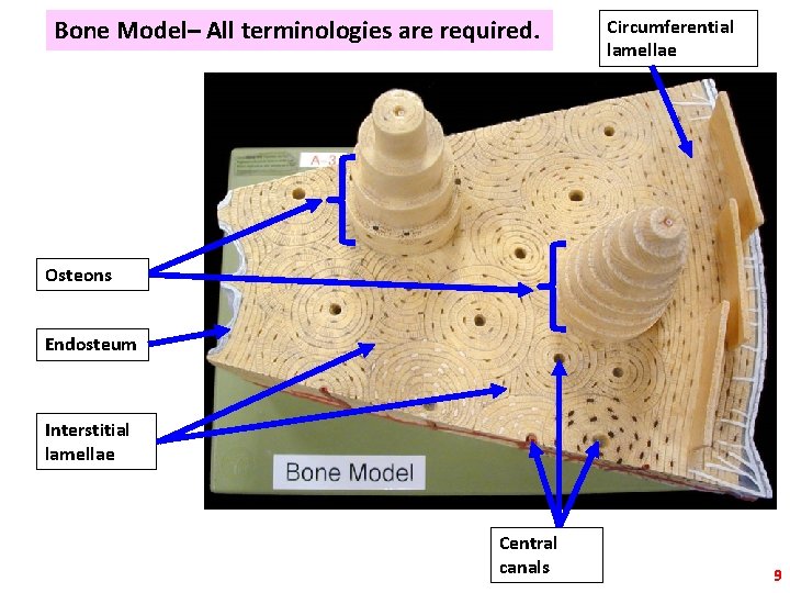
Bone Model– All terminologies are required. Circumferential lamellae Osteons Endosteum Interstitial lamellae Central canals 9
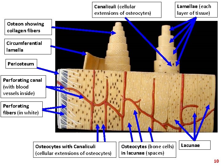
Canaliculi (cellular extensions of osteocytes) Lamellae (each layer of tissue) Osteon showing collagen fibers Circumferential lamella Periosteum Perforating canal (with blood vessels inside) Perforating fibers (in white) Osteocytes with Canaliculi (cellular extensions of osteocytes) Osteocytes (bone cells) in lacunae (spaces) Lacunae 10
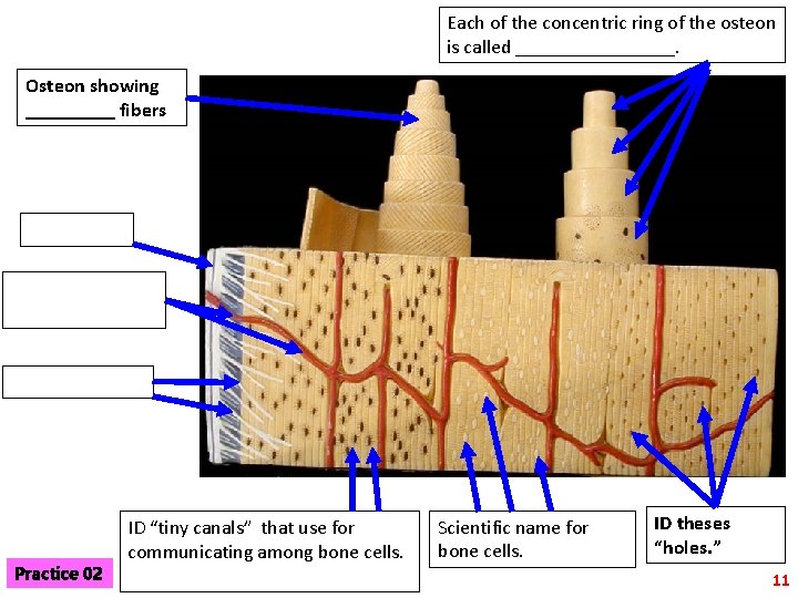
Each of the concentric ring of the osteon is called ________. Osteon showing _____ fibers Practice 02 ID “tiny canals” that use for communicating among bone cells. Scientific name for bone cells. ID theses “holes. ” 11
Lab#4– Ex. 8: Axial skeleton 12
Ex. 8: Axial skeleton; A- The cranial bones 13
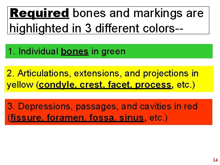
Required bones and markings are highlighted in 3 different colors-1. Individual bones in green 2. Articulations, extensions, and projections in yellow (condyle, crest, facet, process, etc. ) 3. Depressions, passages, and cavities in red (fissure, foramen, fossa, sinus, etc. ) 14
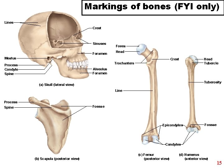
Markings of bones (FYI only) Lines Crest Sinuses Foramen Fovea Head Meatus Crest Trochanters Process Condyle Spine Head Tubercle Alveolus Foramen Tuberosity (a) Skull (lateral view) Line Process Fossae Spine Epicondyles Fossae Condyles (b) Scapula (posterior view) (c) Femur (posterior view) (d) Humerus (anterior view) 15
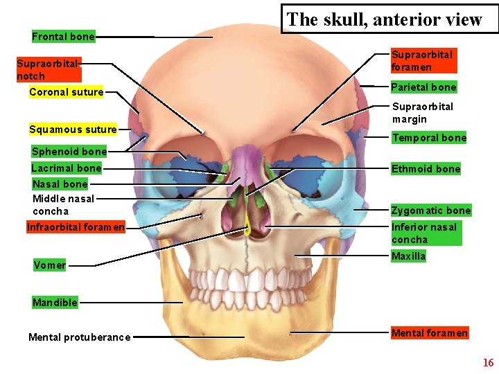
The skull, anterior view Frontal bone Supraorbital notch Coronal suture Squamous suture Supraorbital foramen Parietal bone Supraorbital margin Temporal bone Sphenoid bone Lacrimal bone Ethmoid bone Nasal bone Middle nasal concha Zygomatic bone Infraorbital foramen Vomer Inferior nasal concha Maxilla Mandible Mental protuberance Mental foramen 16
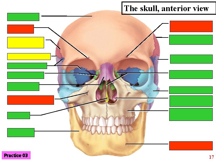
The skull, anterior view Practice 03 17
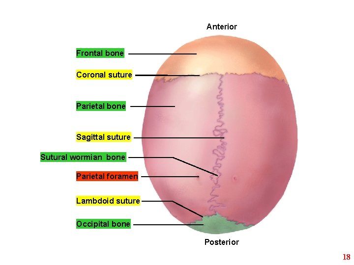
Anterior Frontal bone Coronal suture Parietal bone Sagittal suture Sutural wormian bone Parietal foramen Lambdoid suture Occipital bone Posterior 18
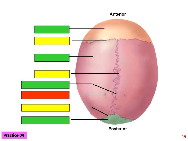
Anterior Posterior Practice 04 19
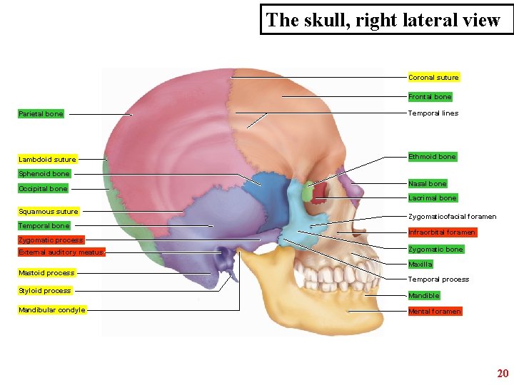
The skull, right lateral view Coronal suture Frontal bone Parietal bone Temporal lines Lambdoid suture Ethmoid bone Sphenoid bone Occipital bone Nasal bone Lacrimal bone Squamous suture Temporal bone Zygomatic process External auditory meatus Zygomaticofacial foramen Infraorbital foramen Zygomatic bone Maxilla Mastoid process Styloid process Mandibular condyle Temporal process Mandible Mental foramen 20
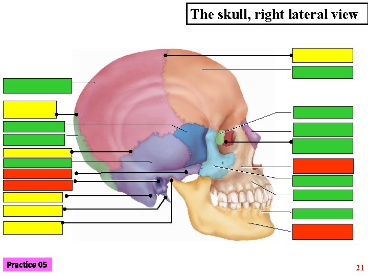
The skull, right lateral view Practice 05 21
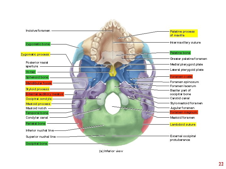
Incisive foramen Palatine process of maxilla Intermaxillary suture Zygomatic bone Palatine bone Zygomatic process Greater palatine foramen Posterior nasal aperture Medial pterygoid plate Lateral pterygoid plate Vomer Sphenoid bone Foramen ovale Mandibular fossa Foramen spinosum Foramen lacerum Basilar part of occipital bone Carotid canal Styloid process External auditory meatus Occipital condyle Mastoid process Mastoid notch Temporal bone Stylomastoid foramen Condylar canal Mastoid foramen Parietal bone Lambdoid suture Jugular foramen Foramen magnum Inferior nuchal line External occipital protuberance Superior nuchal line Occipital bone (a) Inferior view 22
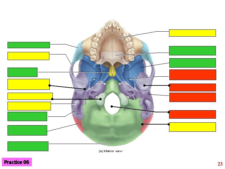
(a) Inferior view Practice 06 23
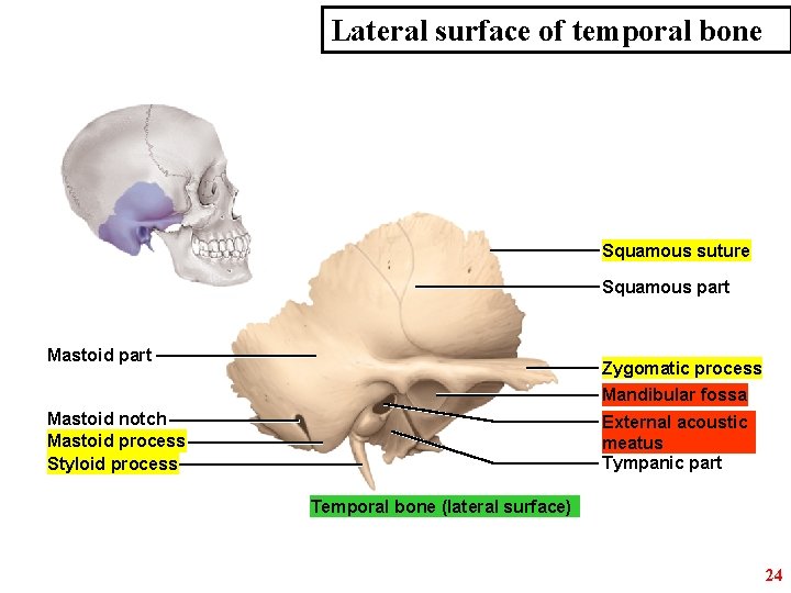
Lateral surface of temporal bone Squamous suture Squamous part Mastoid part Zygomatic process Mandibular fossa Mastoid notch Mastoid process Styloid process External acoustic meatus Tympanic part Temporal bone (lateral surface) 24
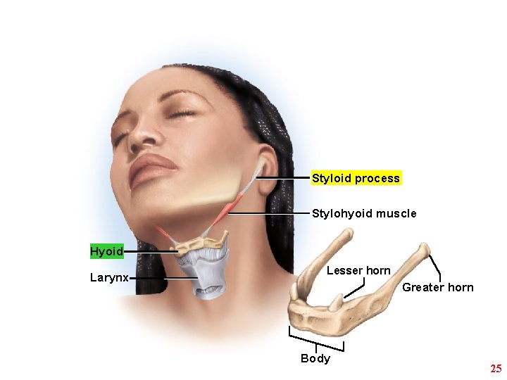
Styloid process Stylohyoid muscle Hyoid Larynx Lesser horn Greater horn Body 25
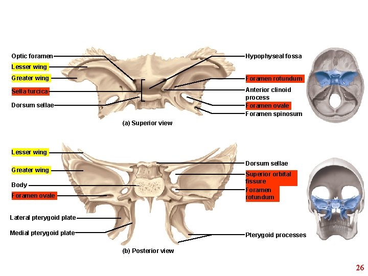
Optic foramen Hypophyseal fossa Lesser wing Greater wing Foramen rotundum Sella turcica Anterior clinoid process Foramen ovale Foramen spinosum Dorsum sellae (a) Superior view Lesser wing Dorsum sellae Greater wing Superior orbital fissure Foramen rotundum Body Foramen ovale Lateral pterygoid plate Medial pterygoid plate Pterygoid processes (b) Posterior view 26
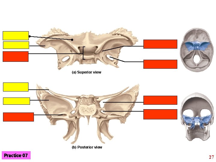
(a) Superior view (b) Posterior view Practice 07 27
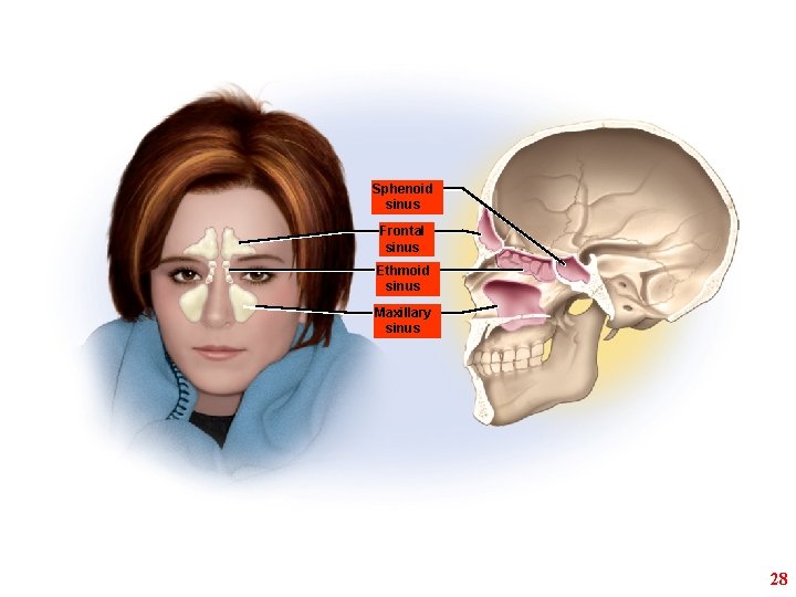
Sphenoid sinus Frontal sinus Ethmoid sinus Maxillary sinus 28
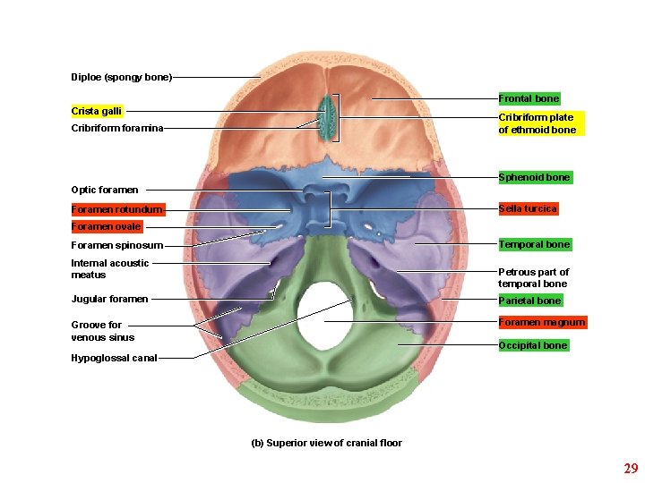
Diploe (spongy bone) Frontal bone Crista galli Cribriform plate of ethmoid bone Cribriform foramina Sphenoid bone Optic foramen Sella turcica Foramen rotundum Foramen ovale Temporal bone Foramen spinosum Internal acoustic meatus Petrous part of temporal bone Jugular foramen Parietal bone Groove for venous sinus Foramen magnum Occipital bone Hypoglossal canal (b) Superior view of cranial floor 29
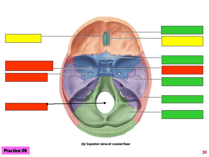
(b) Superior view of cranial floor Practice 08 30
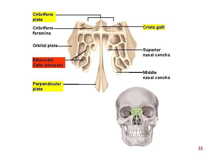
Cribriform plate Cribriform foramina Orbital plate Crista galli Superior nasal concha Ethmoidal Cells (sinuses) Middle nasal concha Perpendicular plate 31
Ex. 8: Axial skeleton; B- The facial bones 32
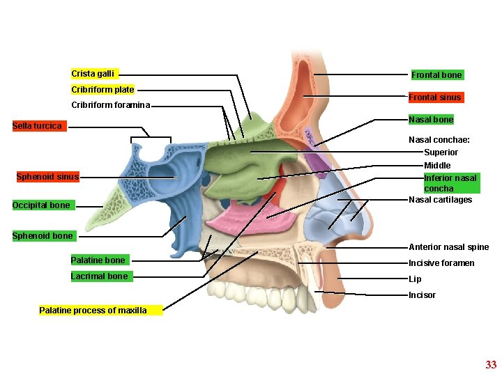
Crista galli Cribriform plate Cribriform foramina Frontal bone Frontal sinus Nasal bone Sella turcica Nasal conchae: Superior Middle Sphenoid sinus Occipital bone Inferior nasal concha Nasal cartilages Sphenoid bone Anterior nasal spine Palatine bone Incisive foramen Lacrimal bone Lip Incisor Palatine process of maxilla 33
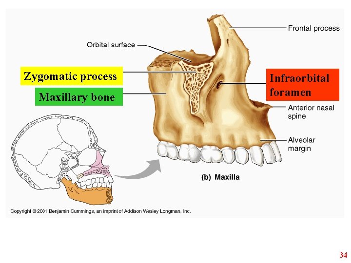
Zygomatic process Maxillary bone Infraorbital foramen 34
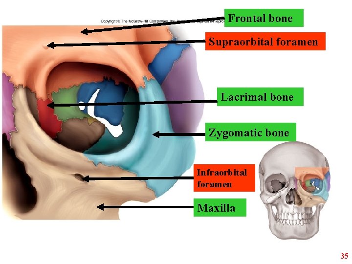
Frontal bone Supraorbital foramen Lacrimal bone Zygomatic bone Infraorbital foramen Maxilla 35
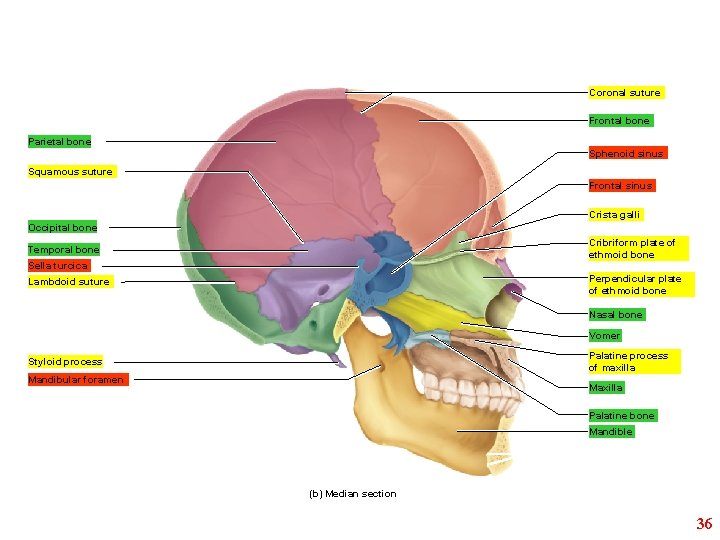
Coronal suture Frontal bone Parietal bone Sphenoid sinus Squamous suture Frontal sinus Crista galli Occipital bone Cribriform plate of ethmoid bone Temporal bone Sella turcica Perpendicular plate of ethmoid bone Lambdoid suture Nasal bone Vomer Palatine process of maxilla Styloid process Mandibular foramen Maxilla Palatine bone Mandible (b) Median section 36
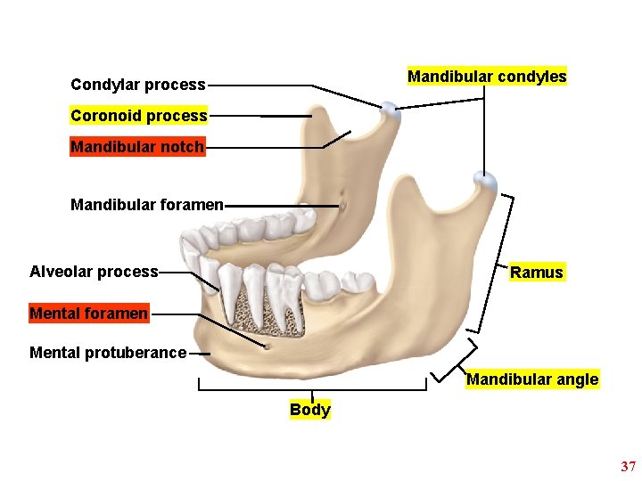
Mandibular condyles Condylar process Coronoid process Mandibular notch Mandibular foramen Alveolar process Ramus Mental foramen Mental protuberance Mandibular angle Body 37
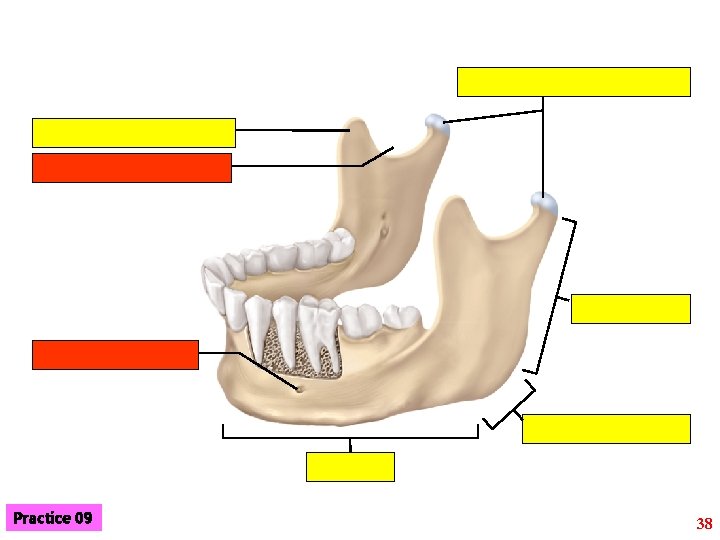
Practice 09 38
Ex. 8: Axial skeleton; C- The vertebral column & the thoracic cage 39
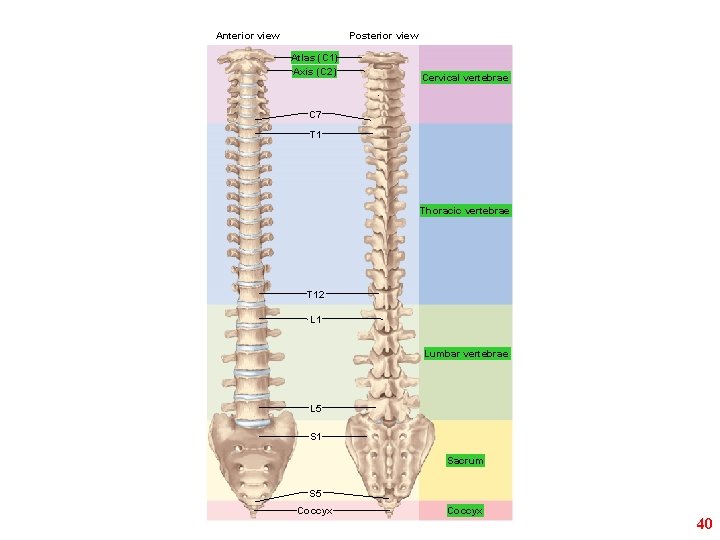
Anterior view Posterior view Atlas (C 1) Axis (C 2) Cervical vertebrae C 7 T 1 Thoracic vertebrae T 12 L 1 Lumbar vertebrae L 5 S 1 Sacrum S 5 Coccyx 40
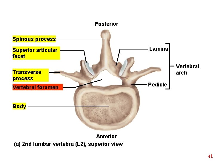
Posterior Spinous process Superior articular facet Lamina Vertebral arch Transverse process Vertebral foramen Pedicle Body Anterior (a) 2 nd lumbar vertebra (L 2), superior view 41
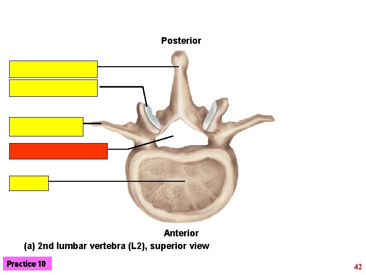
Posterior Anterior (a) 2 nd lumbar vertebra (L 2), superior view Practice 10 42
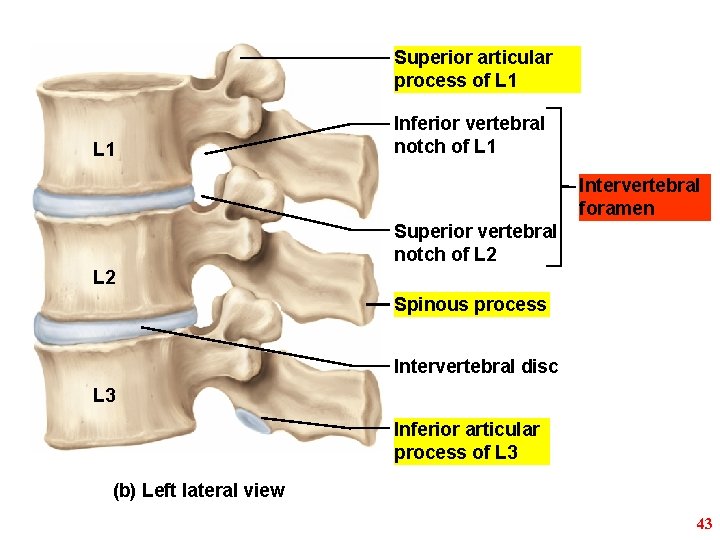
Superior articular process of L 1 Inferior vertebral notch of L 1 Intervertebral foramen Superior vertebral notch of L 2 Spinous process Intervertebral disc L 3 Inferior articular process of L 3 (b) Left lateral view 43
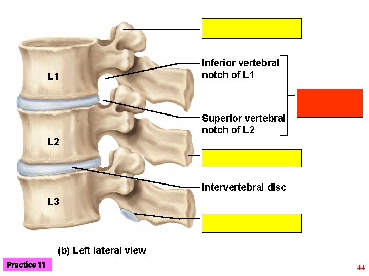
L 1 Inferior vertebral notch of L 1 Superior vertebral notch of L 2 Intervertebral disc L 3 (b) Left lateral view Practice 11 44
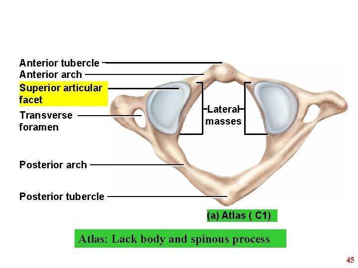
Anterior tubercle Anterior arch Superior articular facet Transverse foramen Lateral masses Posterior arch Posterior tubercle (a) Atlas ( C 1) Atlas: Lack body and spinous process 45
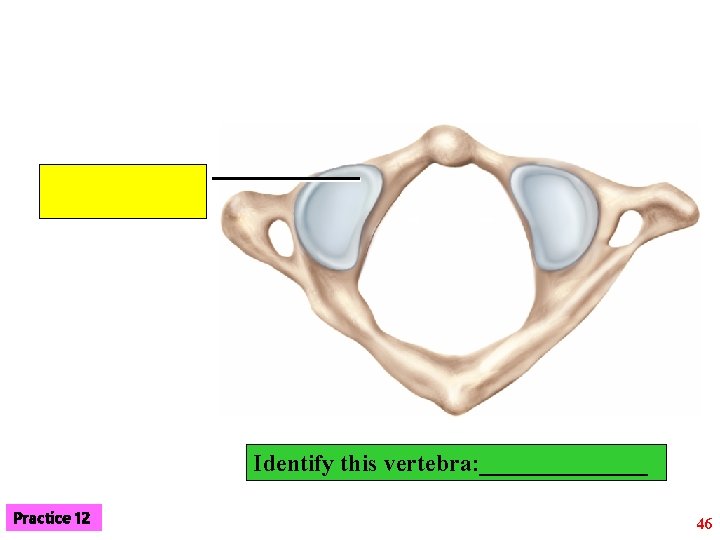
Identify this vertebra: _______ Practice 12 46
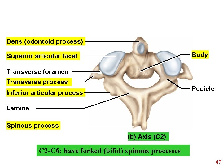
Dens (odontoid process) Body Superior articular facet Transverse foramen Transverse process Pedicle Inferior articular process Lamina Spinous process (b) Axis (C 2) C 2 -C 6: have forked (bifid) spinous processes 47
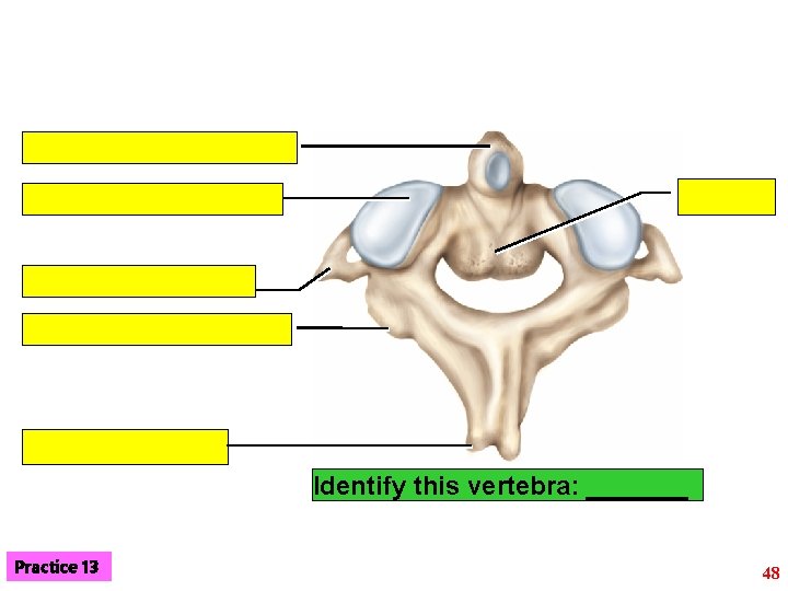
Identify this vertebra: _______ Practice 13 48
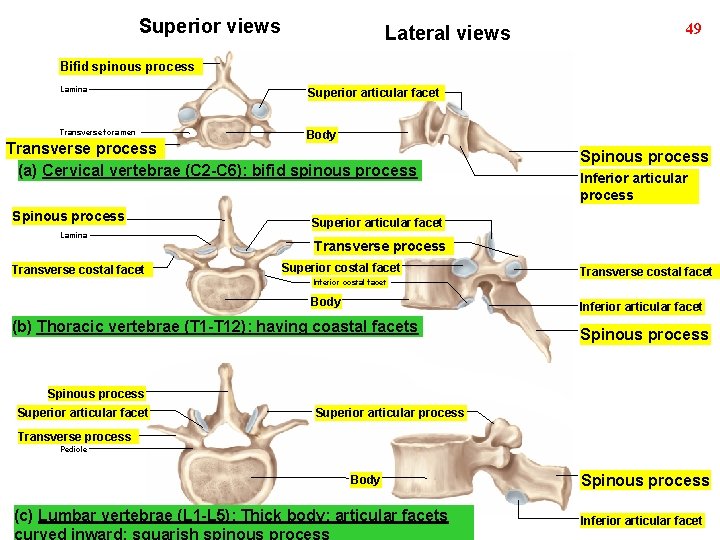
Superior views Lateral views 49 Bifid spinous process Lamina Superior articular facet Transverse foramen Body Transverse process (a) Cervical vertebrae (C 2 -C 6): bifid spinous process Spinous process Lamina Transverse costal facet Spinous process Inferior articular process Superior articular facet Transverse process Superior costal facet Inferior costal facet Body Transverse costal facet Inferior articular facet (b) Thoracic vertebrae (T 1 -T 12): having coastal facets Spinous process Superior articular facet Superior articular process Transverse process Pedicle Body (c) Lumbar vertebrae (L 1 -L 5): Thick body; articular facets Spinous process Inferior articular facet
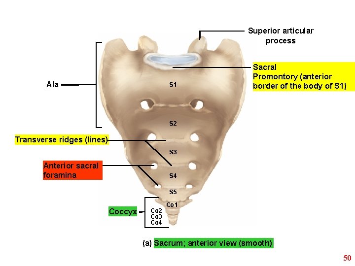
Superior articular process Ala S 1 Sacral Promontory (anterior border of the body of S 1) S 2 Transverse ridges (lines) S 3 Anterior sacral foramina S 4 S 5 Coccyx Co 2 Co 3 Co 4 Co 1 (a) Sacrum; anterior view (smooth) 50
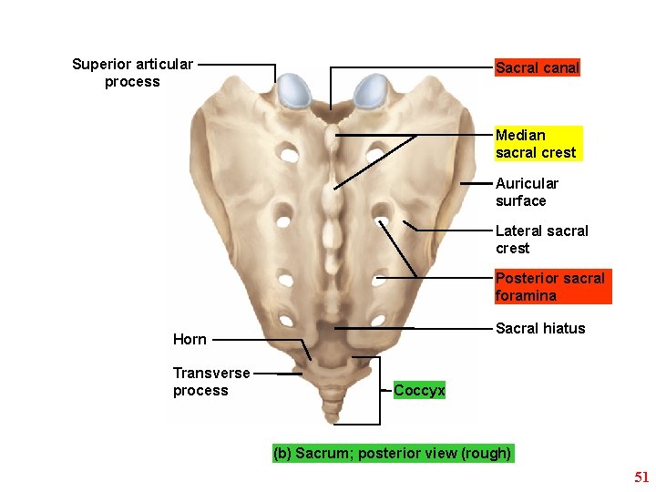
Superior articular process Sacral canal Median sacral crest Auricular surface Lateral sacral crest Posterior sacral foramina Sacral hiatus Horn Transverse process Coccyx (b) Sacrum; posterior view (rough) 51
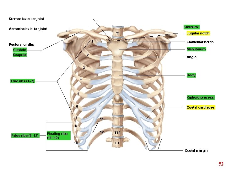
Sternoclavicular joint Sternum: Acromioclavicular joint T 1 1 Pectoral girdle: Jugular notch Clavicular notch Manubrium Clavicle Scapula 2 Angle 3 Body 4 True ribs (1– 7) 5 Xiphoid process 6 7 Costal cartilages 11 8 False ribs (8– 12) Floating ribs (11– 12) 9 10 12 T 12 L 1 Costal margin 52
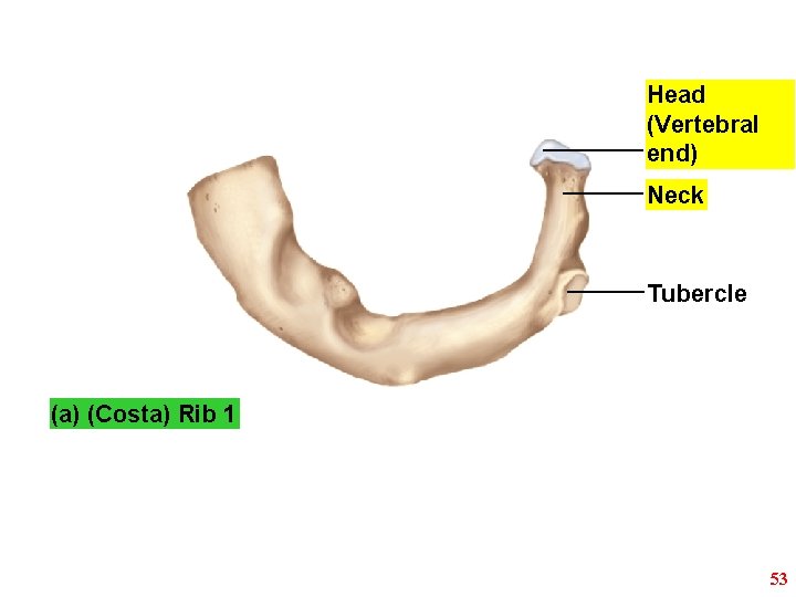
Head (Vertebral end) Neck Tubercle (a) (Costa) Rib 1 53
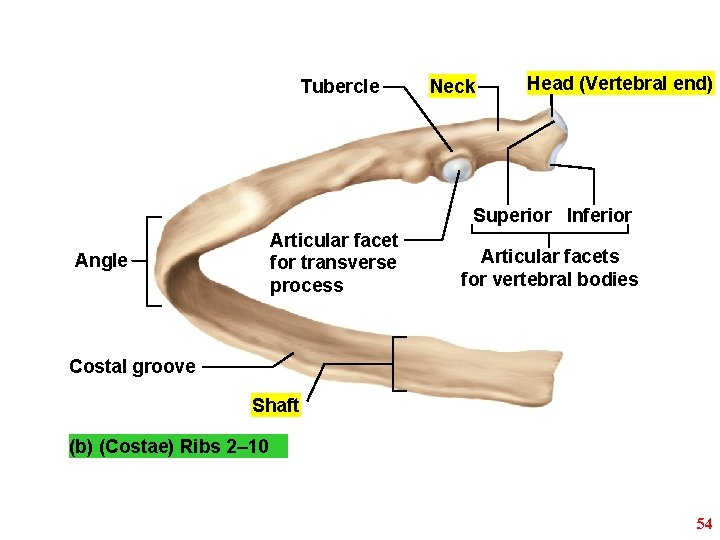
Tubercle Neck Head (Vertebral end) Superior Inferior Articular facet for transverse process Angle Articular facets for vertebral bodies Costal groove Shaft (b) (Costae) Ribs 2– 10 54
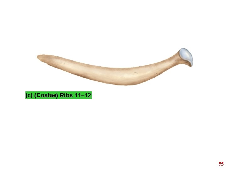
(c) (Costae) Ribs 11– 12 55
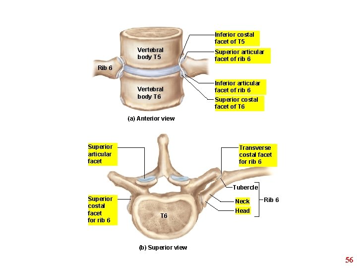
Inferior costal facet of T 5 Vertebral body T 5 Superior articular facet of rib 6 Rib 6 Vertebral body T 6 Inferior articular facet of rib 6 Superior costal facet of T 6 (a) Anterior view Superior articular facet Transverse costal facet for rib 6 Tubercle Superior costal facet for rib 6 Neck T 6 Rib 6 Head (b) Superior view 56
- Slides: 56