Pediatric Dermatology Potpourri A Variety of Cases Teresa


 • Most common congenital midline nasal lesion • 1")


 • Most common acquired AI blistering disorder of")


 • • Congenital absence of skin Localized or widespread Superficial")


 1. 2. 3. 4. 5. 6. 7. 8. 9. Scalp ACC")
 • Etiology – Ischemic and/or thrombotic events in the placenta and")


 • “Hashimoto-Pritzker Disease” • Rare variant of LCH •")


 – Nail dystrophy – Subungual hyperkeratosis –")


 • Rare vascular tumor – Extremities, trunk, retroperitoneum • Labs –")
 • Severe thrombocytopenic coagulopathy • Typically seen with rare vascular tumors")


 • Rarely possible – Other: • Radiation •")


- Slides: 80
Pediatric Dermatology Potpourri: A Variety of Cases Teresa S. Wright, MD, FAAP Le. Bonheur Children’s Hospital Memphis, TN
Disclosures • Sanofi Genzyme- Advisory Board
Learning Objectives • Recognize features of several unusual skin conditions in children. • Describe the key features that support the correct diagnosis. • List one important lesson demonstrated by each case.
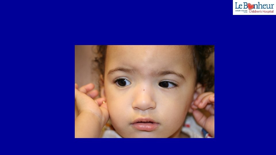
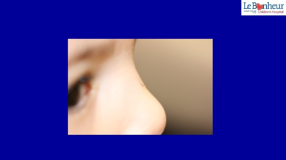
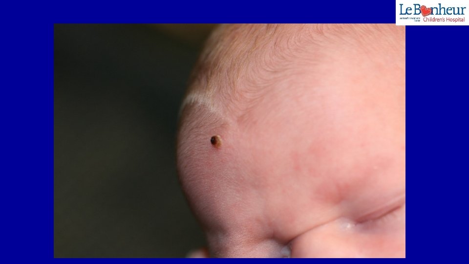
Case #1 • • • 14 mo old female Pit on the nasal bridge With hairs No hx of redness, pain, swelling, drainage Mom advised to “pluck the hairs”
Nasal Dermoid Sinus Cyst (NDSC) • Most common congenital midline nasal lesion • 1 -3% of all dermoids • 4 -12% of head/neck dermoids Kim HK et al. Clinical and Experimental Otorhinolaryngology. Vol 3(1): 48 -51, March 2010.
NDSC • • Cystic mass or sinus opening Midline nasal dorsum Between glabella and columella At birth or during early childhood
NDSC • Potential Complications – Nasal deformity – Infection • Meningitis • Intracranial abscess
NDSC • Imaging – CT and/or MRI • Complete surgical excision – Plastic surgery – Neurosurgery
Lesson • A tiny lesion can have potential for serious complications.
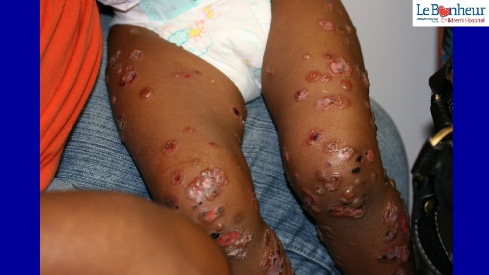
Case #2 • • • 21 mo old female 2 weeks of blistering rash No fever or other systemic symptoms No new medications + pruritus
Case #2 • Physical exam: – tense vesicles and large tense annular bullae (many with hemorrhagic centers) – serous and hemorrhagic crusts – superficial erosions – scattered diffusely over face, trunk, groin, buttocks, and bilateral upper and lower extremities – mucous membranes are clear
Chronic Bullous Disease of Childhood (CBDC) • Most common acquired AI blistering disorder of children • 6 mo – 10 yr (mean = 4. 5 yr) Mintz EM and Morel KD. Dermatol Clin 29(2011) 459 -462.
CBDC • Presentation – Abrupt onset – Clear/hemorrhagic tense vesicles/annular bullae • String of pearls • Cluster of jewels • Rosette
CBDC • Distribution – Face, trunk, extremities • Lower trunk, groin, medial thighs – +/- mucous membranes • Symptoms – Variable • Asymptomatic to severe pruritus • +/- burning
CBDC • DDx – Bullous impetigo • Superficial, fragile bullae – EBA – Other AI bullous
CBDC • Diagnosis – Biopsy • Subepidermal • Neutrophilic infiltrate – DIF • Linear Ig. A at BMZ
CBDC • Pathogenesis – Not well understood • Component of BMZ becomes antigenic – Often idiopathic – Genetic susceptibility • haplotypes HLA-B 8, Cw 7, DR 3 – Triggers • Infections, drugs, vaccinations, UV radiation, malignancy
CBDC • Course – Remits spontaneously – Several mos to 4 years • By puberty – Non-scarring • dyspigmentation
CBDC • Treatment – Dapsone* – Topical steroids – Systemic steroids – Other IS agents • MMF, Cs. A – Antibiotics • Anti-inflammatory Mintz EM and Morel KD. Dermatol Clin 29(2011) 699 -700.
Lesson • Can be difficult to control • Multiple agents may be needed • Will generally remit
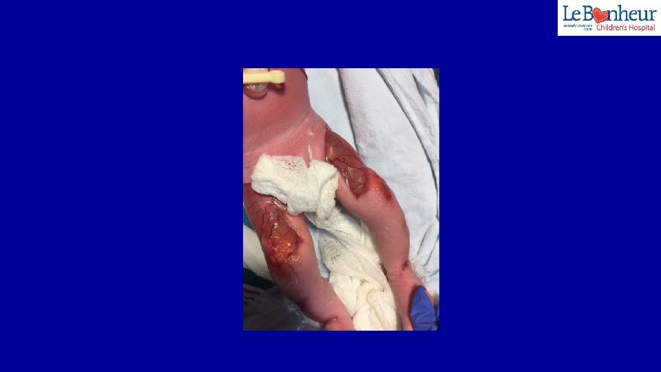
Case #3 • • 37 week male Triplet gestation Fetal demise at 17 weeks Remaining sister healthy
Aplasia Cutis Congenita (ACC) • • Congenital absence of skin Localized or widespread Superficial or deep Typically presents with eroded, absent, or scarred areas of skin at birth
ACC • Etiology – No single cause – Disruption of skin development has occurred • Multi factorial – Genetic factors – Teratogens – Decreased blood supply – Trauma
ACC • Scalp is most common site – 86% of solitary lesions – 80% at parietal hair whorl – Size ranges from small to large • Large may extend to dura or meninges • ? Graft for >3 -4 cm 2 – Shape varies • Round, oval, linear, stellate Frieden, IJ. J of Am Acad Derm. Volume 14(4): 646 -656.
ACC (9 Groups) 1. 2. 3. 4. 5. 6. 7. 8. 9. Scalp ACC without multiple anomalies Scalp ACC with assoc limb abnormalities Scalp ACC with assoc epidermal and organoid nevi ACC overlying embryologic malformations ACC with assoc fetus papyraceus or placental infarcts ACC assoc with EB ACC localized to the extremities, without blistering ACC caused by specific teratogens ACC assoc with syndromes
ACC (Group 5) • Etiology – Ischemic and/or thrombotic events in the placenta and fetus – Sporadic • Management – Conservative • Gentle cleansing • Vaseline gauze dressings
Lesson • Severe lesions will often heal well with minimal intervention.
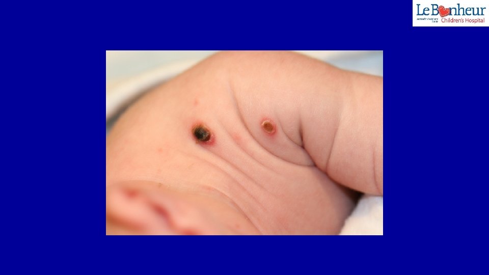
Case #4 • FT healthy male • Several crusted papules on face and trunk at birth
Congenital Self Healing Reticulohistiocytosis (CSHR) • “Hashimoto-Pritzker Disease” • Rare variant of LCH • Limited to skin only
CSHR • Birth or neonatal period – Red brown papules/nodules – +/- ulceration – Single or multiple – Involute w/in weeks to months
CSHR • Incidence? – Not clear • Likely underreported – Spontaneous resolution – Lack of recognition
CSHR • True CSHR is a benign process. • However: – cannot predict systemic involvement based on skin findings – thorough evaluation and follow up are indicated Stein SL et al. Arch Pediatr Adolesc Med 2001; 155: 778 -783.
CSHR • Up to 53% of pts with multisystem LCH present with skin lesions • 35 patients referred for presumed skin limited LCH – 40% had multisystem dz • Bone (70%) • Risk organ (liver, spleen, bone marrow) (50%) Simko SJ et al. J Pediatr Nov 2014; Vol 165(5): 990 -996.
Lesson • Babies with “CSRH” should have a thorough evaluation for multisystem disease.
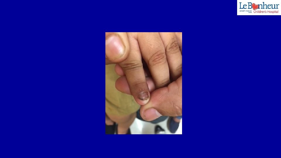
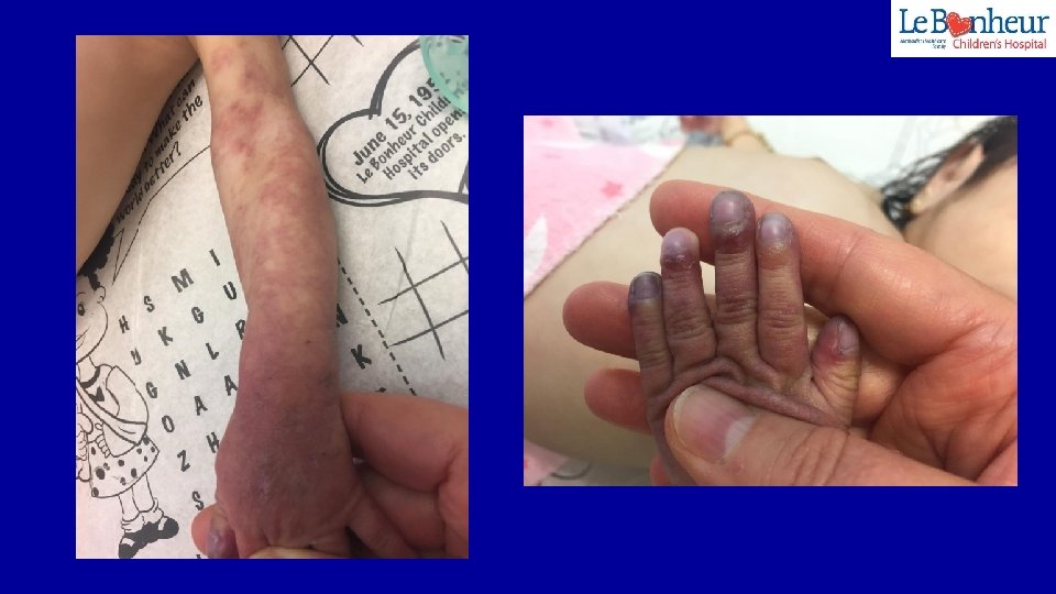
Case #5 • 2 yr old male • p/w “bumps on the skin” and an abnormal fingernail
Case #5 • History – “bumps” on arms present for months – Nail change present for months – Chronic dermatitis at the base of penis – Recent “abscess” in left thigh • I&D with negative cultures
Case #5 • PExam – Few small waxy dome shaped papules on arms • Molluscum contagiosum – Left index fingernail • Subungual hyperkeratosis, onycholysis – Faint pink patch at base of penis – Non tender nodule in left thigh with well healed scar
Langerhans Cell Histiocytosis! • Nail changes (rare) – Nail dystrophy – Subungual hyperkeratosis – Purpuric striae – Subungual pustules – Onycholysis – Longitudinal grooves – Paronychia – Loss of nail plate Kumar V et al. Pediatr Dermatol. 2017; 34: 732 -734.
Nail Changes in LCH • DDx: – Psoriasis, LP, AA, Darier’s, Reiter syndrome – Infection • Bacterial (esp with paronychia) • Fungal
Nail Changes in LCH • Prognostic factor? – Controversial • ? Correlation with multisystem disease – Poor prognosis • Spontaneous resolution – even a severe case Prayogo RL et al. Pediatr Dermatol. 2020; 37: 180 -183.
“Molluscum” • “LCH mimicking molluscum contagiosum: A case series” – Fernandez Armenteros et al. Pediatr Blood Cancer. 2018; 65: e 27047. • Up to 80% have skin involvement – Wide range of features – Classic: mimics seb derm • Erythematous scaly papules/plaques on scalp and diaper area – “molluscum” type is rare
Lesson • LCH can present with nail dystrophy. • LCH can mimic molluscum. • Have a high index of suspicion when patient has multiple unusual skin findings.
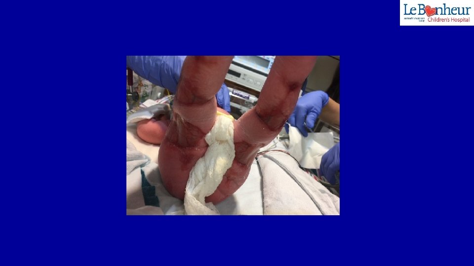
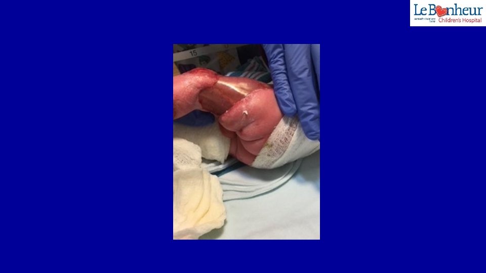
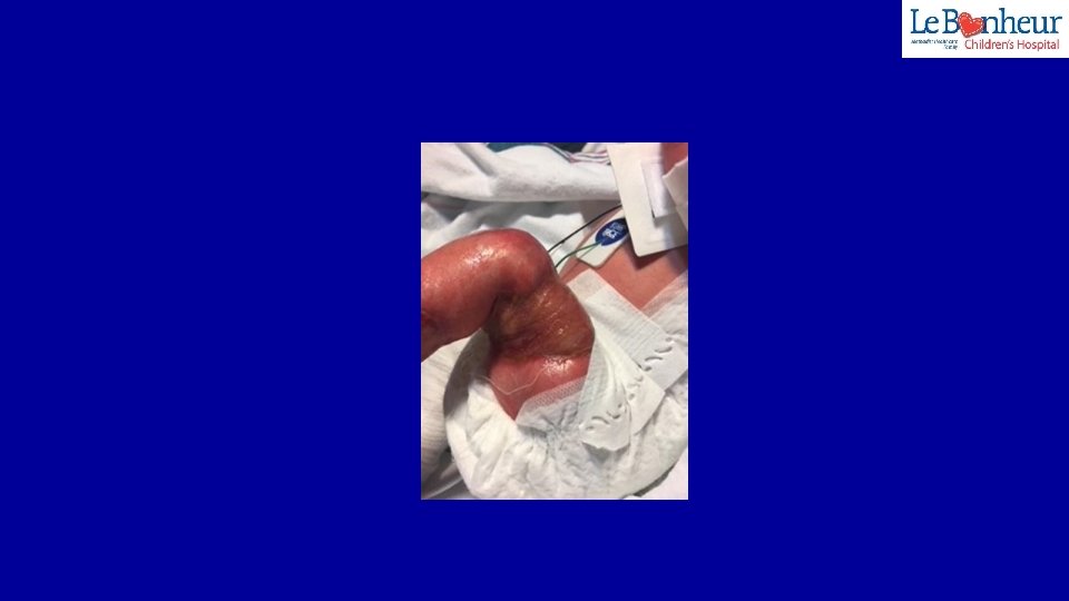
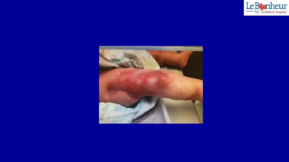
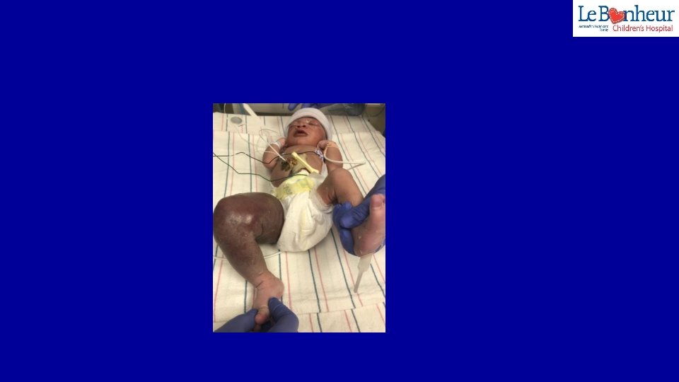
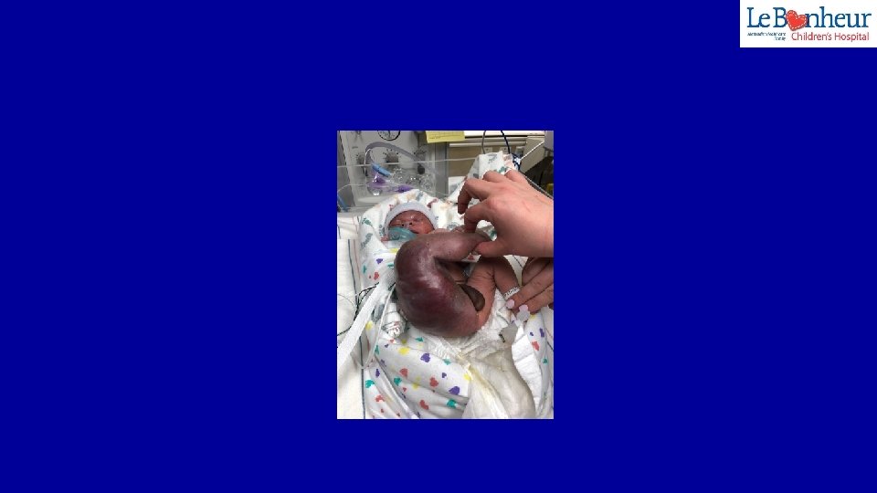
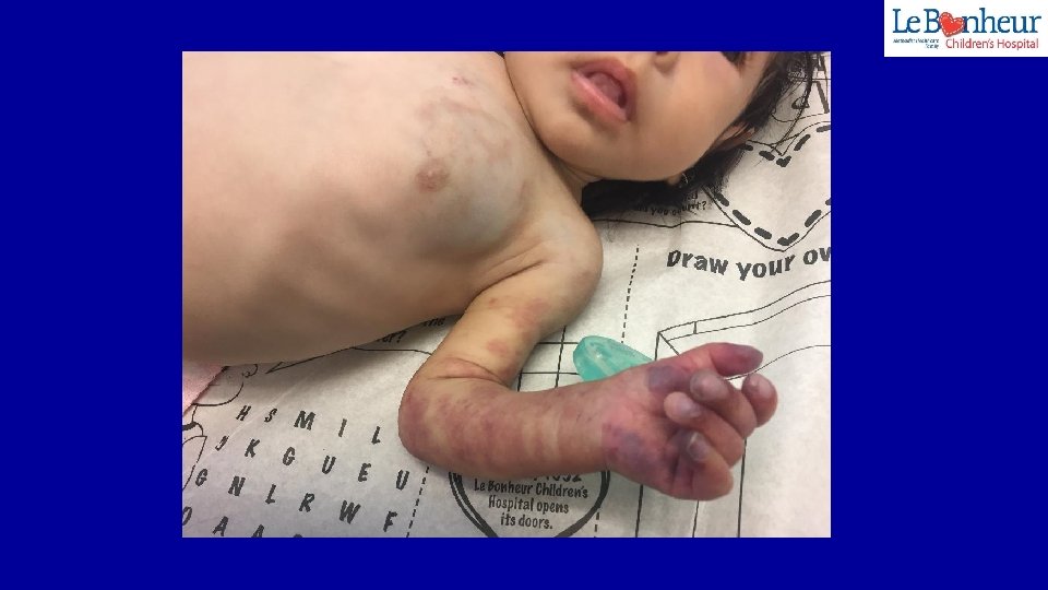
Case #6 • FT male • Uncomplicated pregnancy • Right leg swollen, hard, purple, tender
Kaposiform Hemangioendothelioma (KHE) • Rare vascular tumor – Extremities, trunk, retroperitoneum • Labs – Platelets low (16, 000) – Fibrinogen low – D-dimer elevated – PT/PTT WNL
Kasabach-Merritt Phenomenon (KMP) • Severe thrombocytopenic coagulopathy • Typically seen with rare vascular tumors – KHE > tufted angioma • Mechanism – Platelet trapping • Clinical – Rapidly enlarging infiltrative vascular tumor – Often painful
KHE/KMP • Management – Avoid platelet transfusion! • Unless active bleeding • Exacerbate platelet trapping – Cryoprecipitate may be given, if needed – PRBC’s for symptomatic anemia only – Pain control
KHE/KMP • Management – Primarily pharmacologic • • Systemic steroids Vincristine Alpha interferon Sirolimus Blatt J et al. Pediatr Blood Cancer 2010; 55: 1396 -1398.
KHE/KMP – Surgery (for small tumors) • Rarely possible – Other: • Radiation • Embolization Mahajan P et al. Clin Med Insights Blood Disord. 2017 Mar 16: 10.
KHE
Lesson • Avoid platelet transfusions! • Sirolimus can be very effective for KHE/KMP.
Case #7 • 2 week old FT female • p/w "red marks" on left chest, arm, and hand – Present at birth – Not visibly changing
Interim History • Delay in follow up • RSV – Anemia – Heme/onc consult – prbc’s • Le. B ED – Swelling, ? tenderness of left chest
Hospital Course • Hgb/HCT= 6. 6/21. 5 – Transfused with prbc’s • Stool guiaic +
Hospital Course • Imaging – Head ultrasound – Abdominal ultrasound • One lesion c/w “hemangioma” – Ultrasound of left chest • c/w deep infantile hemangioma – MRI of chest/arm • c/w “venous malformation” – Echocardiogram • WNL
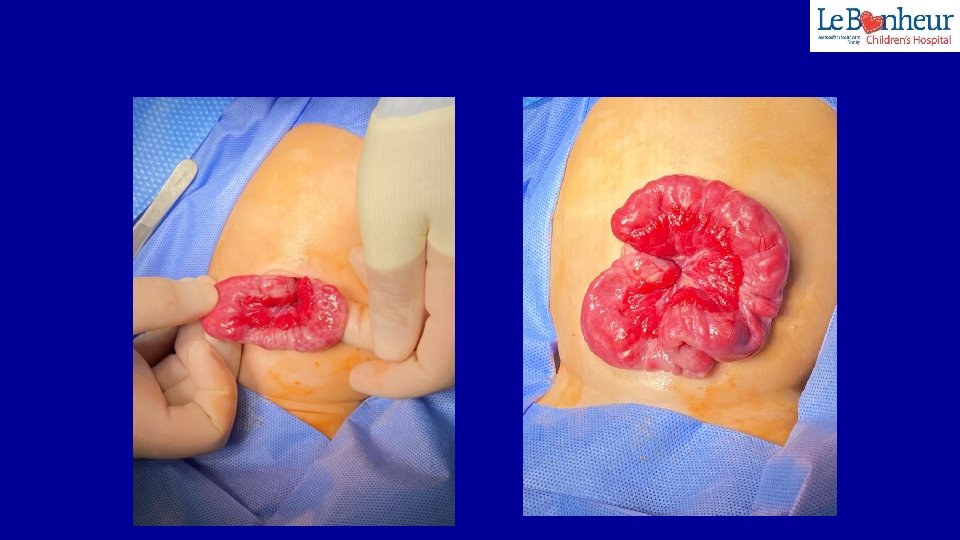
Hospital Course • Anemia continues • GI work up – EGD and colonoscopy negative • OR: – Laparotomy with push endoscopy – Biopsy of left chest lesion
Hospital Course • Biopsies of small intestine and chest lesions – c/w infantile hemangioma • GLUT-1 positive • MRI/MRA of head/neck – negative
Treatment • Propranolol • Prednisone • Close follow up – prbc’s
Segmental Hemangioma • Increased risk – Complications – Associated abnormalities • PHACE, LUMBAR syndrome – Visceral lesions
Segmental Hemangioma – Assoc of non facial segmental HOI • Visceral lesions –Liver> GI tract> brain Metry DW et al. Arch Dermatol May 2004. Vol 140: 591 -596.
GI Bleeding as a Complication of Segmental HOI • Report of 10 patients – Female – Hispanic or Caucasian – Large segmental HOI • Facial (8) and non-facial (2) – +/- PHACE syndrome (9/10) Drolet BA et al. , J Pediatr. Vol 160(60): 1021 -1029.
Segmental HOI of GI Tract – Lower GI bleeding • Multiple rbc transfusions • Difficult to diagnose • Located in small intestine/mesentery – Segmental – Assoc with arterial anomalies » Vascular dysplasia of the aorta
Lessons • Atypical morphology of segmental hemangioma • Risk of visceral HOI – Even with non-facial segmental HOI • Localized • Segmental – Present with GI bleeding
Thank You! • twrigh 43@uthsc. edu