Nuclear Medicine RADIOPHARMACEUTIC AL CHEMISTRY Positron Emission Tomography
- Slides: 19
Nuclear Medicine RADIOPHARMACEUTIC AL CHEMISTRY
Positron Emission Tomography q What is PET ? q By which principle it work ? q How can be performed ? q Benefits and risks ? q Common isotopes used? q Main differences between PET and CT scan ?
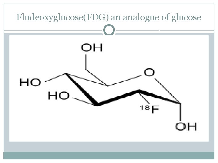
PET scan is a functional imaging technique that is used to observe metabolic processes in the body. Positron emission tomography (PET): uses small amounts of radioactive materials called radiotracers(the biological active molecule chosen Fludeoxyglucose(FDG) an analogue of glucose) a special camera and a computer to help evaluate your organ and tissue functions. By identifying body changes at the cellular level , recently use of PET to detect the cancer tumours, may also used in cardiology and neurology.
PET principle �PET use x-ray in order to present a 3 D picture �short line radioactive tracer isotope injected into the living subject usually in blood circulation , the tracer is chemically incorporated into a biological active molecule �There is a waiting period while the active molecule become concentrated in tissues of inert �Radioisotope undergo a PE decay it will emit a positron
Fludeoxyglucose(FDG) an analogue of glucose
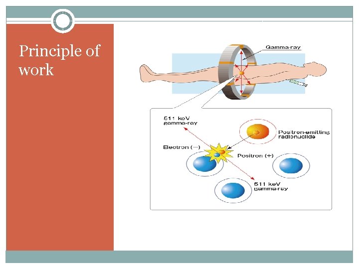
PET principle � Travelling few(milli meter)the positron encounter an electron �The encounter annihilates them both produce a pair of (gamma) photon moving in opposite directions �These are detected when they reach scintillators in the scanning device create a burst of light �The technicians can the create an image of the parts of brain for example which are overactive
Principle of work
How can we performed it ? �If you’re pregnant or believe you could be pregnant, tell your doctor. The test may be unsafe for your baby. You should also tell your doctor about any medical conditions you have. If you have diabetes, you’ll get special instructions for test preparation because fasting beforehand could affect your blood sugar levels.
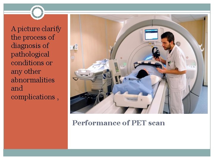
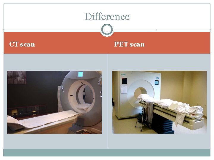
How can we performed it ? �Before the scan, you’ll get tracers through a vein in your arm, through a solution you drink, or in a gas you inhale. Your body needs time to absorb the tracers, so you’ll wait about an hour before the scan begins. �Next, you’ll undergo the scan. This involves lying on a narrow table attached to a PET machine, which looks like a giant letter “O. ” The table glides slowly into the machine so that the scan be conducted.
How can we performed it ? �You’ll need to lie still during the scan �You may be asked to hold your breath for short periods. �You’ll hear buzzing and clicking noises during the test. When all the necessary images have been recorded, you will slide out of the machine. The test is then complete.
A picture clarify the process of diagnosis of pathological conditions or any other abnormalities and complications , Performance of PET scan
Benefits Ø medicine examinations provide unique information—including details on both function and anatomic structure of the body. Ø nuclear medicine scans yield the most useful information needed to make a diagnosis or to determine appropriate treatment. Ø Nuclear medicine is less expensive and may yield more information than surgery by identifying changes in the body at the cellular level.
Risks A radioactive drug (tracer) will be put into body. The amount of radiation was exposed, and the risk of negative effects 1. Cause a major allergic reaction. 2. Expose unborn baby to radiation if pregnant 3. Expose your child to radiation if breast-feeding
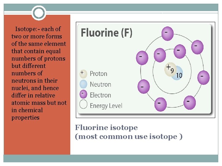
1. Isotope: - each of two or more forms of the same element that contain equal numbers of protons but different numbers of neutrons in their nuclei, and hence differ in relative atomic mass but not in chemical properties Fluorine isotope (most common use isotope )
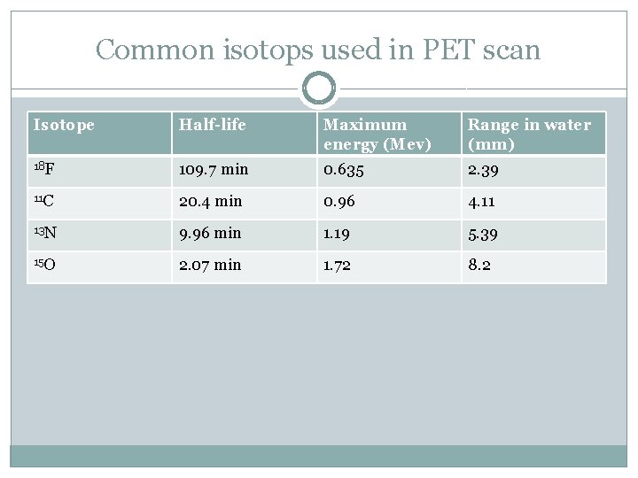
Common isotops used in PET scan Isotope Half-life Maximum energy (Mev) Range in water (mm) 18 F 109. 7 min 0. 635 2. 39 11 C 20. 4 min 0. 96 4. 11 13 N 9. 96 min 1. 19 5. 39 15 O 2. 07 min 1. 72 8. 2
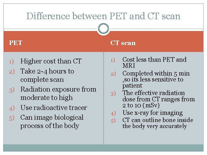
Difference between PET and CT scan PET 1) 2) 3) 4) 5) Higher cost than CT Take 2 -4 hours to complete scan Radiation exposure from moderate to high Use radioactive tracer Can image biological process of the body CT scan 1) 2) 3) 4) 5) Cost less than PET and MRI Completed within 5 min , so its less sensitive to patient The effective radiation dose from CT ranges from 2 to 10 (m. Sv) Use x-ray for imaging CT can outline bone inside the body very accurately
Difference CT scan PET scan
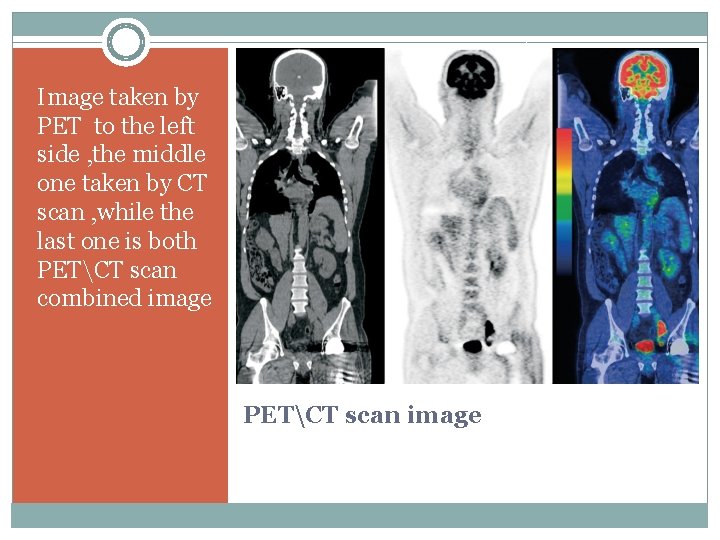
Image taken by PET to the left side , the middle one taken by CT scan , while the last one is both PETCT scan combined image PETCT scan image
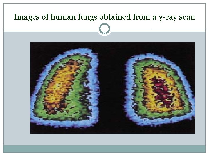
Images of human lungs obtained from a γ-ray scan