
Positron Emission Tomography Instrumentation Present Status and Future




- Slides: 52
Positron Emission Tomography Instrumentation: Present Status and Future Directions William W. Moses Lawrence Berkeley National Laboratory October 11, 2016 Outline: • Overview • Present Status • Future Directions
Nuclear Medicine Gamma Camera • Patient injected with small amount of radioactive drug. • Drug localizes in patient according to metabolic properties of that drug. • Radioactivity decays, emitting gamma rays. • Gamma rays that exit the patient are imaged. • Well Established Clinical Technique • 10 Million Studies Annually
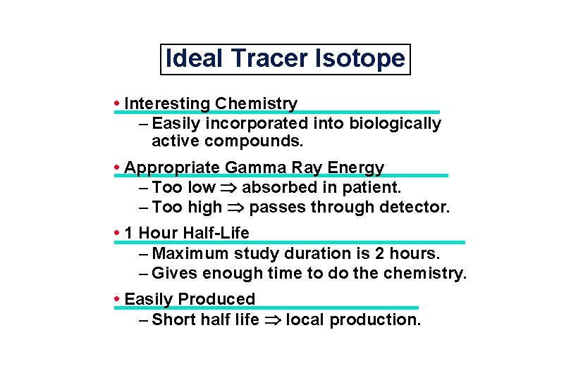
Ideal Tracer Isotope • Interesting Chemistry – Easily incorporated into biologically active compounds. • Appropriate Gamma Ray Energy – Too low absorbed in patient. – Too high passes through detector. • 1 Hour Half-Life – Maximum study duration is 2 hours. – Gives enough time to do the chemistry. • Easily Produced – Short half life local production.
Common Positron-Emitting Tracer Isotopes + 2 hour half-life. 18 F ± Chemically very good (replaces OH). – Cyclotron produced. + Chemically excellent. 15 O, 11 C, 13 N – 2 to 20 minute half-life. – Cyclotron produced. + Generator produced. 82 Rb – 2 minute half-life. – Chemically OK (acts like Na and K).
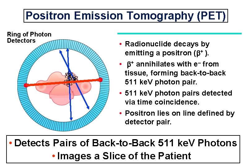
Positron Emission Tomography (PET) Ring of Photon Detectors • Radionuclide decays by emitting a positron ( + ). • + annihilates with e– from tissue, forming back-to-back 511 ke. V photon pair. • 511 ke. V photon pairs detected via time coincidence. • Positron lies on line defined by detector pair. • Detects Pairs of Back-to-Back 511 ke. V Photons • Images a Slice of the Patient
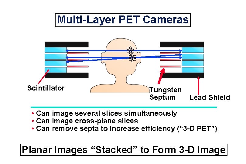
Multi-Layer PET Cameras Scintillator Tungsten Septum Lead Shield • Can image several slices simultaneously • Can image cross-plane slices • Can remove septa to increase efficiency (“ 3 -D PET”) Planar Images “Stacked” to Form 3 -D Image
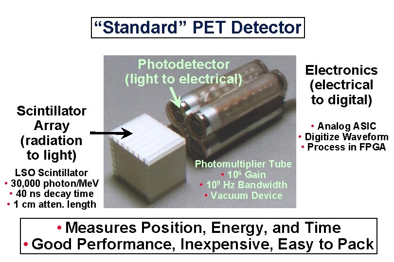
“Standard” PET Detector Photodetector (light to electrical) Scintillator Array (radiation to light) LSO Scintillator • 30, 000 photon/Me. V • 40 ns decay time • 1 cm atten. length Electronics (electrical to digital) • Analog ASIC • Digitize Waveform • Process in FPGA Photomultiplier Tube • 106 Gain • 109 Hz Bandwidth • Vacuum Device • Measures Position, Energy, and Time • Good Performance, Inexpensive, Easy to Pack
Computed Tomography Planar X-Ray Computed Tomography Separates Objects on Different Planes Images courtesy of Robert Mc. Gee, Ford Motor Company
Principle of Computed Tomography 2 -Dimensional Object 1 -Dimensional Vertical Projection 1 -Dimensional Horizontal Projection By measuring all 1 -dimensional projections of a 2 -dimensional object, you can reconstruct the object
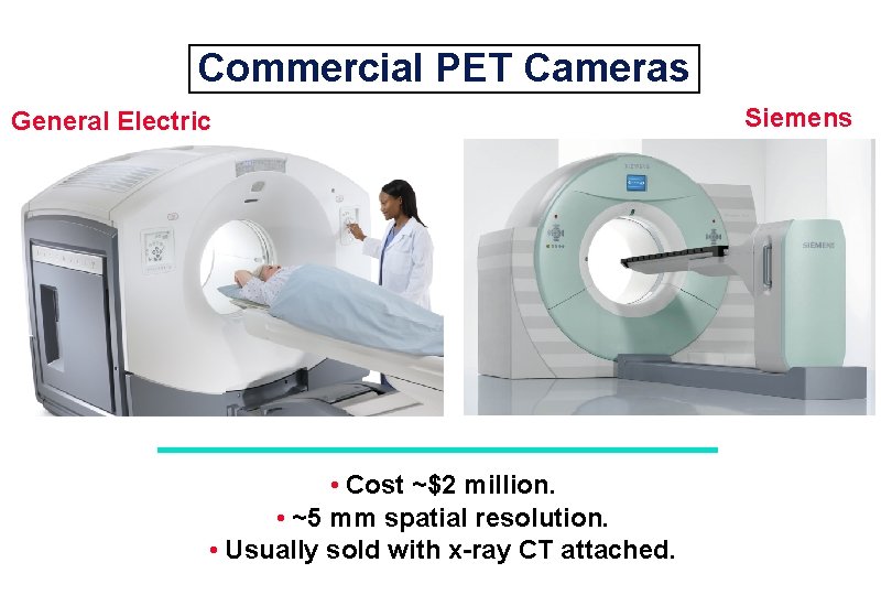
Commercial PET Cameras General Electric • Cost ~$2 million. • ~5 mm spatial resolution. • Usually sold with x-ray CT attached. Siemens
Common Clinical Uses of PET • Cancer / Oncology • Heart Tissue Viability • Brain Dysfunction – Stroke – Epilepsy – Alzheimer’s Disease Images Function, Not Structure!
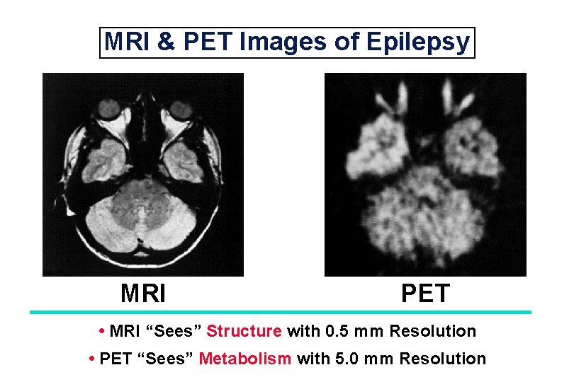
MRI & PET Images of Epilepsy MRI PET • MRI “Sees” Structure with 0. 5 mm Resolution • PET “Sees” Metabolism with 5. 0 mm Resolution
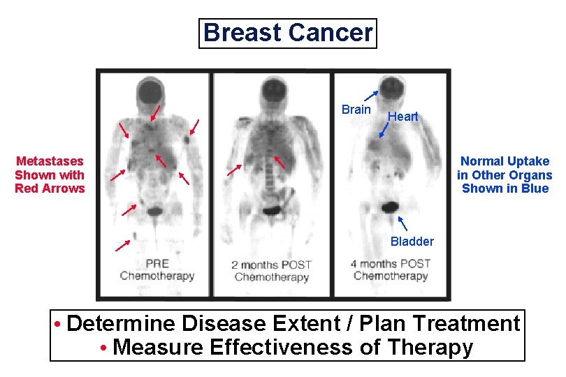
Breast Cancer Brain Heart Metastases Shown with Red Arrows Normal Uptake in Other Organs Shown in Blue Bladder • Determine Disease Extent / Plan Treatment • Measure Effectiveness of Therapy
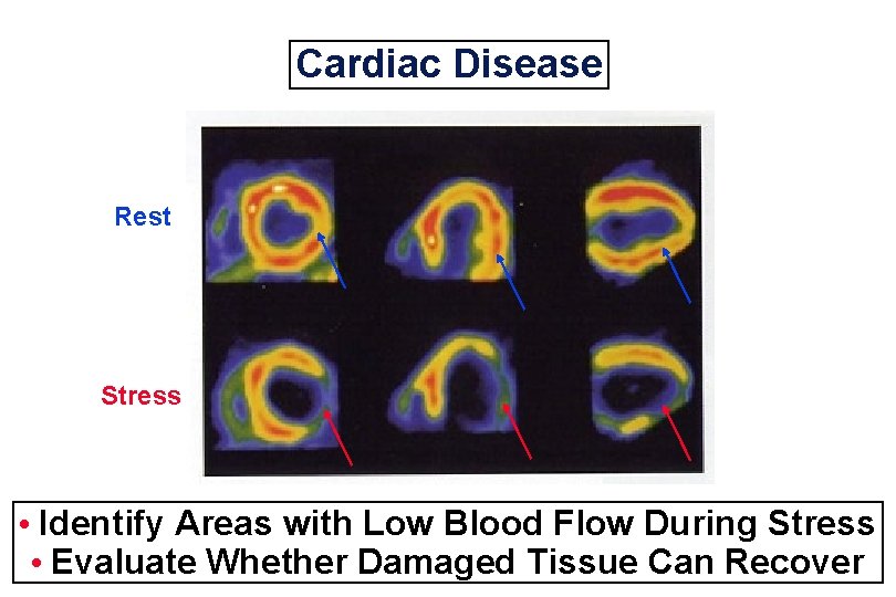
Cardiac Disease Rest Stress • Identify Areas with Low Blood Flow During Stress • Evaluate Whether Damaged Tissue Can Recover
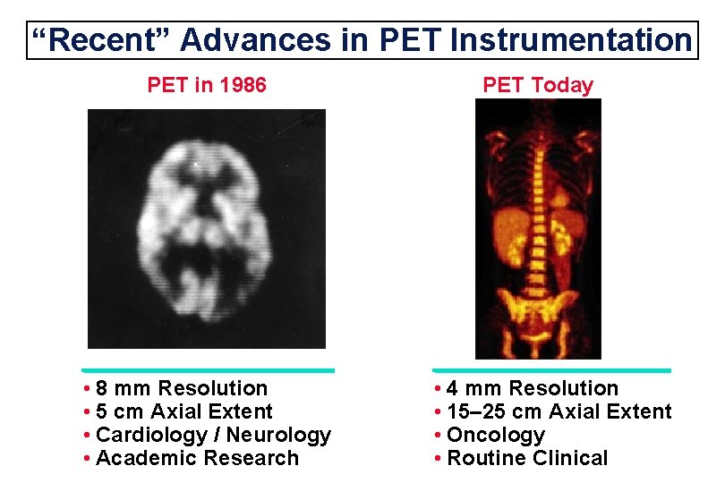
“Recent” Advances in PET Instrumentation PET in 1986 • 8 mm Resolution • 5 cm Axial Extent • Cardiology / Neurology • Academic Research PET Today • 4 mm Resolution • 15– 25 cm Axial Extent • Oncology • Routine Clinical
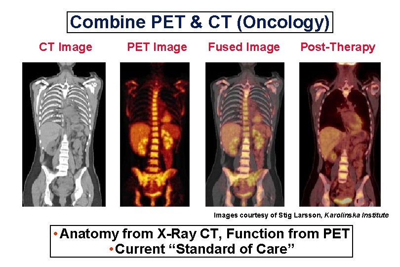
Combine PET & CT (Oncology) CT Image PET Image Fused Image Post-Therapy Images courtesy of Stig Larsson, Karolinska Institute • Anatomy from X-Ray CT, Function from PET • Current “Standard of Care”
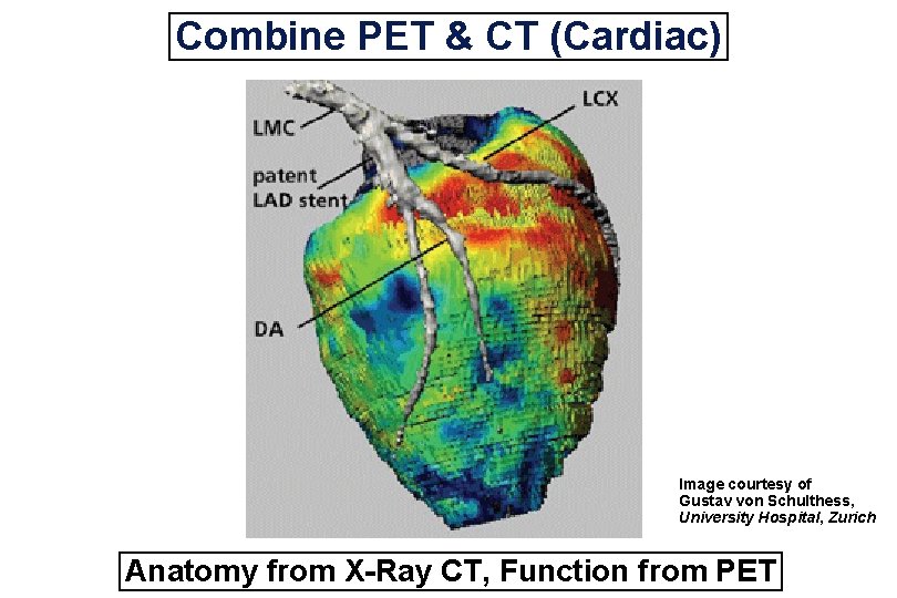
Combine PET & CT (Cardiac) Image courtesy of Gustav von Schulthess, University Hospital, Zurich Anatomy from X-Ray CT, Function from PET
Research Directions in PET • Small Animal • Organ Specific Cameras • PET / MRI
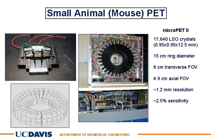
Small Animal (Mouse) PET micro. PET II 17, 640 LSO crystals (0. 95 x 12. 5 mm) 15 cm ring diameter 8 cm transverse FOV 4. 9 cm axial FOV ~1. 2 mm resolution ~2. 5% sensitivity DEPARTMENT OF BIOMEDICAL ENGINEERING
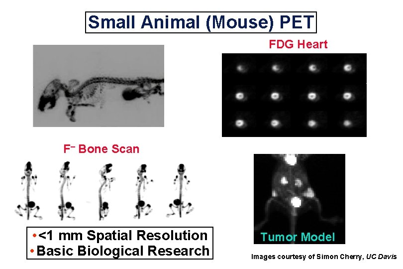
Small Animal (Mouse) PET FDG Heart F– Bone Scan • <1 mm Spatial Resolution • Basic Biological Research Tumor Model Images courtesy of Simon Cherry, UC Davis
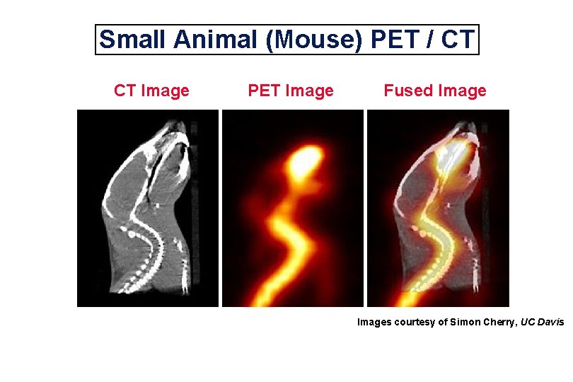
Small Animal (Mouse) PET / CT CT Image PET Image Fused Images courtesy of Simon Cherry, UC Davis
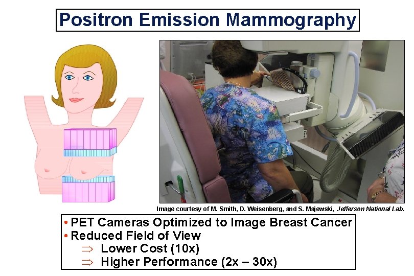
Positron Emission Mammography Image courtesy of M. Smith, D. Weisenberg, and S. Majewski, Jefferson National Lab. • PET Cameras Optimized to Image Breast Cancer • Reduced Field of View Þ Lower Cost (10 x) Þ Higher Performance (2 x – 30 x)
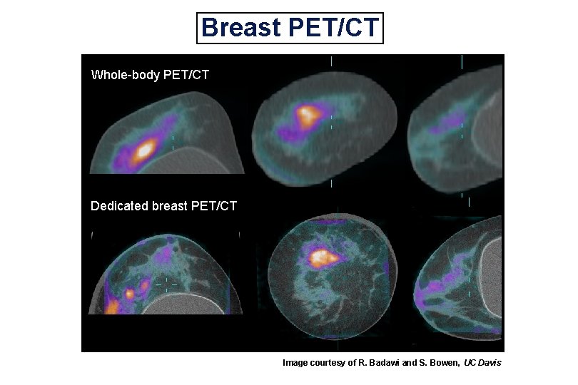
Breast PET/CT Whole-body PET/CT Dedicated breast PET/CT Image courtesy of R. Badawi and S. Bowen, UC Davis
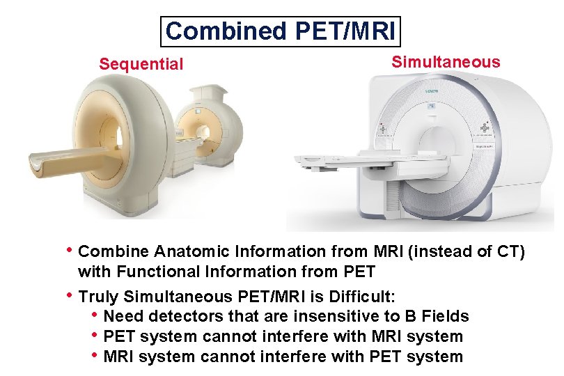
Combined PET/MRI Sequential Simultaneous • Combine Anatomic Information from MRI (instead of CT) • with Functional Information from PET Truly Simultaneous PET/MRI is Difficult: • Need detectors that are insensitive to B Fields • PET system cannot interfere with MRI system • MRI system cannot interfere with PET system
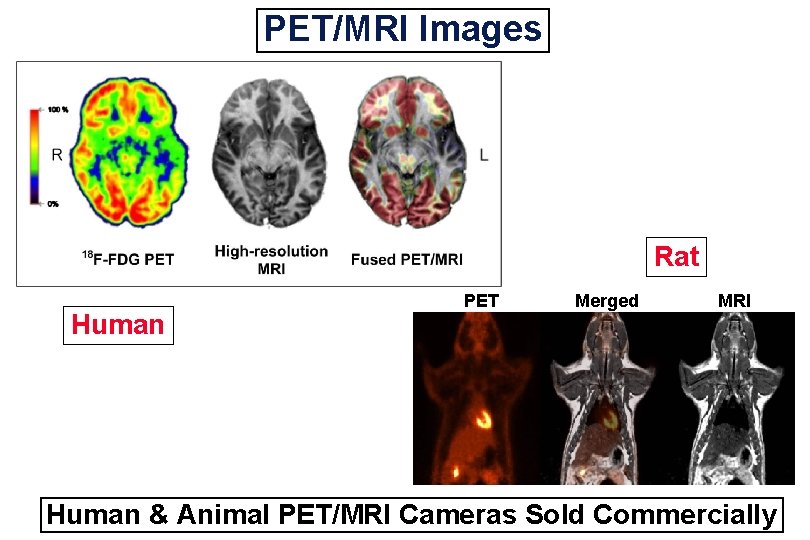
PET/MRI Images Rat Human PET Merged MRI Human & Animal PET/MRI Cameras Sold Commercially
What Can Be Improved in PET? • Signal to Noise Ratio – Dominated by Statistical Noise • Spatial Resolution – Near Fundamental Limit
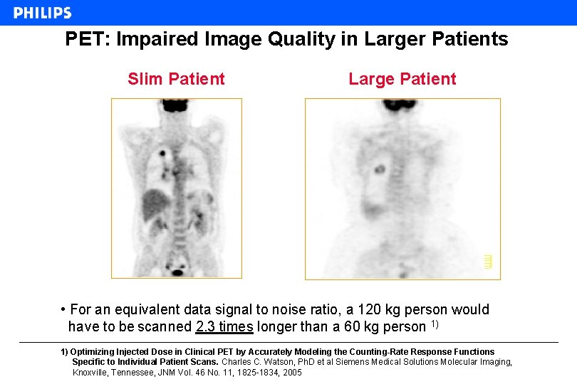
PET: Impaired Image Quality in Larger Patients Slim Patient Large Patient • For an equivalent data signal to noise ratio, a 120 kg person would have to be scanned 2. 3 times longer than a 60 kg person 1) 1) Optimizing Injected Dose in Clinical PET by Accurately Modeling the Counting-Rate Response Functions Specific to Individual Patient Scans. Charles C. Watson, Ph. D et al Siemens Medical Solutions Molecular Imaging, Knoxville, Tennessee, JNM Vol. 46 No. 11, 1825 -1834, 2005
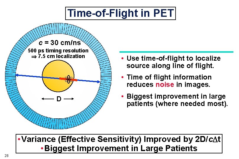
Time-of-Flight in PET c = 30 cm/ns 500 ps timing resolution 7. 5 cm localization • Use time-of-flight to localize source along line of flight. • Time of flight information reduces noise in images. D • Biggest improvement in large patients (where needed most). • Variance (Effective Sensitivity) Improved by 2 D/c t • Biggest Improvement in Large Patients 28
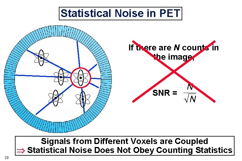
Statistical Noise in PET If there are N counts in the image, SNR = Signals from Different Voxels are Coupled Statistical Noise Does Not Obey Counting Statistics 29
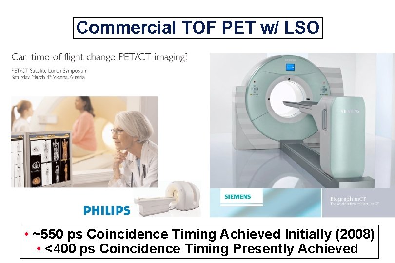
Commercial TOF PET w/ LSO • ~550 ps Coincidence Timing Achieved Initially (2008) • <400 ps Coincidence Timing Presently Achieved
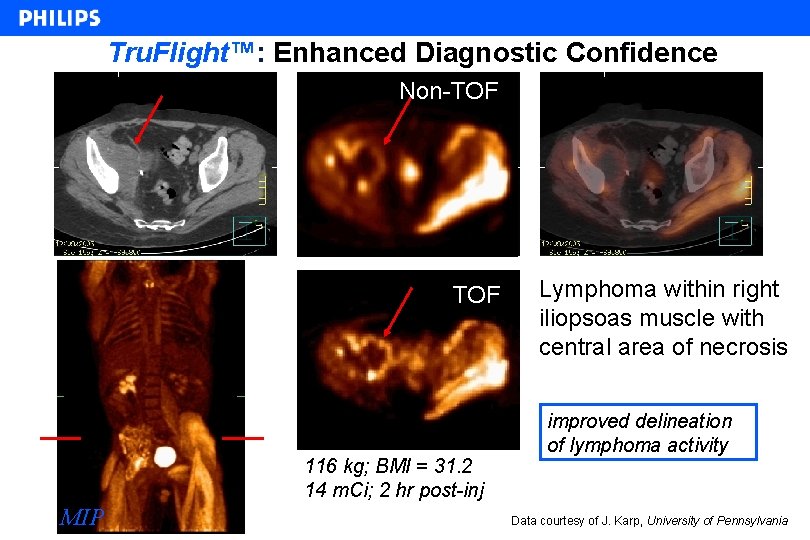
Tru. Flight™: Enhanced Diagnostic Confidence Non-TOF 116 kg; BMI = 31. 2 14 m. Ci; 2 hr post-inj MIP Lymphoma within right iliopsoas muscle with central area of necrosis improved delineation of lymphoma activity Data courtesy of J. Karp, University of Pennsylvania
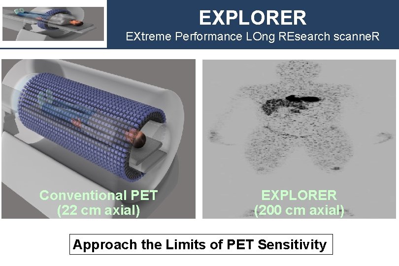
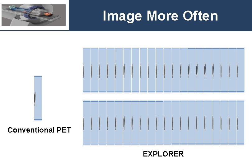
EXPLORER EXtreme Performance LOng REsearch scanne. R Conventional PET (22 cm axial) EXPLORER (200 cm axial) Approach the Limits of PET Sensitivity
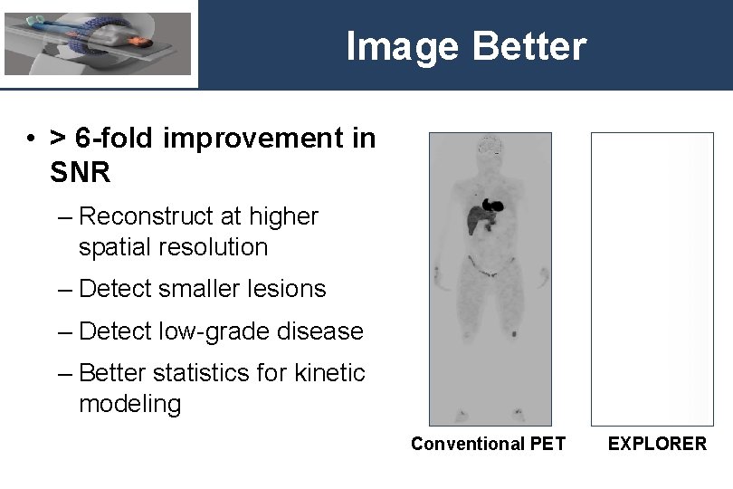
Image Better • > 6 -fold improvement in SNR – Reconstruct at higher spatial resolution – Detect smaller lesions – Detect low-grade disease – Better statistics for kinetic modeling Conventional PET EXPLORER
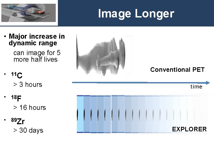
Image Longer • Major increase in dynamic range can image for 5 more half lives • 11 C > 3 hours Conventional PET time • 18 F > 16 hours • 89 Zr > 30 days EXPLORER
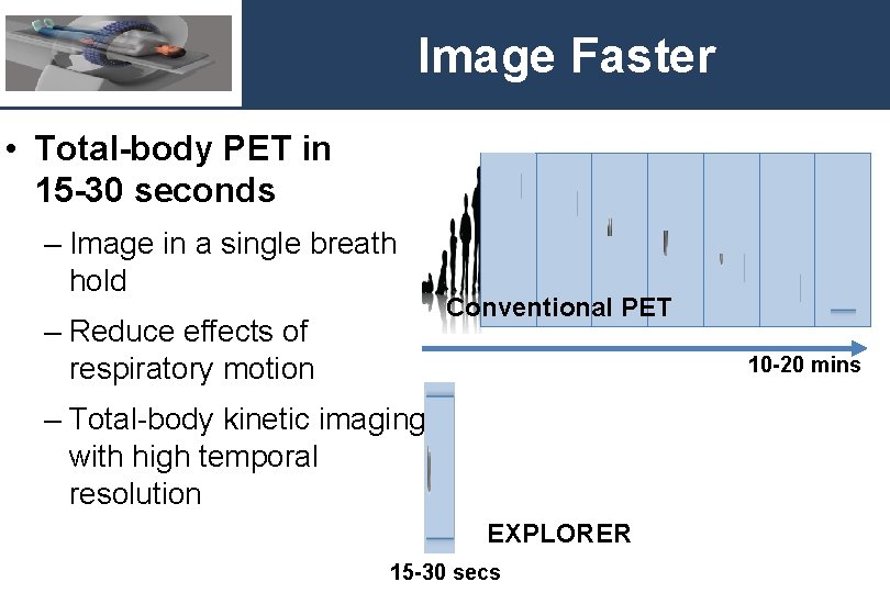
Image Faster • Total-body PET in 15 -30 seconds – Image in a single breath hold – Reduce effects of respiratory motion Conventional PET 10 -20 mins – Total-body kinetic imaging with high temporal resolution EXPLORER 15 -30 secs
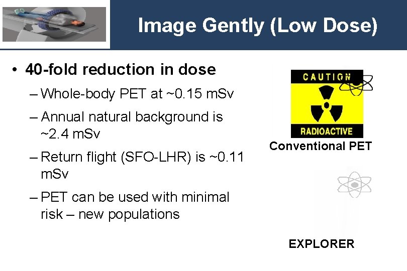
Image Gently (Low Dose) • 40 -fold reduction in dose – Whole-body PET at ~0. 15 m. Sv – Annual natural background is ~2. 4 m. Sv – Return flight (SFO-LHR) is ~0. 11 m. Sv Conventional PET – PET can be used with minimal risk – new populations EXPLORER
Image More Often Conventional PET EXPLORER
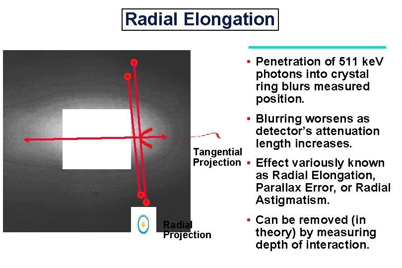
Radial Elongation • Penetration of 511 ke. V photons into crystal ring blurs measured position. Tangential Projection Radial Projection • Blurring worsens as detector’s attenuation length increases. • Effect variously known as Radial Elongation, Parallax Error, or Radial Astigmatism. • Can be removed (in theory) by measuring depth of interaction.
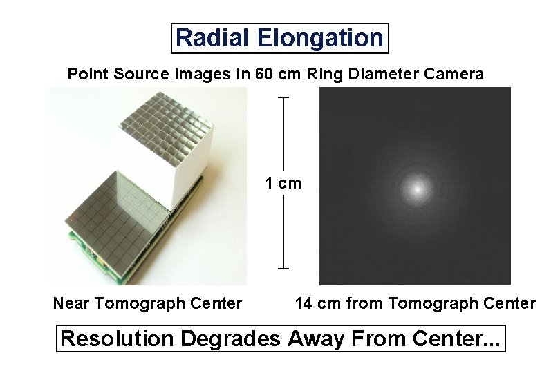
Radial Elongation Point Source Images in 60 cm Ring Diameter Camera 1 cm Near Tomograph Center 14 cm from Tomograph Center Resolution Degrades Away From Center. . .
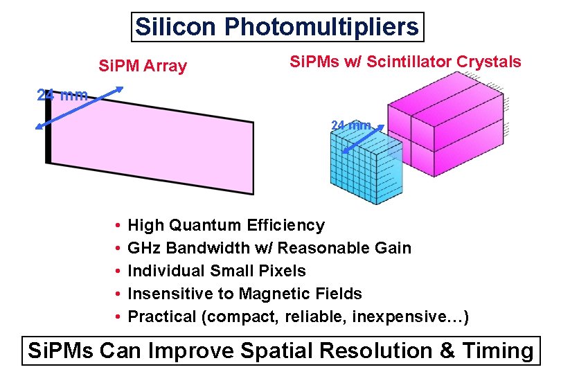
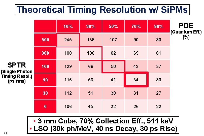
Silicon Photomultipliers Si. PM Array Si. PMs w/ Scintillator Crystals 24 mm • • • High Quantum Efficiency GHz Bandwidth w/ Reasonable Gain Individual Small Pixels Insensitive to Magnetic Fields Practical (compact, reliable, inexpensive…) Si. PMs Can Improve Spatial Resolution & Timing
Theoretical Timing Resolution w/ Si. PMs 10% SPTR (Single Photon Timing Resol. ) (ps rms) 41 30% 50% 70% 90% 500 245 138 107 90 80 300 188 106 82 69 61 100 129 66 50 42 37 50 116 56 41 34 30 30 112 51 38 31 27 0 106 45 32 26 22 PDE (Quantum Eff. ) (%) • 3 mm Cube, 70% Collection Eff. , 511 ke. V • LSO (30 k ph/Me. V, 40 ns Decay, 30 ps Rise)
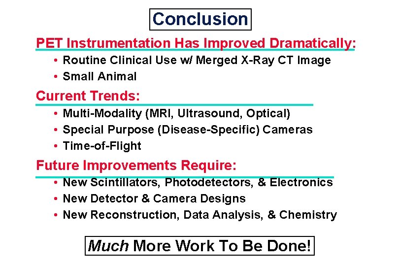
Conclusion PET Instrumentation Has Improved Dramatically: • Routine Clinical Use w/ Merged X-Ray CT Image • Small Animal Current Trends: • Multi-Modality (MRI, Ultrasound, Optical) • Special Purpose (Disease-Specific) Cameras • Time-of-Flight Future Improvements Require: • New Scintillators, Photodetectors, & Electronics • New Detector & Camera Designs • New Reconstruction, Data Analysis, & Chemistry Much More Work To Be Done!
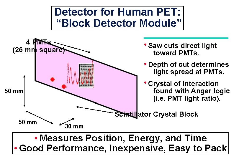
Detector for Human PET: “Block Detector Module” 4 PMTs (25 mm square) Saw cuts direct light toward PMTs. Depth of cut determines light spread at PMTs. Crystal of interaction found with Anger logic (i. e. PMT light ratio). 50 mm Scintillator Crystal Block 50 mm 30 mm • Measures Position, Energy, and Time • Good Performance, Inexpensive, Easy to Pack
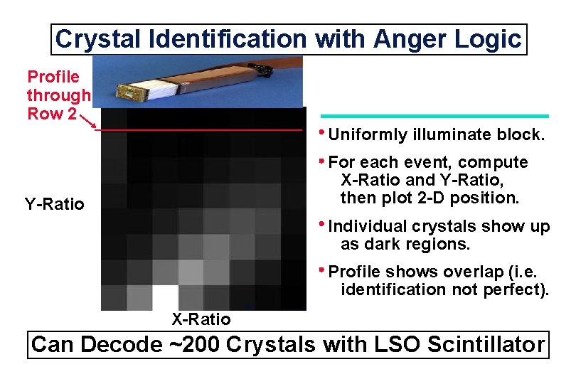
Crystal Identification with Anger Logic Profile through Row 2 Uniformly illuminate block. For each event, compute X-Ratio and Y-Ratio, then plot 2 -D position. Y-Ratio Individual crystals show up as dark regions. Profile shows overlap (i. e. identification not perfect). X-Ratio Can Decode ~200 Crystals with LSO Scintillator
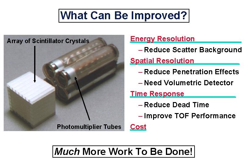
What Can Be Improved? Array of Scintillator Crystals Energy Resolution – Reduce Scatter Background Spatial Resolution Photomultiplier Tubes – Reduce Penetration Effects – Need Volumetric Detector Time Response – Reduce Dead Time – Improve TOF Performance Cost Much More Work To Be Done!
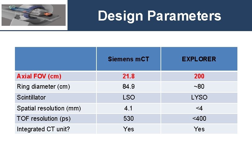
Design Parameters Siemens m. CT EXPLORER Axial FOV (cm) 21. 8 200 Ring diameter (cm) 84. 9 ~80 Scintillator LSO LYSO Spatial resolution (mm) 4. 1 <4 TOF resolution (ps) 530 <400 Integrated CT unit? Yes
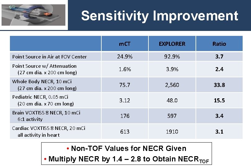
Sensitivity Improvement m. CT EXPLORER Ratio Point Source in Air at FOV Center 24. 9% 92. 9% 3. 7 Point Source w/ Attenuation (27 cm dia. x 200 cm long) 1. 6% 3. 9% 2. 4 Whole Body NECR, 10 m. Ci (27 cm dia. x 200 cm long) 75. 7 2, 560 33. 8 Pediatric NECR, 0. 05 m. Ci (20 cm dia. x 70 cm long) 3. 12 48. 0 15. 5 Brain VOXTISS 8 NECR, 10 m. Ci 6: 1 activity 176 597 3. 4 Cardiac VOXTISS 8 NECR, 20 m. Ci all activity in heart 613 1910 3. 1 • Non-TOF Values for NECR Given • Multiply NECR by 1. 4 – 2. 8 to Obtain NECRTOF
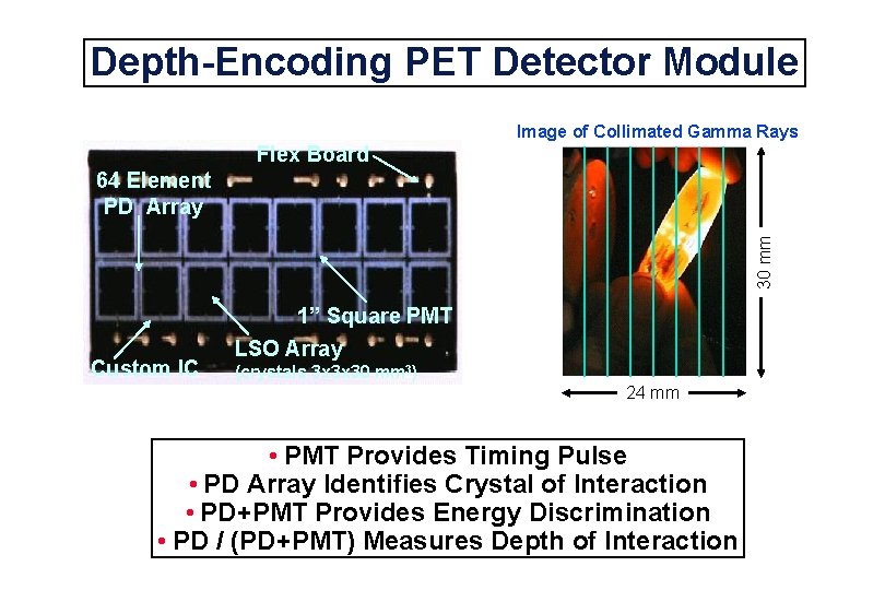
Depth-Encoding PET Detector Module Image of Collimated Gamma Rays Flex Board 30 mm 64 Element PD Array 1” Square PMT Custom IC LSO Array (crystals 3 x 3 x 30 mm 3) 24 mm • PMT Provides Timing Pulse • PD Array Identifies Crystal of Interaction • PD+PMT Provides Energy Discrimination • PD / (PD+PMT) Measures Depth of Interaction
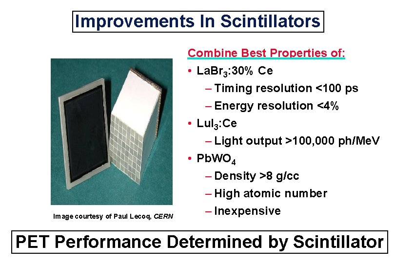
Improvements In Scintillators Combine Best Properties of: • La. Br 3: 30% Ce Image courtesy of Paul Lecoq, CERN – Timing resolution <100 ps – Energy resolution <4% • Lu. I 3: Ce – Light output >100, 000 ph/Me. V • Pb. WO 4 – Density >8 g/cc – High atomic number – Inexpensive PET Performance Determined by Scintillator
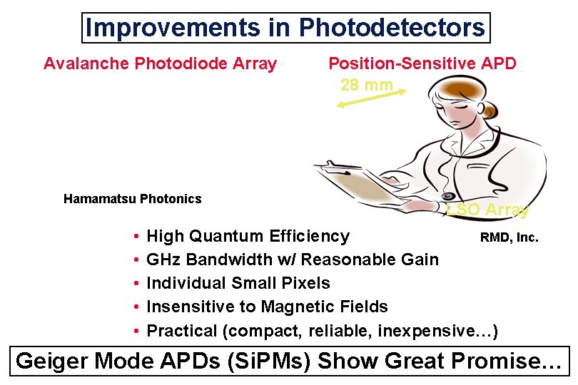
Improvements in Photodetectors Avalanche Photodiode Array Hamamatsu Photonics • • • Position-Sensitive APD 28 mm LSO Array High Quantum Efficiency RMD, Inc. GHz Bandwidth w/ Reasonable Gain Individual Small Pixels Insensitive to Magnetic Fields Practical (compact, reliable, inexpensive…) Geiger Mode APDs (Si. PMs) Show Great Promise…
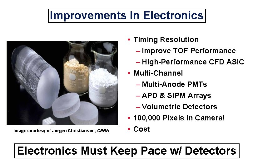
Improvements In Electronics Image courtesy of Jorgen Christianson, CERN • Timing Resolution – Improve TOF Performance – High-Performance CFD ASIC • Multi-Channel – Multi-Anode PMTs – APD & Si. PM Arrays – Volumetric Detectors • 100, 000 Pixels in Camera! • Cost Electronics Must Keep Pace w/ Detectors
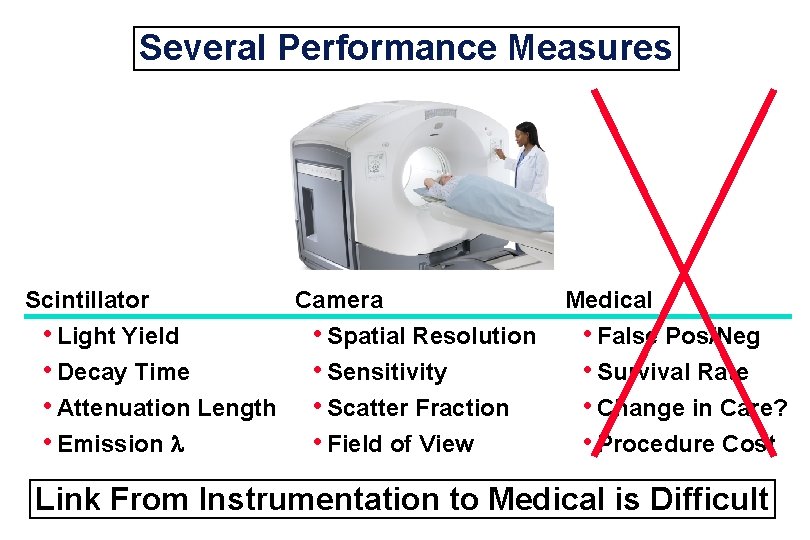
Several Performance Measures Scintillator • Light Yield • Decay Time • Attenuation Length • Emission Camera • Spatial Resolution • Sensitivity • Scatter Fraction • Field of View Medical • False Pos/Neg • Survival Rate • Change in Care? • Procedure Cost Link From Instrumentation to Medical is Difficult