Nervous Sensory systems Ch 38 2 parts of


 Receptors (sensory neurons) Integrators (interneurons) motor neurons Effectors (muscles, glands)")


 scala")


- Slides: 39
Nervous & Sensory systems Ch 38
2 parts of Nervous System Ø CNS = Brain and main nerve cords Ø PNS = paired cranial and spinal nerves
Peripheral Nervous System Ø Somatic nerves l l Motor functions Skeletal muscle voluntary (Shown in green) Ø Autonomic nerves l l Visceral functions smooth & cardiac muscle involuntary (Shown in red)
Fig. 34 -4, p. 575
Two Types of Autonomic Nerves Ø Sympathetic Ø Parasympathetic Ø Most organs receive input from both Ø Usually have opposite effects on organ
Sympathetic Nerves Ø Promote responses that prepare the body for stress or physical activity (fight-or-flight response) Ø Adrenaline secretion = increase heart rate, glucose released into blood, slow down of housekeeping activities
Parasympathetic Nerves Ø Promote housekeeping responses such as digestion Ø Lower heart rate Ø Glucose uptake
Communication Lines Stimulus (input) Receptors (sensory neurons) Integrators (interneurons) motor neurons Effectors (muscles, glands) Figure 34. 5 Page 575 Response (output)
Brains…. . Ø A. Most simple = cerebral ganglia Ø collection of nerves near head Ø B. Vertebrate brains Ø 1. gray matter: neuron cell bodies, non myelinated axons & glia Ø 2. white matter: axons covered in myelin Ø 3. cerebrospinal fluid fills ventricles in brain Ø a. filtered from arteries returned to veins Ø b. supplies nutrients/ carries hormones, removes waste
4. Neuroglia cells Ø astrocytes – provide support, direct formation of blood-brain barrier (tight junctions) Ø Oligodendrocytes – make myelin
5. Regions of the Brain Ø A. Brain Stem: conducts signals from spinal cord to brain, controls homeostasis Ø (breathing, heart rate, sleep) Ø 1. Hind Brain Ø a. medulla oblongata Ø b. Pons Ø 2. Mid Brain (hearing)
Ø B. Cerebellum – part of hind brain Ø 1. balance Ø 2. motor skills Ø 3. hand-eye coordination
Ø C. Thalamus – sensory info Ø D. Hypothalamus – homeostasis Ø heat Ø hunger/thirst Ø hormones Ø E. Cerebrum – Ø information processing
Function of the Spinal Cord Ø Expressway for signals btwn brain & peripheral nerves Ø Sensory & motor neurons make direct reflex connections in the spinal cord Ø Spinal reflexes do not involve the brain
Reflexes Ø Automatic movements in response to stimuli Ø Reflex arcs: sensory neurons synapse directly on motor neurons Ø (skip integrators)
Stretch Reflex STIMULUS Biceps stretches. sensory neuron motor neuron Response Biceps contracts. Figure 34. 16 Page 585
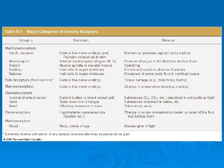
Assessing a Stimulus Ø Action potentials don’t vary in amplitude Ø Brain tells nature of stimulus by: Ø 1) Particular pathway that carries the signal Ø 2) Frequency of action potentials (how quickly is signal repeated) Ø 3) Number of receptors signalling
Taste Ø Chemoreceptors Ø Five primary sensations: l sweet, sour, salty, bitter, and umami Figure 35. 8 Page 604
Smell Ø Olfactory receptors Ø Receptor axons lead to olfactory lobe Ø Binding molecules triggers action potential olfactory bulb receptor cell Figure 35. 7 Page 604
Anatomy of Human Ear stirrup anvil auditory nerve hammer auditory canal eardrum cochlea Fig. 35. 11 a Page 614
Sound Reception Ø Sound waves make the eardrum vibrate Ø Vibrations are transmitted to the bones of the middle ear Ø The stirrup transmits force to the oval window of the fluid-filled cochlea
Sound Reception Ø Movement of oval window causes waves in the fluid inside cochlear ducts oval window (behind stirrup) eardrum round window Figure 35. 11 c Page 606 scala vestibuli scala tympani
scala vestibuli cochlear duct organ of Corti sensory neurons (to the auditory nerve) scala tympani Fig. 35 -11 d, p. 607
Sound Reception hair cells in organ of Corti basilar membrane tectorial membrane lumen of cochlear duct lumen of scala tympani to auditory nerve Figure 35. 11 e Page 607
Fig. 35 -12 a, p. 607
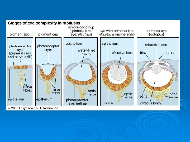
Vision Ø Sensitivity to light does not equal vision Ø Vision requires two components l Eyes l Capacity for image formation in the brain
Invertebrate Eyes Limpet ocellus ommatidium cuticle epidermis lens Compound eye of a deerfly sensory neuron Land snail eye Figures 35. 13 & 35. 14 Pages 608 & 609
vitreous body lens cornea retina optic tract Fig. 35 -15, p. 609
Human Eye sclera retina choroid iris lens pupil cornea aqueous humor fovea optic disk part of optic nerve ciliary muscle vitreous body Figure 35. 17 Page 610
Organization of Retina Ø Photoreceptors lie at the back of the retina, in front of a pigmented epithelium Ø For light to reach the photoreceptors, it must pass layers of neurons involved in visual processing
Organization of Retina Ø Signals from photoreceptors are passed to bipolar sensory neurons, then to ganglion cells Figure 35. 20 Page 612
Ø Reference: Ø http: //www. bio. davidson. edu/people/midor cas/animalphysiology/websites/2003/Mulle r/development%20 of%20 the%20 cephalop od%20 eye. htm Ø video Ø https: //vimeo. com/23085747
a Light rays from an object converge on the retina, form an inverted, reversed image. muscle contracted b When a ciliary muscle contracts, the lens bulges, bending the light rays from a close object so that they become focused on the retina. close object slack fibers muscle relaxed c When the muscle relaxes, the lens flattens, focusing light rays from a distant object on the retina. distant object taut fibers Fig. 35 -18, p. 611
cone cell stacked, pigmented membrane rod cell Fig. 35 -19, p. 612
The Photoreceptors Ø Rods l l Contain the pigment rhodopsin Detect very dim light, changes in light intensity Ø Cones l Three kinds; detect red, blue, or green l Provide color sense and daytime vision
Retina to Brain optic retina nerve lateral geniculate nucleus visual cortex Figure 35. 23 Page 613