Mycology Review Identification of Common Dermatophytes Sandy Arduin


- Slides: 23

Mycology Review: Identification of Common Dermatophytes Sandy Arduin, MT (ASCP) Bruce Palma, MT (ASCP) Mycology Unit Bureau of Laboratories Michigan Department of Community Health This project was supported in part by Grant/Cooperative Agreement Number. U 10/CCU 523395 -01 from the Centers for Disease Control and Prevention (CDC) Its contents are solely the responsibility of Michigan Department of Community Health and do not necessarily represent the official views of CDC

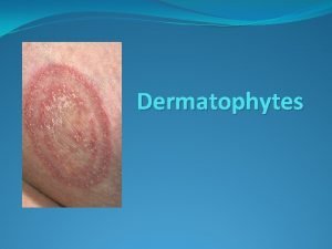
Dermatophytes

Index Trichophyton rubrum Trichophyton mentagrophytes Hair Perforation Test Trichophyton tonsurans Trichophyton verrucosum Trichophyton terrestre Epidermophyton floccosum Microsporum canis Microsporum gypseum Microsporum nanum Differentiation Table Test Your Knowledge Unknown 1 Unknown 2 Unknown 3 Unknown 4 Unknown 5 Unknown 6 Double click on any of the words listed above to go directly to the slide on that organism. To return to the index, click on the home button. To return to the last slide viewed, click on the return button. You must be in slide show mode to use these functions. Macroscopic colony morphology descriptions are based on cultures grown on SAB agar. Colony morphology may vary on other culture media.

Trichophyton rubrum Colony growth is slow to moderate, downy, white on the surface with a red to brown reverse. Microconidia are club-shaped to pyriform and are formed along the sides of the hyphae. Macroconidia are pencil-shaped to cigar-shaped. Lab tests: hair perforation test negative, urease negative, growth at 37°C. Infection is typically found on the feet, hands, nails, or groin.

Trichophyton mentagrophytes Colony growth is moderately rapid, powdery to granular, white to cream colored on the surface with a yellowish, brown or red-brown reverse. Microconidia are numerous, unicellular, round to pyriform and found in grape like clusters. Spiral hyphae are often present. Macroconidia are multiseptate, clubshaped and often absent. Lab tests: hair perforation test positive, urease positive, growth at 37°C. Infection is typically found on the feet, hands, or groin, but can also be associated with inflammatory lesions of the scalp, nails, and beard.

Hair Perforation Test Perforations Trichophyton mentagrophytes, Hair perforation test is positive. Trichophyton rubrum, Hair perforation test is negative.

Trichophyton tonsurans Colony growth is slow, suede-like to powdery, white, beige, pale yellow to sulphur yellow on the surface with a yellow to dark brown reverse. Microconidia are numerous, varying in shape and size (pyriform, clubshaped to balloon- shaped). Macroconidia are rare. When present they are sinuous with smooth walls. Lab tests: hair perforation test typically negative, urease positive, growth at 37°C. Growth is enhanced on thiamine. Infections are primarily of the scalp. Occasionally the glabrous skin or nails are infected.

Trichophyton verrucosum Colony growth is very slow, glabrous to lightly downy, white, sometimes yellow or grey on the surface without any characteristic pigment on the reverse. Microconidia are club-shaped, but are rare or absent. Typically, chlamydospores in chains are seen. Macroconidia have a “rat tail” appearance, but are rarely seen. Lab tests: hair perforation test negative, urease negative, growth at 37°C. Growth is enhanced on media with thiamine and inositol, and is more rapid at 37ºC than at 25ºC. Infection is more common on cattle or other farm animals. Infection in humans is typically found on the scalp, beard or glabrous skin.

Trichophyton terrestre Colony growth is rapid, powdery to velvety, white to cream on the surface with a pale, slightly yellow reverse. Occasionally, isolates may have a pink, red-brown, or wine-colored reverse. Microconidia are numerous, clubshaped, with a squared-off base, often borne on short pedicels. Macroconidia are 2 -8 celled and generally borne at right angles to the hyphae. Lab tests: hair perforation test positive, urease positive and will not grow at 37°C. This is a geophilic fungus, very common in soil. It can also be isolated from the fur of small mammals.

Epidermophyton floccosum Colony growth is slow, powdery, with a yellow to khaki surface color and chamois to brown reverse. Macroconidia are club shaped, with thin smooth walls and can be solitary or grouped in clusters. Chlamydospores are often produced in large numbers. Microconidia are absent. Lab tests: hair perforation test negative, urease positive, growth at 37°C. Infections are commonly cutaneous, especially of the groin or feet.

Microsporum canis Colony growth is rapid, downy to wooly, cream to yellow on the surface with a yellow to yelloworange reverse. Microconidia are club-shaped but typically are absent. Macroconidia are fusoid, verrucose, and thick walled. They have a recurved apex and contain 5 -15 cells. Lab tests: hair perforation test positive and urease positive. Infection in humans occurs on the scalp and glabrous skin. It is also a cause of ringworm in cats and dogs.

Microsporum gypseum Colony growth is rapid, downy, becoming powdery to granular, cream, tawny-buff, or pale cinnamon on the surface with a beige to redbrown reverse. Microconidia are moderately abundant and club-shaped. Macroconidia are abundant, ellipsoidal to fusiform, sometimes verrucose, and thin walled. They typically contain 3 -6 cells. Lab tests: hair perforation test positive and urease positive. Infection in humans is found on the scalp and glabrous skin; it is more frequently isolated from the soil and from the fur of small rodents.

Microsporum nanum Colony growth is rapid, downy to powdery, white to buff on the surface, with a red-brown reverse. Microconidia, if present, occur in small numbers. Macroconidia are numerous, 1 -3 celled, and have a characteristic pear or egg shape. Typically macroconidia are 2 celled. Conidia are solitary on the ends of short conidiophores. Lab tests: hair perforation test positive and urease positive. Infection is rarely transmitted to humans; it is the principal cause of tinea in pigs.

Dermatophyte Differentiation Table: Hair Perforation Test Urease Test Growth at 37°C Macro-conidia Micro-conidia Distinguishing Characteristics Trichophyton rubrum Negative Positive Pencil shaped/cigar shaped Club shaped to pyriform, along the sides of the hyphae Red reverse pigment Hair perf. test neg. Club shaped microconidia Trichophyton mentagrophytes Positive Club shaped when present Numerous Unicellular to round in grape like clusters Round microconidia in grape like clusters Spiral hyphae Trichophyton tonsurans Usually (-) Occasionally + Positive Cylindrical to cigar shaped and sinuous, if present Numerous, varying in shape and size, club shaped to balloon shaped Microconidia varying in shape and size Growth enhanced by thiamine Trichophyton verrucosum Negative Positive “Rat-tailed” if present Rare or Absent Chlamydospores in chains typically seen Chlamydospores in chains Growth better on media with thiamine and inositol Trichophyton terrestre Positive Negative 2 -8 celled borne at right angles to hyphae Club shaped with squared-off base on pedicels Microconidia with squared -off base on short pedicels Epidermophyton floccosum Negative Positive Club shaped, often in clusters Absent Khaki colored colony with brown reverse Microconidia absent Microsporum canis Positive NA Fusoid, thick, rough walled with recurved apex Typically absent Club shaped if present Fusoid, rough walled macroconidia with recurved apex Microsporum gypseum Positive NA Ellipsoidal to fusiform, thin, Rough walled Moderately abundant Club shaped Thin walled macroconidia Tawny-buff granular colony Microsporum nanum Positive NA Typically 2 celled Pear or egg shaped Rough walled Clavate when present 2 celled pear shaped macroconidia

Test Your Knowledge Each unknown slide has the following navigation buttons to help you: View dermatophyte differentiation table View index slide Return to previously viewed slide Answer View correct answer

Unknown 1 Colony growth is rapid, downy to wooly, cream to yellow on the surface with a yellow to yellow- orange reverse. Answer

Unknown 2 Colony growth is moderately rapid, powdery to granular, white to cream colored on the surface with a yellowish, brown or red -brown reverse. Answer

Unknown 3 Colony growth is rapid, downy to powdery, white to buff on the surface, with a redbrown reverse. Answer

Unknown 4 Colony growth is very slow, glabrous to lightly downy, white, sometimes yellow or grey on the surface without any characteristic pigment on the reverse. Growth is enhanced on media with thiamine and inositol, and is more rapid at 37ºC than at 25ºC. Answer

Unknown 5 Colony growth is slow to moderate, downy, white on the surface with a red to brown reverse. Answer

Unknown 6 Colony growth is rapid, downy, becoming powdery to granular, cream, tawny-buff, or pale cinnamon on the surface with a beige to redbrown reverse. Answer

Glossary Anthropophilic A fungus (dermatophyte) which grows preferentially on humans, rather than on animals or in soil. Clavate Club-shaped. Conidium A unicellular or multicellular fungal element which serves as an asexual reproductive structure. Dermatophyte A mould belonging to the genera: Epidermophyton, Microsporum, Trichophyton; typically infecting skin, hair and nails. Fusoid Spindle shaped; ellipsoidal with two tapered ends. Glabrous Smooth, lacking hairs. Geophilic A fungus (dermatophyte) which grows preferentially on substrates found in the soil, rather than on animals or humans. Macroconidia The larger of two types of conidia produced by the same fungus. May be multicellular. Microconidia The smaller of two types of conidia produced by the same fungus. Typically unicellular. Onychomycosis Fungal infection of the nails. Spiral hyphae Hyphae curved into a spiral. Typically seen in Trichophyton mentagrophytes, but may be seen in other dermatophytes as well Verrucose Having many warts Zoophilic A fungus (dermatophyte) which grows preferentially on animals, rather than on humans or in soil.

Bibliography de Hoog, G. S. , Guarro, J. , Figueras, Gene & M. J. 2000. Atlas of Clinical Fungi, 2 nd ed. Centraalbureau voor Schimmelcultures. Utrecht, The Netherlands. Benecke, E. S. , and Rogers, A. L. 1996. Medical Mycology and Human Mycoses. Star Publishing Company, Belmont, California. Kane, Julius, Summerbell, Richard, Sigler, Lynn, Krajden, Sigmund, and Land, Geoffrey. 1997. Laboratory Handbook of Dermatophytes. Star Publishing Co. , Belmont, CA. Larone, Davise H. 1995. Medically Important Fungi, A Guide to Identification, 3 rd ed. , ASM Press, Washington, D. C. Mc. Ginnis, M. R. 1980. Laboratory Handbook of Medical Mycology, Academic Press, New York. Mc. Ginnis, M. R. , D'Amato, RF. , Land, GA. 1982. Pictorial Handbook of Medically Important Fungi and Aerobic Actinomycetes. Praeger Publishing. Murray, P. R. , Brown, E. J. , Pfallen, M. A. , Tenover, F. C. , Yolken, R. H. , Manual of Clinical Microbiology, 7 th Edition, ASM Press, Washington, D. C. Rebell, Gerbert, Taplin, David. 1974. Dermatophytes, Their Recognition and Identification. University of Miami Press, Coral Gables, Florida. Rippon, J. W. , 1974. Medical Mycology The Pathogenic Fungi and The Pathogenic Actinomycetes. W. B. Saunders, Philadelphia, PA. St-Germain, G. , Summerbell, R. 1996. Identifying Filamentous Fungi, Star Publishing Company. Belmont, CA.
 Presumptive identification vs positive identification
 Mycology
 Tease mount
 Medical mycology ppt
 Mycology
 Mycology
 Mycology test
 Aspergillus
 Fungi motile
 Dimorphic fungi examples
 S i n g u l a r
 Mycology
 Industrial mycology
 Mycology
 Mycology lecture
 Mycology and lichens
 White piedra
 Ascomycota
 Mycology lecture
 Hardwood and softwood trees
 Andy sometimes read comics
 Sandy tatiana rivas
 Sandy miller real estate
 Sandy irani

