Medial Patellar Ligament Desmotomy Preoperation Indications Upward patellar

 occurs when the patellar fibrocartilage and the medial")


- Slides: 16
Medial Patellar Ligament Desmotomy Pre-operation
Indications • Upward patellar fixation (UPF) occurs when the patellar fibrocartilage and the medial patellar ligament fix over the medial trochlear ridge of the femur, inhibiting flexion of the hock and stifle. • Some predisposing factors are poor muscle tone and condition, straight hind limb conformation, stifle trauma and hereditary factors.
Indications • Depending on the duration and severity for the recurrent UPF, the medial patellar ligament desmotomy should be considered as the last resort, since many cases respond to more conservative therapy. Example young horses will improve after appropriate conditioning and development of quadriceps muscle tone through training.
Contraindication • Long-term osteoarthritis may result due to maltracking of the patella and/or altered gait. • May predispose to patella fragmentation.
Welfare consideration • http: //www. thehorse. com/articles/31656/equine -field-anesthesia-considerations • Please refer to the link for field anaesthesia considerations. • Also refer to the pdf for general welfare considerations.
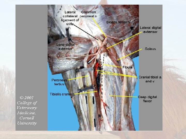
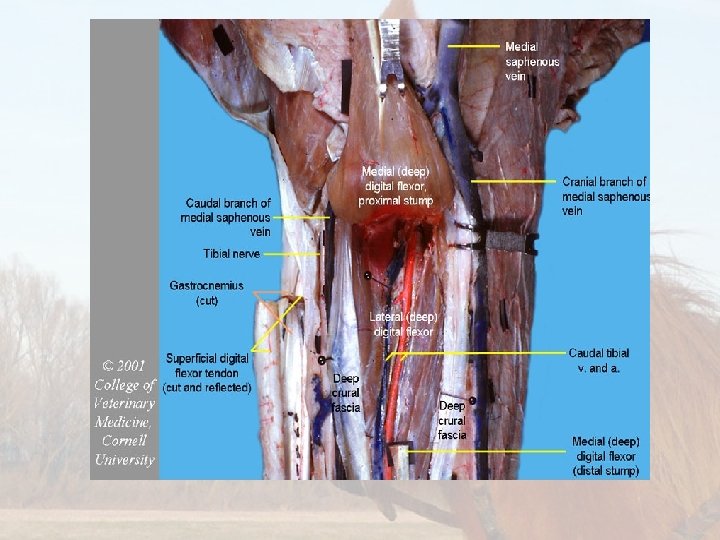
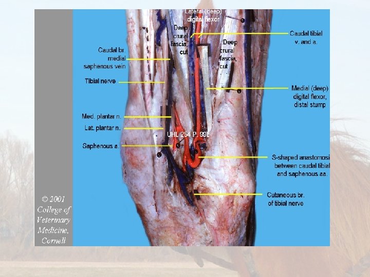
Anatomy
Equipment • • Halters Chlorhexidine Iodine solution Alcohol Cotton/ gauze Sterile pack Radiography/ Ultrasound Equipment • Blunt-ended bistoury (tenotomy) knife. • Scalpel blade • Non-absorbable suture(polypropylene/ nylon) • Needle and syringes • Surgical drapes • Sterile instruments.
Physical Examination https: //www. youtube. com/watch? v=0 t 5 jfk 4 ain. U • Please refer to the link that shows distance and physical examination of a horse. • Also note the ASA grade of a horse must be 1 to preform the surgery.
Drugs •
Drugs
Preparation for the surgery Ø Ensure the surgery room/stall in clean thoroughly to avoid any contamination during surgery. Ø Further more pre-surgery radiographs should be done before the animal is prep. Ø Administer Tetanus antitoxin by injecting 1 m. L dose intramuscularly using aseptic technique. Administer a second 1 m. L dose 4 to 8 weeks after the first dose. Revaccinate annually using one 1 m. L dose. Ø The surgical procedure is performed with the animal standing. Ø The horse is sedated using sedatives Ø Depending on the horse’s temperament tranquilizers can be used.
Preparation for the surgery Ø Next the horses tail is wrapped to avoid contamination of the surgical site. Ø After the horse is sedated, the area of the middle and medial patellar ligament is clipped. Ø Following that the site is surgically prepared. This is done by scrubbing the area with alcohol (removing any heavy debris) and then wiping the area with iodine in a unilateral manner starting from the center of the site. Ø 2 ml of the local anesthetic are injected subcutaneously over the medial border of the middle patellar ligament.
Preparation for the surgery Ø A 1 inch, needle is then inserted through this bleb, and the subcutaneous area around the distal part of the medial patellar ligament is infiltrated with the local anesthetic of choice.