Lower Leg and Ankle Injuries Shin Splints Medial


 l l l Rest. The sooner you rest the sooner it will")


 l l l X-ray MRI CT scans")
 Unusual")


 l l l AAFP (see table 3) R. I. C. E. Ice")


- Slides: 31
Lower Leg and Ankle Injuries
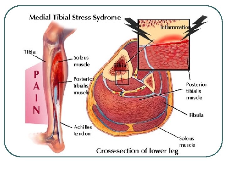
Shin Splints Medial l Anterior l
Medial Tibial Stress Syndrome l l Tenderness is usually found between 3 and 12 centimeters above the tip of the medial malleolus at the posterio-medial aspect of the tibia. Inflammation of the periostium (periostitis) Most frequently involved is the Tibalis Posterior tendon and muscle, but the Flexor Digitorum Longus and Flexor Hallucis Longus may also be involved. Stress fractures can also occur in this area.
Anterior Compartment Syndrome l l Soft tissue injuries at the muscular origin and bony or periosteal interface of the bone and muscle origin. Due to micro tears of the Tibialis Anterior either at the origin or in the fibers themselves. Or microtrauma to the bone structure itself. Stress fractures can also occur in this area.
Exertional Compartment Syndrome l l Caused by the muscles swelling within a closed compartment with a resultant increase in pressure in the compartment. The blood supply can be compromised and muscle injury and pain may occur. Abnormal compartment pressure: • • • A resting pressure greater than 20 mm Hg; or An exertional pressure greater than 30 mm Hg; or A pressure of 25 mm Hg or higher 5 minutes after stopping exercise. This may require surgical decompression of the compartment.
Key Causes l l l Tight posterior muscles Imbalance between the posterior and anterior muscles Running on concrete or other hard surfaces Improper Shoes - inadequate shock protection Overtraining
Treatment (FYI) l l l Rest. The sooner you rest the sooner it will heal. Apply ice 10 -15 minutes for 2 -3 x per day in the early stages when it is very painful. Anti inflammatory drugs Wear shock absorbing insoles in shoes. Maintain fitness with other non weight bearing exercises. Apply heat and use a heat retainer after the initial acute stage, particularly before training.
Stress Fractures l l l Bone remodeling Repetitive stress weakens the bone 10 -20% of injuries to athletes Most common locations: tibia, fibula and metatarsals. Tibial and fibular stress fractures can develop from “shin splints”
Causes of Stress Fractures l l l l Training errors Abnormal limb length Low body weight (< 75% of ideal) Eating disorders Previous inactivity White race Female
Diagnosis (FYI) l l l X-ray MRI CT scans
Metatarsal Stress Fracture l l CAUSES: Decreased density of the bones (eg. osteoporosis) Unusual stress on a metatarsal due to mal position or another forefoot deformity (eg. bunion) Abnormal foot structure or mechanics (eg. flatfoot, over inversion)
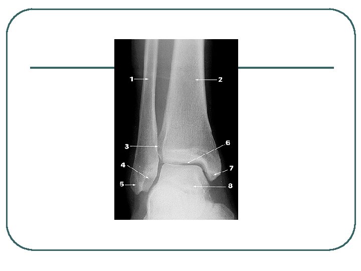
Ankle Sprains l l Most common athletic injury. 25% of all injuries. The risk of ankle sprains varies with the sport • 21 -53% basketball, 17 -29% soccer, 25% volleyball. Ankle sprains account for 10% to 15% of all lost playing time The medial malleolus is shorter than the lateral mallelous so there is naturally more inversion than eversion.
Ankle Sprains l l l Greater inversion increases the potential for overstretching of the lateral ligaments. Most sprains involve the lateral ligaments from excessive inversion. Deltoid ligament is sprained less often (25% of ankle sprains) Of the lateral ligments, the ATFL is sprained the most often followed by the CFL Sprains ocur most often with the foot in plantar flexion and inversion.
Lateral Collateral Ligament
Ankle Sprains
Classification of Sprains l l 1 st Degree: • • Stretching of the ATFL little or no edema tenderness maintain function. 2 nd Degree • • • Partial tear of the ATFL and/or CFL moderate edema some function loss l 3 rd Degree • • • Complete tear ATFL, CFL, and/or PTFL total loss of function significant edema
Ankle Sprains by Grade Sign/symptom Grade III Tendon No tear Partial tear Complete tear Loss of functional ability Minimal Some Great Pain Minimal Moderate Severe Swelling Minimal Moderate Severe Ecchymosis Usually not Frequently Yes Difficulty bearing weight No Usually Almost always
Treatment (FYI) l l l AAFP (see table 3) R. I. C. E. Ice for 20 minutes on and 20 minutes off for the first two hours. After that, 20 min intervals over the next 48 -72 hours, Compression wrap with donut or horse shoes to fill in gaps around malleolus from 24 -36 hours; after 48 -72 hours contrasts baths with ROM exercises for 4 minutes in warm and 1 min in ice water.
Achilles Tendonitists l l Causes • • Rapidly increasing training effort Adding hills or stair climbing to training • Starting too quickly after a layoff • Poor footwear • Excessive pronation • Tight posterior leg muscles If left untreated, it may progress to a complete rupture.
Achilles Tendon Rupture l l l Most frequently ruptured tendon Complete ruptures are due to eccentric loading during abrupt stopping, landing from a jump. Usually a popping sound is heard with a complete tear. There may or may not be an obvious gap 2 to 6 cm from the calcaneus attachment. Treatment may or may not include surgery but both require immobilized for 3 months.
Plantar Fasciitis l l l The plantar fascia runs from the calcaneus to the metatarsals. This tight band acts like a bow string to maintain the arch of the foot. Plantar fasciitis refers to an inflammation of the plantar fascia.
Plantar Fasciitis l l l Inflammation is usually due to repeated trauma to where the tissue attaches to the calcaneus. The trauma results in microscpic tears at the calcaneus attachment site. This may produce heal spurs
Plantar Fasciitis l l Pain is worse in the morning or after a period of inactivity Causes • High arch • Excessive pronation • Footwear (worn out, stiff) • Increase in intensity
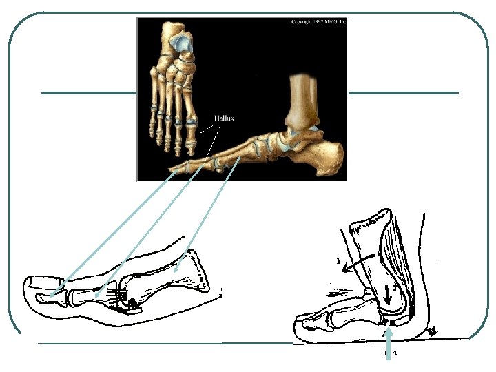
Turf Toe l l Turf toe is really a bruise or sprain that occurs at the base of the big toe at the joint called the metatarsal phalangeal joint. It usually occurs when the toe is jammed forcibly into the ground or, more commonly, when the toe is bent backward too far (hyperextended) It causes significant pain and swelling at the base of the big toe. It can be a significant problem because players use the toe when they run and plant and push off.
Ankle Exercises l Calf stretch l Soleus stretch l Resisted dorsal and plantar flexion l Heel raises l Step-up l Jump rope
Ankle Exercises Wobble Boards