INGUINAL REGION Dr Aiman Qais Afar Surgical Anatomist


- Slides: 30

INGUINAL REGION Dr. Aiman Qais Afar Surgical Anatomist College of Medicine / University of Mutah 2020 -2021 Tuesday 2 March 2021 1

Inguinal Region The inguinal region (groin) extending between the ASIS and pubic tubercle, is an important area anatomically and clinically. v Anatomically because it is a region where structures exit and enter the abdominal cavity and v Clinically because the pathways of exit and entrance are potential sites of herniation. Tuesday 2 March 2021 Dr. Aiman Qais Afar 2

Inguinal Region In fact, the majority of abdominal hernias occur in this region, with inguinal hernias in particular accounting for 75% of all abdominal hernias, (approximately 86%) occur in males because of the passage of the spermatic cord through the inguinal canal. Tuesday 2 March 2021 Dr. Aiman Qais Afar 3

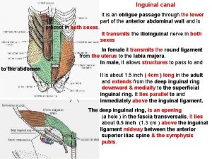
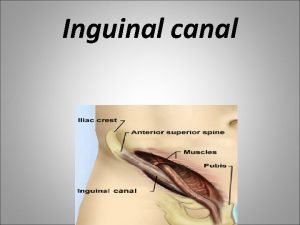
Inguinal Canal v. The inguinal canal is an oblique passage through the lower part of the anterior abdominal wall. In the males, it allows structures to pass to and from the testis to the abdomen. v. In females it allows the round ligament of the uterus to pass from the uterus to the labium majus. Tuesday 2 March 2021 Dr. Aiman Qais Afar 4

Inguinal Canal The canal is about 1. 5 in. (4 cm) long in the adult and extends from the deep inguinal ring, a hole in the fascia transversalis downward and medially to the superficial inguinal ring, a hole in the aponeurosis of the external oblique muscle Tuesday 2 March 2021 Dr. Aiman Qais Afar 5

Inguinal Canal q. The deep inguinal ring, an oval opening in the fascia transversalis, lies about 0. 5 in. (1. 3 cm) above the inguinal ligament midway between the anterior superior iliac spine and the symphysis pubis v. Related to it medially are the inferior epigastric vessels, which pass upward from the external iliac vessels v. The margins of the ring give attachment to the internal spermatic fascia Tuesday 2 March 2021 Dr. Aiman Qais Afar 6

Inguinal Canal q. The superficial inguinal ring is a triangular-shaped defect in the aponeurosis of the external oblique muscle and lies immediately above and medial to the pubic tubercle v. The margins of the ring, sometimes called the crura, give attachment to the external spermatic fascia Tuesday 2 March 2021 Dr. Aiman Qais Afar 7

Walls of the Inguinal Canal üAnterior wall üPosterior wall üRoof üFloor Tuesday 2 March 2021 Dr. Aiman Qais Afar 8

Walls of the Inguinal Canal Anterior wall: External oblique aponeurosis, reinforced laterally by the origin of the internal oblique from the inguinal ligament Posterior wall: Conjoint tendon medially, fascia transversalis laterally. Roof or superior wall: Arching lowest fibers of the internal oblique and transversus abdominis muscles Floor or inferior wall: Upturned lower edge of the inguinal ligament and, at its medial end, the lacunar ligament Tuesday 2 March 2021 Dr. Aiman Qais Afar 9

Walls of the Inguinal Canal Dr. Aiman Qais Afar Tuesday 2 March 2021 üAnterior wall is therefore strongest where it lies opposite the weakest part of the posterior wall, namely, the deep inguinal ring. üPosterior wall is therefore strongest where it lies opposite the weakest part of the anterior wall, namely, the superficial inguinal ring 10

Spermatic Cord Is a collection of structures that pass through the inguinal canal to and from the testis. It begins at the deep inguinal ring lateral to the inferior epigastric artery and ends at the testis. Structures of the Spermatic Cord v. Vas deferens v. Testicular artery v. Testicular veins (pampiniform plexus) v. Testicular lymph vessels v. Autonomic nerves v. Remains of the processus vaginalis v. Genital branch of the genitofemoral nerve, which supplies the cremaster muscle Tuesday 2 March 2021 Dr. Aiman Qais Afar 11

v. Vas Deferens (Ductus Deferens) Dr. Aiman Qais Afar Tuesday 2 March 2021 üIs a cordlike structure that can be palpated between finger and thumb in the upper part of the scrotum. ü It is a thick-walled muscular duct that transports spermatozoa from the epididymis to the urethra Vasectomy ? ? ? 12

v. Testicular Artery q. A branch of the abdominal aorta (at the level of the second lumbar vertebra), the testicular artery is long and slender and descends on the posterior abdominal wall. q. It traverses the inguinal canal and supplies the testis and the epididymis Tuesday 2 March 2021 Dr. Aiman Qais Afar 13

v. Testicular Veins Dr. Aiman Qais Afar Tuesday 2 March 2021 q. An extensive venous plexus, the pampiniform plexus, leaves the posterior border of the testis as the plexus ascends, it becomes reduced in size so that at about the level of the deep inguinal ring, a single testicular vein is formed. q. This runs up on the posterior abdominal wall and drains into the left renal vein on the left side and into the inferior vena cava on the right side 14

v. Processus Vaginalis The remains of the processus Vaginalis are present within the cord Hydrocele of Spermatic Cord and/or Testis ? ? Tuesday 2 March 2021 Dr. Aiman Qais Afar 15

Coverings of the Spermatic Cord (the Spermatic Fasciae) üExternal spermatic fascia derived from the external oblique aponeurosis and attached to the margins of the superficial inguinal ring üCremasteric fascia derived from the internal oblique muscle üInternal spermatic fascia derived from the fascia transversalis and attached to the margins of the deep inguinal ring Dr. Aiman Qais Afar Tuesday 2 March 2021 16

Scrotum Dr. Aiman Qais Afar Tuesday 2 March 2021 The scrotum is an outpouching of the lower part of the anterior abdominal wall and contains the testes, the epididymides, and the lower ends of the spermatic cords. 17

Scrotum The wall of the scrotum has the following layers: v. Skin v. Superficial fascia; üThe dartos muscle, replaces the fatty (camper fascia), and üThe membranous layer (Scarpa's fascia) is now called Colles' fascia. v. External spermatic fascia derived from the external oblique v. Cremasteric fascia derived from the internal oblique 18 Dr. Aiman Qais Afar Tuesday 2 March 2021

Scrotum The wall of the scrotum: v. Internal spermatic fascia derived from the fascia transversalis v. Tunica vaginalis, which is a closed sac that covers the anterior, medial, and lateral surfaces of each testis Tuesday 2 March 2021 Dr. Aiman Qais Afar 19

Testis Dr. Aiman Qais Afar Tuesday 2 March 2021 ØThe testis is a firm, mobile organ lying within the scrotum. ØThe left testis usually lies at a lower level than the right. ØEach testis is surrounded by a tough fibrous capsule, the tunica albuginea 20

Testis üExtending from the inner surface of the capsule is a series of fibrous septa that divide the interior of the organ into lobules. ü Lying within each lobule are one to three coiled seminiferous tubules. üThe tubules open into a network of channels called the rete testis. üSmall efferent ductules connect the rete testis to the upper end of the epididymis Tuesday 2 March 2021 Dr. Aiman Qais Afar 21

Epididymis q The epididymis is a firm structure lying posterior to the testis, with the vas deferens lying on its medial side. q It has an expanded upper end, the head, a body, and a pointed tail inferiorly. q Laterally, a distinct groove lies between the testis and the epididymis, which is lined with the inner visceral layer of the tunica vaginalis and is called the sinus of the epididymis. Tuesday 2 March 2021 Dr. Aiman Qais Afar 22

Blood Supply of the Testis and Epididymis üThe testicular artery is a branch of the abdominal aorta. üThe testicular veins emerge from the testis and the epididymis as a venous network, the pampiniform plexus. üThis becomes reduced to a single vein as it ascends through the inguinal canal. üThe right testicular vein drains into the inferior vena cava, and the left vein joins the left renal vein. Tuesday 2 March 2021 Varicocele ? ? Dr. Aiman Qais Afar 23

Formation of the inguinal canals and descent of testes Dr. Aiman Qais Afar Tuesday 2 March 2021 24

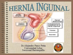
Dr. Aiman Qais Afar q A hernia is the protrusion of part of the abdominal contents beyond the normal confines of the abdominal wall. v Indirect Inguinal Hernia : The sac enters the inguinal canal through the deep inguinal ring lateral to the inferior epigastric vessels. (the most common 85%) v Direct Inguinal Hernia : The sac of a direct hernia bulges directly anteriorly through the posterior wall of the inguinal canal medial to the inferior epigastric vessels (about 15%) Tuesday 2 March 2021 25

The inguinal hernia form about 75% of all abdominal wall hernias( Femoral, Umbilical, Incisional , Epigastric…etc. ) Tuesday 2 March 2021 Dr. Aiman Qais Afar 26

Indirect Inguinal Hernia ■■ It is the remains of the processus vaginalis and therefore is congenital in origin. ■■ It is more common than a direct inguinal hernia. ■■ It is much more common in males than females. ■■ It is more common on the right side. ■■ It is most common in children and young adults. ■■ The hernial sac enters the inguinal canal through the deep inguinal ring and lateral to the inferior epigastric vessels. The neck of the sac is narrow. ■■ The hernial sac may extend through the superficial inguinal ring above and medial to the pubic tubercle. (Femoral hernia is located below and lateral to the pubic tubercle) ■■ The hernial sac may extend down into the scrotum or labium majus Tuesday 2 March 2021 Dr. Aiman Qais Afar 27

Direct Inguinal Hernia A direct inguinal hernia can be summarized as follows: ■■ It is common in old men with weak abdominal muscles and is rare in women. ■■ The hernial sac bulges forward through the posterior wall of the inguinal canal medial to the inferior epigastric vessels. ■■ The neck of the hernial sac is wide. Tuesday 2 March 2021 Dr. Aiman Qais Afar 28

Tuesday 2 March 2021 Dr. Aiman Qais Afar 29

Dr. Aiman Qais Afar Tuesday 2 March 2021 Dr. Aiman Qais Afar 30
 Qais marji
 Myrrh is mine its bitter perfume
 Does god know our thoughts
 Roof of inguinal canal
 Inguinal canal boundaries mnemonic
 Floor of the inguinal canal
 Oblique passage
 Aiman hanna concordia
 Habic benjamin joel
 Michelle chong company
 Aimanhanna
 Aiman
Aiman Aiman hanna
 Ece
Ece Vertebral region and scapular region
 Hernias inguinales
 Malgaigne’s bulgings
 Spermatic cord layers mnemonic
 Gastoschisis
 Triangulo de hasselbach
 Incision subcostal izquierda
 Annulus inguinalis superficialis
 Epitrochlear lymph nodes location
 Spermatic cord
 Conjoint tendon
 Campers fascia
 Canalis femoralis
 Inguinal üçgen
 Inguinal canal
 Fisiopatologia hernia inguinal
 Invagination test


