Infectious diseases 45 th Semester Classes on Infectious
")


 Dengue +/- warning symptoms Severe Dengue 1. Severe plasma leakage")


 Group B (any")


- Slides: 29
Infectious diseases 4/5 th Semester Classes on Infectious Diseases, 8 -9 AM, Tuesdays (LT-1) Topics 1 1 Approach to Infectious Diseases and their prevention 2 Antibiotic stewardship practices 3 Community-Acquired Infections 4 Health Care–Associated Infections 5 Gram-Positive Bacteria (part-1) 6 Gram-Positive Bacteria (part-2) 7 Gram-Negative Bacteria (part-1) 8 Gram-Negative Bacteria (part-2) 9 Spirochetal Diseases 10 Diseases Caused by Atypical/Miscellaneous Bacterial Infections 11 Revision-cum-exam on bacteria (Must to know type) 12 Infections Due to DNA Viruses 13 Infections Due to RNA Viruses (part 1) 14 Infections Due to RNA Viruses (part 2) 15 HIV/AIDS – part 1 16 HIV/AIDS – part 2 17 Fungal Infections 18 Parasitic Infections (part 1) 19 Parasitic Infections (part 2) 20 Revision-cum-exam on Virus, Fungal, and Parasite (Must to know type) Dr. P. K. Panda, Asst. Professor Department of Medicine AIIMS, Rishikesh
Family Common virus Picornaviridae Polio Entero Calciviridae Noro Reoviridae Rota Orthomyxviridae Influenza Paramyxoviridae Measle Mumps Parainfluenza Coronaviridae SARS MARS Togaviridae Rubella Chikungunya Flaviviridae Dengue West nile Filoviridae Ebola Rhabdoviridae Rabies Bunyaviridae Hanta Nairo (CCHF) Retroviridae HIV
RABIES • Zoonotic infection that occurs in a variety of mammals throughout the world except in Antarctica and on some islands • Canine rabies continues to be a threat to humans; others – bat/raccoon rabies • IP: 20– 90 days but in rare cases is as short as a few days or >1 year • usually transmitted to humans by the bite; rarely aerosol, transplantation, human possibly • Neuronal dysfunction—rather than neuronal death—is responsible; microglial nodules called Babes nodules & Negri bodies (eosinophilic cytoplasmic inclusions) • Disease usually presents as atypical encephalitis with relative preservation of consciousness • Prodromal – nonspecific, sometimes earliest specific neurologic symptoms that include paresthesias, pain, or pruritus near the site of the exposure • Acute neurologic – encephalitic (furious) in 80% and paralytic in 20%. • Comatose -
• Autonomic dysfunction is common and may result in hypersalivation, gooseflesh, cardiac arrhythmia, and priapism • Episodes of hyperexcitability are typically followed by periods of complete lucidity that become shorter as the disease progresses • Early brainstem involvement (hydrophobia, aerophobia) • In paralytic type, commonly misdiagnosed as Guillain-Barre syndrome
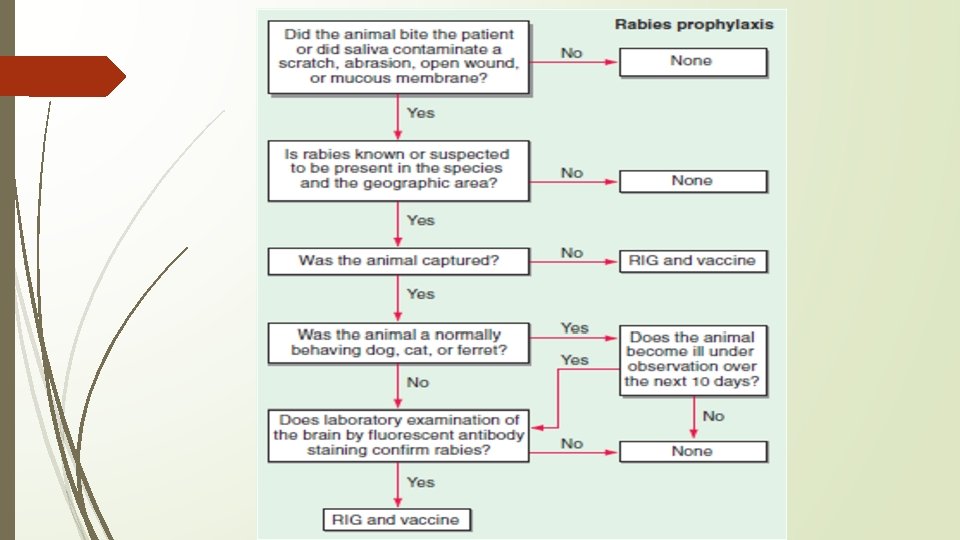
• Diagnosis should be considered in patients presenting with acute atypical encephalitis or acute flaccid paralysis, including those in whom Guillain-Barre syndrome is suspected • Diagnostically useful specimens include serum, CSF, fresh saliva, skin biopsy samples from the neck, and brain tissue • Presence of rabies virus–specific antibodies in the CSF suggests rabies encephalitis, regardless of immunization status • Other methods: RT-PCR, Direct fluorescent antibody • DD: Anti-N-methyl-d-aspartate receptor (anti-NMDA) encephalitis, Postinfectious (immunemediated) encephalomyelitis, psychiatric disorder (Rabies hysteria) • There is no established treatment for rabies • There are seven well-documented cases of survival from rabies • Postexposure Prophylaxis (PEP) – • Healthy dogs, cats, or ferrets may be confined and observed for 10 days • If an animal escapes after an exposure, it must be considered rabid, and PEP must be initiated • Includes local wound care and both active and passive immunization • If anatomically feasible, the entire dose of RIG (20 IU/kg) should be infiltrated at the site of the bite • Vaccines; Four 1 -m. L doses of rabies vaccine should be given IM in the deltoid area (NOT gluteal) – 0, 3, 7, and 14
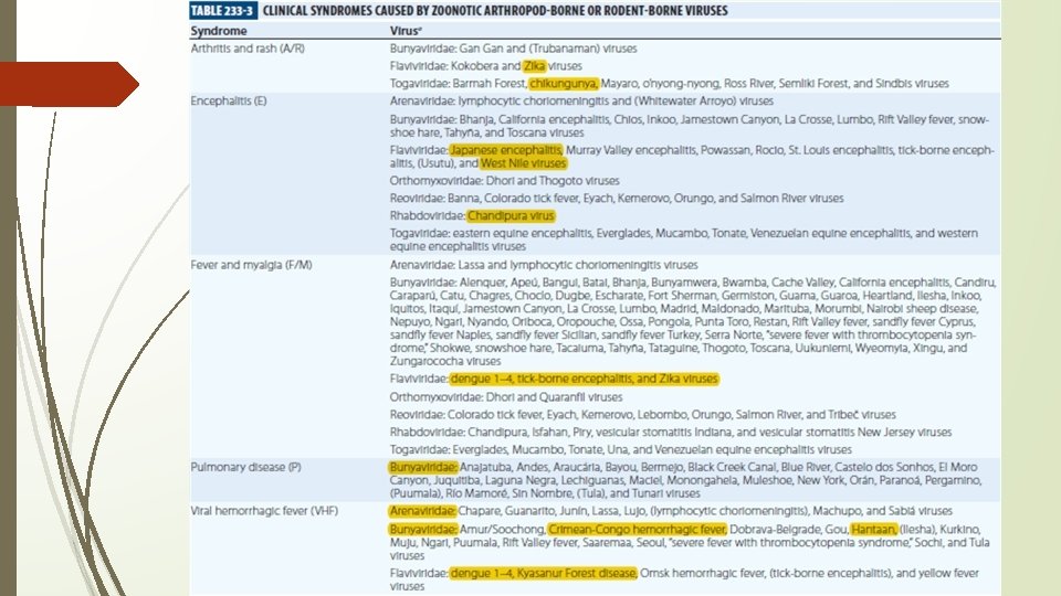
Zoonotic viruses: Arthropod-Borne and Rodent-Borne Virus Infections • Extrinsic incubation, typically lasts 1– 3 weeks in mosquitoes; Arboviruses infect their vectors after ingestion of a blood meal from vertebrate; some arthropods by saliva-activated transmission, Rarely transovarial transmission • Intrinsic incubation, as per type of infections • Seven families: Arenaviridae, Bunyaviridae, Flaviviridae, Orthomyxoviridae, Reoviridae, Rhabdoviridae, and Togaviridae • Arena and hanta viruses are rodent borne viruses • Diagnosis; recognized history of mosquito bite or tick bite (more diagnostic) or rodent exposure; serology; PCR; Hantavirus infections differ from other viral infections in that severe acute disease is immunopathologic; SYNDROMES - grouped into one of five broad categories
ARTHRITIS AND RASH • D/D - hepatitis B, parvovirus B 19 infection, and rubella, and occasionally due to adenoviruses, enteroviruses, herpesviruses, and mumps virus • Chikungunya: Ø Aedes albopictus was identified as the major vector with IP – 2 -10 DAYS Ø Abrupt onset of Fever (often severe) with a saddleback pattern and severe arthralgia accompanied by chills and constitutional symptoms and signs, such as abdominal pain, anorexia, conjunctival injection, headache, nausea, and photophobia Ø Migratory polyarthritis mainly affects the small joints of the ankles, feet, hands, and wrists; rarely large joints Ø Rashes often coincides with defervescence; Children also often develop a bullous rather than a maculopapular/petechial rash Ø Maternal–fetal transmission has been reported Ø petechiae are occasionally seen and epistaxis is not rare, but chikungunya virus should not be considered a VHF agent Ø Mildly decreased platelet counts may be seen Ø Nonsteroidal anti-inflammatory drugs and sometimes chloroquine for refractory arthritis
ENCEPHALITIS • Seasonal diseases, commonly occurring in the warmer months • Japanese encephalitis is the most important viral encephalitis in Asia • The virus is particularly common in areas of irrigated rice fields (attract the natural avian vertebrate hosts and provide abundant breeding sites for mosquitoes such as Culex tritaeniorhynchus) • Additional amplification host by pigs and horses • Unspecific febrile presentation (nausea, vomiting, diarrhea, cough) • aseptic meningitis, • meningoencephalitis, • acute flaccid paralysis, • severe encephalitis.
• Common findings in JE are cerebellar signs, cranial nerve palsies, and cognitive and speech impairments • Diagnosis by CSF/serum PCR study along with clinical features • Symptomatic treatment only • Usually two intramuscular doses of the vaccine are given 28 days apart Chandipura virus seems to be an emerging in India • It is transmitted among hedgehogs by mosquitoes and sandflies • It is characterized by high lethality in children West Nile virus is the primary cause of arboviral encephalitis in the United States • Few cases are reported from India
FEVER AND MYALGIA • Typically begins with the abrupt onset of fever, chills, intense myalgia, and malaise; “influenza-like” symptoms • The most clinically important flaviviruses that cause this syndrome are dengue viruses 1– 4 Hantavirus syndrome: It was in 1966 that Thottapalayam virus, the first indigenous hantavirus species was isolated. The Old World hantaviruses cause haemorrhagic fever with renal syndrome (HFRS) in Asia and Europe while the New World hantaviruses cause hantavirus cardiopulmonary syndrome (HCPS) in the America.
Dengue Agent- Dengue Virus • • Single stranded RNA Virus Family: Flaviviridae Genus: Flavivirus 4 serotypes: DENV-1, DENV-2, DENV-3 and DENV-4 Vector- Mosquito • Aedes aegypti , Aedes albopictus • Day feeders, Recurrent biter, Anthropophilic 1 • Fresh water mosquitoes • White bands or scale patterns on its legs and thorax DENV- Dengue virus Gubler Djet al. New York: CAB International; 1997. p. 1– 22.
Dengue- An emerging disease Mosquito Travel Temperature Urbanization Source: WHO-TDR Guidelines for diagnosis, management, prevention and control of dengue 2009 Ref: Gubler DJ, Trop Med Health. 2011 December; 39(4 Suppl): 3– 11
Dengue- Seasonal Trends 2013 -14 2014 -15 2015 -16 Source: Dengue Trends, https: //www. google. org/denguetrends/intl/en_us/
Man-Mosquito-Man Cycle Mosquito with no dengue virus Intrinsic incubation period within the mosquito 8 -12 days Mosquito bites and gets dengue virus in blood meal Dengue infected mosquito bites healthy person and transmits the virus Extrinsic incubation period in the infected person 3 -14 days Febrile viremia in a boy infected with dengue virus Ref: WHO and TDR, Handbook for clinical management of dengue 2012.
Immune-pathogenesis The Original Antigenic Sin The First Dengue Infection T and B memory cells Reinfection B cells- Ab production & Antibody dependent enhanced replication Ag-Ab complex formation with complement activation Deposition on various tissues, vessels and platelets Thrombocytopenia bleeding T cell activation Chemical mediators Mast cell mediated vascular pathology Cytokine Storm/Tsunami Increased vascular pathology Vasculopathy capillary leakage Ref: Mongkolsapaya J et al. 2003. Nat Med 9: 921– 927 Mathew A et al. 2008. Immunol Rev 225: 300– 313
Day of illness 0 9 10 Course of Dengue 40ᴼC Temperature 38ᴼC 1 2 Febrile 4 5 Critical Dehydration Potential clinical problems 3 Bleeding Shock 6 7 8 Recovery Reabsorption and Fluid Overload Organ Dysfunction Capillary permeability Platelet WBC Laboratory parameters Hematocrit Virology & Serology Viremia Ig. M/Ig. G Ref: WHO-TDR Guidelines for diagnosis, management, prevention and control of dengue 2009
Dengue Case classification (2009) Dengue +/- warning symptoms Severe Dengue 1. Severe plasma leakage 2. Severe haemorrhage 3. Severe organ impairment with warning signs Without Criteria for Dengue +/- warning symptoms Probable dengue live in /travel to dengue endemic area. Fever and 2 of the following criteria: • Nausea, vomiting • Rash • Aches and pains • Tourniquet test positive • Leukopenia • Any warning sign Laboratory-confirmed dengue Warning signs* • Abdominal pain or tenderness • Persistent vomiting • Clinical fluid accumulation • Mucosal bleed • Lethargy, restlessness • Liver enlargement >2 cm • Laboratory: increase in HCT concurrent with rapid decrease in platelet count Criteria for Severe Dengue Severe plasma leakage leading to: • Shock (DSS) • Fluid accumulation with respiratory distress Severe bleeding as evaluated by clinician Severe organ involvement • Liver: AST or ALT >=1000 • CNS: Impaired consciousness • Heart and other organs *(requiring strict observation and (important when no sign of plasma medical intervention) leakage) DSS-Dengue shock syndrome Ref: WHO-TDR Guidelines for diagnosis, management, prevention and control of dengue 2009
Clinical Features Tourniquet test • +ve when 10 or more petechia per 1 square inch area over forearm • Midpoint between SBP and • Definite positive test with 20 DBP • 5 minutes SBP- systolic blood pressure DBP- diastolic blood pressure petechiae or more Ref: National guidelines for clinical management of Dengue , NVBDCP, 2014.
Confirming a case of Dengue Isolation of Virus Up to 6 days of onset of illness Have to process the sample without delay. Definite test Takes 7 -10 days PCR Up to 6 days of onset of illness RT-PCR, one step nested RTPCR, NASBA, real time RTPCR ELISA and Dot blot for EM and NS 1 Ag Up to 6 days of onset of illness MAC ELISA From day 5 till day 60 Ig. G ELISA Represents past infection Hemagglutination Inhibition Test Not commonly used Neutralisation test Not commonly used Rapid diagnostic tests For anti dengue Ig. M, Ig. G, NS 1. high false positive PCR- polymerase chain reaction, ELISA- Enzyme linked immunosorbent assay, NASBA-
Management of Dengue Group A- Sent Home (all of the following) Group B (any of the following) Group C (any of the following) • Getting adequate volumes of • Has warning signs oral fluids • Has coexisting conditions • Passing urine at least once diabetes mellitus, renal every six hours failure, pregnant, infant or • No warning signs elderly • Stable hematocrit • Has social circumstances: • Hemodynamically stable Living alone or living far away without reliable methods of transport • Severe plasma leakage leading to dengue shock and/or fluid accumulation with respiratory distress • Severe haemorrhages • Severe organ impairment (hepatic damage, renal impairment, cardiomyopathy, encephalopathy or encephalitis) 1. Oral fluids- ORS, fruit juices 2. Paracetamol 3. Anticipatory guidance to caregivers 4. Follow up daily 5. Serial hemograms 6. Identify Warning signs 1. Emergency treatment with intesive care facility and blood transfusion 2. Fluid resuscitation 1. Inpatient care 2. Monitor Hct and hemodynamic stability 3. Use IV fluids judiciously 4. Correct acidosis and electrolyte disturbances Ref: WHO and TDR, Handbook for clinical management of dengue 2012.
Use of Hematocrit should be interpreted alongside clinical condition of the patient Vitals stable No requirement of IV fluids Observe closely for 24 hrs, Hct should start to fall as plasma leakage resolve Rising Hemodynamic instability Active plasma leakage Further fluid replacement Hematocrit Vitals stable Hemodilution ? Reabsorption of extravasated fluids Reduce IV fluids in a stepwise manner to prevent pulmonary edema Falling Hemodynamic instability Major Hemorrhage Ref: WHO and TDR, Handbook for clinical management of dengue 2012. Immediate blood transfusion
IV Fluids • When to start? • In critical phase for 24 -48 hrs • In presence of features of shock • In febrile phase if oral fluids are insufficient • What fluids to be used? • Isotonic solutions like Ringer lactate and Normal saline • Colloids used to restore blood pressure immediately • Which IV fluids to be avoided? • Hypotonic saline, FFP, Dextrose solution, albumin solutions • How much fluids to give and how fast? • Compensated shock: 5 to 10 ml/kg over one hour • Hypotensive shock: 10 to 20 ml/kg over 15 -30 minutes • Maintenance fluids according to Holliday- Segar formula • 4 ml/kg/hour for first 10 kg body weight • 2 ml/kg/hour for next 10 kg body weight • 1 ml/kg/hour for onward each kg body weight FFP- Fresh frozen plasma Ref: WHO and TDR, Handbook for clinical management of dengue 2012.
Discharge criteria • All of the following conditions must be present: • Clinical • No fever for 48 hours • Improvement in clinical status (general well-being, appetite, haemodynamic status, urine output) • No respiratory distress • Laboratory • Increasing trend of platelet count • Stable haematocrit without intravenous fluids
Fever patient with history, symptoms and signs of Dengue Natural course of Dengue fever- Temperature, Potential clinical problems, Lab parameters, Virology/ Serology Criteria for Dengue: Confirm the case, ? Warning signs and coexisting conditions Classify into Groups A, B, C for management Management according to protocol IV fluids <48 hrs
Dengue Vaccine • Most promising candidate is CYD-TDV vaccine/Dengvaxia • Approval by WHO in Dec 2015 • Each engineered to express surface envelope and membrane proteins of 4 serotypes of dengue virus • Administered as 3 doses (0/1/6 months) • Striking benefit of reduction in hospitalizations among children > 9 years of age • Short term safety profile encouraging • Recently withdrawn from Philippines after 14 children death
Cure for Dengue?
THANK YOU