Bovine Warts Bovine Papillomavirus Papilloma is a common
")
Bovine Warts (Bovine Papillomavirus)

• Papilloma is a common neoplastic viral disease that affects cattle worldwide. • Calves are most susceptible; few cases of warts seen in cattle over 2 years of age. • Occasionally, warts are found on the teats of lactating dairy cows.
 are epitheliotropic viruses that cause benign")
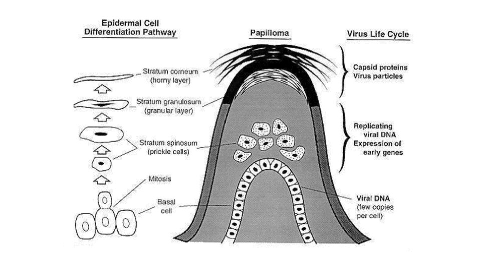
Etiology • Papillomaviridae • DNA • Papillomaviruses (PVs) are epitheliotropic viruses that cause benign proliferative lesions in the skin (warts or papillomas) and mucous membranes of their natural hosts. • In bovines specifically, 13 types of bovine papillomaviruses (BPVs) are currently well characterized, although there may actually be more than 20. https: //talk. ictvonline. org/ictv-reports/ictv_9 th_report/dsdna-viruses-2011/w/dsdna_viruses/122/papillomaviridae-figures

• BPVs are classified into four genera based on homology within the genomic regions of the L 1 ORF, the most conserved sequence. • These are • • Deltapapillomavirus (BPV-1, 2 and 13), Xipapillomavirus (BPV-3, 4, 6, 9, 10, 11, and 12), Epsilonpapillomavirus (BPV-5 and 8) Dyoxipapillomavirus (BPV-7) • They are the etiologic agent of papillomatosis and neoplasia of the upper gastrointestinal tract and urinary bladder. • Virus has a predilection for epithelial tissues. • Group A - cause fibropapillomas and usually do not yield infectious virus. BPV-1, BPV-2 and BPV-5 • Group B -Cause epithelial papillomas from which infectious virus can be recovered. BPV-3, BPV-4 and BPV-6

Transmission • The virus is transmitted by direct contact, fomites, and possibly by insects. • Calves are easily infected the papillomavirus entering the cut or abraded skin. • Bovine papillomavirus DNA has been identified in blood, milk, urine, and other biologic fluids obtained from infected animals.

Pathogenesis • Papillomas occur on the body locally and singularly. • It causes proliferation in the deep fibroblasts. • Papillomas have primer nodules before they grow and have keratinized cauliflower-like appearance. • Papillomatosis depends on age, environmental and genetic factors.

Venuti, A. , Paolini, F. , Nasir, L. , Corteggio, A. , Roperto, S. , Campo, M. S. , & Borzacchiello, G. (2011). Papillomavirus E 5: the smallest oncoprotein with many functions. Molecular cancer, 10(1), 140.

Clinical Signs • Warts appear about two months after exposure and may last up to a year. • Papillomatosis (generalised papillomas) are often seen in calves on the head, neck, and shoulders, and occasionally on the back and abdomen. • The extent and duration of the lesions depend on the type of virus, area affected, and degree of susceptibility. • Papillomitosis becomes a herd problem when a large group of young, susceptible animals become infected. • Urinary tumours have also been reported.

• Some lesions develop into papulonodules with a warty surface. Such fibropapillomas may involve the venereal regions where they can cause pain, disfigurement, infection of the penis of young bulls, and dystocia when the vaginal mucosa of heifers is affected. • A form of persistent cutaneous papillomatosis with smaller numbers of papillomas may be seen in herds of older cattle. A bovine papilloma virus has also been demonstrated in bladder tumors associated with bracken fern poisoning.

1 - Fungiform Skin Papillomotosis: Fungiform papillomas are formed in the breast and nipples in different regions of the skin, mostly in young cattle. by BPV-6. 2 - Filiform Papillomatosis: It is formed in the shape of yarn (rice) at the beginning of the breast and nipple, by BPV-5. 3 -Mucosa Papillomatosis: These occur as Genital Papillomas. Prepusium, Glans Penis and Vaginal Mucosa and in the form of cauliflower, BPV-2. 4 -Visceral Papillomatosis: papilloma occurs in bladder, in the stomach and pharynx and can be malignant. BPV-4

http: //calfology. com/sites/default/files/imagecache/370 x 260_slideshow/feature-photo/warts_2. jpg Dagalp, S. B. , Dogan, F. , Farzanı, T. A. , Salar, S. , & Bastan, A. (2017). The genetic diversity of bovine papillomaviruses (BPV) from different papillomatosis cases in dairy cows in Turkey. Archives of Virology, 162(6), 1507 -1518.

Diagnosis • Diagnosis is primarily based on presenting clinical signs augmented with laboratory determination of viral antigens using PCR assays. Immunity • Immunity usually develops three four weeks after initial infection, but papillomitosis occasionally recurs, probably due to loss of immunity.

Prevention and Control • Vaccines are available but due to the small incidence of this disease in cattle are rarely warranted. • In many instances, treatment of warts is not necessary, but frond-like lesions that interfere with milking may require excision. • The use of autogenous vaccines and virucidal teat dips may be recommended in herd outbreaks. • Instruments and tack used on infected animals should be disinfected before use on other animals. The infected animal may not have visible warts, but they may still contaminate equipment. • Tattoo or tagging pliers can be disinfected between use on calves, with a 2 to 4% solution of formaldehyde.

EQUINE PAPILLOMAVIRUSES • It is usually seen in horses between 1 and 3 years old. • Papillomas are found on the lips, at the tip of the nose, on the skin, on the back. • They are numerous and 1 cm in size. • Those that show wider spread in the body are called sarcoids. • It spreads only among horses. • Clinical diagnosis is easy. http: //www. ckequinehospital. com/blog/62/Horse-Sense-Myth-Buster-Horse-Warts-Pliers

CANINE PAPILLOMAVIRUSES • Dog papillomas are of the multiple papilloma type. It occurs sporadically in the oral mucosa, lips, tongue and pharynx of young dogs. • wart-like occurrences are observed In the inner part of the ear, in the penis and vagina. • Papillomas originate from epithelial cells, infected cells contain inclusion bodies. http: //www. vetfolio. com/compendium/applied-dermatology-the-cutaneous-viral-dermatoses-in-dogs-and-cats http: //www. vetbook. org/wiki/dog/index. php? title=Canine_papillomavirus

PAPILLOMATOSIS IN SHEEP AND GOATS • In sheep and goats, papillomas are more common in feet, around the mouth and in the nipples. • Ear cancer is caused by BPV-2. • In sheep and goats, papillomas are in the shape of Adenopapillomas. • Sheep at all ages are susceptible to infection. Initially, a serous nasal discharge, voice anxiety, weakening, and then tumor formation are seen.

References • Dagalp, S. B. , Dogan, F. , Farzanı, T. A. , Salar, S. , & Bastan, A. (2017). The genetic diversity of bovine papillomaviruses (BPV) from different papillomatosis cases in dairy cows in Turkey. Archives of Virology, 162(6), 1507 -1518. • http: //www. msdvetmanual. com/reproductive-system/udderdiseases/diseases-of-bovine-teats-and-skin • http: //vetbook. org/wiki/cow/index. php? title=Papilloma_virus • https: //manoa. hawaii. edu/ctahr/tpalm/pdfsmarianas/pdfs/vol_one/5_Livestock%20 Health%20 and%20 Managem ent/warts_on_cattle. pdf
 • Infectious canine hepatitis (ICH) is a worldwide,")
Infectious Canine Hepatitis (Hepatitis Contagiosa Canis) • Infectious canine hepatitis (ICH) is a worldwide, contagious disease of dogs with signs that vary from a slight fever and congestion of the mucous membranes to severe depression, marked leukopenia, and coagulation disorders. • It also is seen in foxes, wolves, coyotes, bears, lynx, and some pinnipeds; other carnivores may become infected without developing clinical illness. • In recent years, the disease has become uncommon in areas where routine immunization is done, but periodic outbreaks, which may reflect maintenance of the disease in wild and feral hosts, reinforce the need for continued vaccination.

Etiology • Adenoviridae ----- Mastadenovirus ------- CAV-1 • nonenveloped DNA virus, • canine adenovirus 1 (CAV-1), which is antigenically related only to CAV-2 (one of the causes of infectious canine tracheobronchitis. Ø Cross-protective immunity between canine adenoviruses 1 and 2. • CAV-1 is resistant to lipid solvents (such as ether), as well as to acid and formalin. • It survives outside the host for weeks or months, but a 1%– 3% solution of sodium hypochlorite (household bleach) is an effective disinfectant.

Transmission CAV-1 infection occurs by inhalation and ingestion of the virus after shedding in the urine, faeces or respiratory secretions. Transmission may be by direct contact, or by indirect contact such as via handlers or infected surfaces. Recovered dogs shed virus in their urine for ≥ 6 mo.

Pathogenesis Initial infection occurs in the tonsillar crypts and Peyer patches, followed by viremia and disseminated infection. Vascular endothelial cells are the primary target, with hepatic and renal parenchyma, spleen, and lungs becoming infected as well. Chronic kidney lesions and corneal clouding (“blue eye”) result from immune -complex reactions after recovery from acute or subclinical disease.

• • • Infection by oronasal route → primary replication in tonsils and regional lymph nodes. • • Direct effects of viral replication → cell lysis/necrosis → clinical signs. Release of virus → viremia; free virions in plasma within 8 -72 hours. Target organs: liver and kidneys → secondary replication in hepatic parenchymal cells and reticuloendothelial system. Focal areas of hepatocellular destruction may coalesce → hepatic necrosis → jaundice. Infection of endothelial cells → effusions and hemorrhagic diathesis. Immune-mediated keratitis → corneal edema. Immune-mediated glomerulonephritis. Either death or recovery with urinary excretion of virus for up to 1 year. Recovered dogs have life-long immunity.

Clinical Signs are apathy, anorexia, thirst, conjunctivitis, serous discharge from the eyes and nose, and occasionally abdominal pain and vomiting. Intense hyperemia or petechiae of the oral mucosa, as well as enlarged tonsils, may be seen. Tachycardia out of proportion to the fever may occur. There may be subcutaneous edema of the head, neck, and trunk. Despite hepatic involvement, there is a notable absence of icterus in most acute clinical cases. In mild cases, transient corneal opacity may be the only sign of disease.

Yellow tint to the whites of the eye indicates liver disease. There are many possible causes, but canine hepatitis virus will be on high on your vet's rule out list if your dog hasn't been vaccinated. Canine hepatitis is often deadly in unvaccinated puppies and young dogs, but older dogs usually tend to have mild symptoms and survive. . . but are carriers of the virus. This young dog with canine hepatitis has "blue eye" which is seen secondary to edema, vascular inflammation, or in the case of canine hepatitis virus, due to an immune response between the eye and the virus. Distemper virus is also associated with having a "blue eye". http: //animalpetdoctor. homestead. com/infectioushepatitis. html http: //www. urdogs. com/what-you-know-and-dont-know-about-adenovirus-1 -in-dogs/

Infection of hepatocytes and endothelial cells with CAV-1 produces characteristic basophilic intranuclear inclusions surrounded by a clear zone that separates them from the marginated chromatin (arrow). H&E stain. (Courtesy Dr. W. Crowell, College of Veterinary Medicine, The University of Georgia and Noah’s Archive, College of Veterinary Medicine, The University of Georgia. In Zachary JF, Mc. Gavin M. Pathologic Basis of Veterinary Disease, 5 ed. St. Louis, MO: Mosby; 2012. ) https: //www. vetstream. com/treat/canis/diseases/canine-adenovirus-type-1 -disease

Diagnosis • Usually, the sudden onset of illness and bleeding suggest ICH, although clinical evidence is not always sufficient to differentiate ICH from distemper. • Definitive antemortem diagnosis is not required before institution of supportive care but can be pursued with commercially available ELISA, serologic, and PCR testing. • PCR or restriction fragment length polymorphism is required to definitively distinguish CAV-1 from CAV-2, if clinically necessary. • Postmortem gross changes in the liver and gallbladder are more conclusive, and diagnosis is confirmed by virus isolation, immunofluorescence, characteristic intranuclear inclusion bodies in the liver, or PCR or fluorescence in situ hybridization studies of infected tissue.
 injectable vaccines are available and are often combined with")
Prevention • Modified-live virus (MLV) injectable vaccines are available and are often combined with other vaccines. • Vaccination against ICH is recommended at the time of canine distemper vaccinations. • Maternal antibody from immune dogs interferes with active immunization in puppies until they are 9– 12 wk old. • Attenuated CAV-1 vaccines have produced transient unilateral or bilateral opacities of the cornea, and the virus may be shed in urine. For these reasons, CAV-2 attenuated live virus strains, which provide cross-protection against CAV-1, are preferentially used because they have very little tendency to produce corneal opacities or uveitis, and the virus is not shed in urine.

References http: //www. msdvetmanual. com/generalized-conditions/infectiouscanine-hepatitis/overview-of-infectious-canine-hepatitis

Infectious Canine Laryngotracheitis Kennel Cough — Canine Respiratory Disease Complex — Infectious Canine Tracheobronchitis — Infectious Canine Tracheitis

A highly contagious acute respiratory disease spread by close contact causing laryngitis, tracheitis, bronchitis and in some cases a rhinitis. Multiple agents are implicated in the disease including Canine Adenovirus 1, Canine Adenovirus 2, Canine Herpes virus, Canine Parainfluenza - 2, Canine Distemper Virus, Mycoplasma species and Bordetella bronchoseptica. Most cases involve a primary viral infection and sometimes with secondary bacterial involvement. Infectious tracheobronchitis results from inflammation of the upper airways. It is a mild, self-limiting disease but may progress to fatal bronchopneumonia in puppies or to chronic bronchitis in debilitated adult or aged dogs. The illness spreads rapidly among susceptible dogs housed in close confinement (eg, veterinary hospitals or kennels).

Etiology ● ● ● Adenoviridae serologically related to CAV-1 Canine adenovirus type 1 disease. 75 nm non-enveloped DNA virus. Moderately resistant: survives days to months in environment. Inactivated by heating to 56°C (steam disinfection). Canine parainfluenza virus, canine adenovirus 2 (CAV-2), or canine distemper virus can be the primary or sole pathogen involved.

Transmission Adenoviruses are spread directly from dog to dog through infected respiratory secretions or by contact with contaminated feces or urine. Affects dogs of all ages. Puppies and immunocompromised dogs are often worst affected.

Pathophysiology Highly infectious by aerosol → localized respiratory tract infection. Replicate in upper and lower respiratory tract → shed in respiratory secretions. Focal necrosis of turbinate/tonsillar epithelium. Primary target site: bronchioles → proliferative, necrotizing bronchiolitis → peribronchiolar inflammation → interstitial pneumonia. ● Secondary bacterial infection, eg. Bordetella bronchiseptica , Pasteurella multocida, Streptococcusspp Streptococcus spp → bronchopneumonia Lung: bacterial pneumonia. ● Uncomplicated infections mild. ● ●

Clinical Signs Incubation period 2 -5 days. Viral shedding for 6 -9 days. Recovery 3 -7 days later unless secondary bacterial infection develops. Respiratory disease (CAV-2) ● Dry, hacking cough ● Retching ● Coughing up white foamy discharge ● Conjunctivitis http: //www. askavetquestion. com/kennelcough. php The veterinarian will feel the trachea to see if this makes the dog cough. Most dogs with kennel cough will have an irritated trachea. https: //www. askjpc. org/wsco/do_slideshow 2. php? pic_series=150404&c. Year=2015&init. Image=2292

Prevention and Control • Dogs should be immunized with modified-live virus vaccines against distemper, parainfluenza, and CAV-2, which also provides protection against CAV-1. • Vaccines can prevent or reduce the severity of disease caused by B. bronchiseptica, parainfluenza virus and adenovirus. • Vaccinated animals can still contract the disease as multiple agents are implicated. • Vaccines are available to be given systemically or intranasally. • Intranasal vaccination provides mucosal Ig. A immunity and the presence of maternal antibodies do not affect actions of the vaccine.

https: //www. northdevonvets. co. uk/kennel-cough-vaccination-only-15 -00 -throughout-may/

It is important to practice good husbandry in areas where groups of dogs mix e. g boarding kennels. Ideally all animals should be vaccinated, any infected animals should be isolated to minimise the spread to unaffected animals and all fomites that have come into contact with an affected animal must be disinfected. Areas should be kept well ventilated and ideally animals should be kept in low population densities.

References • https: //en. wikivet. net/Canine_Infectious_Tracheobronchitis • http: //www. vetstreet. com/care/canine-adenovirus-type-2 -cav-2 • https: //www. merck-animal-health-usa. com/dp/3 • http: //www. msdvetmanual. com/respiratory-system/respiratorydiseases-of-small-animals/tracheobronchitis-in-smallanimals#v 3295117

BOVINE ADENOVIRUS Bovine adenovirus has been associated with a wide spectrum of diseases, with bovine adenovirus type 3 being the serotype most often associated with BRD. Most adenovirus infections in cattle involve either the respiratory or gastrointestinal tracts. In addition there have been reports of adenovirus associated with conjunctivitis, keratoconjunctivitis, and weak calf syndrome. It is now also thought to contribute to the disease complex Enzootic pneumonia of calves. Bovine adenoviruses have also been isolated from healthy cattle.

Etiology • Adenoviruses are DNA viruses that are divided into 10 serotypes. • Bovine adenoviruses are found worldwide and are particularly widespread in Central America and Africa. • Cattle are often infected without showing any signs of disease. • Respiratory and gastrointestinal disease signs can appear when the animal enters a stressful situation. • However, it is more often the case that animals comingled during shipping of movement seroconvert (develop antibodies to the virus due to exposure) without developing signs of disease.

Pathogenesis The respiratory and enteric tracts are the primary targets for adenovirus infection which usually results in cell lysis and virus shedding, but some cells accumulate virus particles in the nucleus establishing persistent infections. Respiratory and faecal shedding usually last for about 10 days and, where the kidney is involved, virus can be excreted for over 10 weeks in urine. With persistent infection, lysis of fragile infected cells produces virus-shedding resulting in infection of susceptible animals that come in contact with the virus.

Clinical Signs • Bovine Adenovirus can infect cattle and zebu of any breed, sex or age, however, younger animals are more at risk as their maternal antibody begins to wane, around the age of 2 weeks to 4 months old. • Interestingly, bovine adenovirus infection is not often associated with any gross changes in the gastrointestinal or respiratory tract. • Bovine adenovirus infection can be associated with a variety of disease conditions including pneumonia, keratoconjunctivitis, enteritis, diarrhea, weak calf syndrome and abortion. • When Adenovirus infection does induce signs of disease, it is often upper or lower respiratory disease accompanied by enteric disease.

Respiratory signs include coughing, serous nasal discharge, dyspnoea and tachypnoea. This may progress to bronchopneumonia if secondary bacterial infection is present. Generalised clinical signs include pyrexia, weight loss, sudden death, lymphadenopathy and generalised weakness.

Diagnosis • Clinical signs and physical examination may add Bovine Adenovirus to the differential list, but definitive diagnosis cannot be achieved. • Adenovirus infection can be diagnosed morphometrically, serologically, and by virus isolation. Rapid presumptive diagnosis can be made either by observation of characteristic virus morphology in intranuclear inclusions by transmission electron microscopy or by immunofluorescent or immunohistochemical labelling of adenovirus antigens in tissues with gross lesions. • Because of the number of types of adenoviruses infecting cattle, virus isolation is necessary to definitively identify the virus. • Virus can be isolated from nasal secretions, tracheal fluids, intestinal contents and tissue homogenates. • PCR

Prevention and Control The main control measure is to ensure adequate colostrum at birth as passive transfer provides immunity to calves. Other control strategies include preventing mixing of calves of different age groups and ensuring good hygiene and ventilation in calf housing.

• Both modified live and inactivated adenovirus vaccines have been developed and evaluated for use in cattle and they should be administered when maternal antibodies have waned, but 2 to 3 weeks before calves from different places are assembled under stressful conditions. • Such vaccines are available in Europe (not UK) and Japan, but not in the USA. Most vaccines are formulated in combination with other agents. • Two to four doses of vaccine administered subcutaneously or intramuscularly are recommended to provide proper protection. Vaccination has not eliminated infection entirely, but has resulted in the reduction in disease incidence and treatment costs.

References https: //en. wikivet. net/Bovine_Adenovirus#cite_note-1 http: //calfology. com/library/wiki/adenovirus
- Slides: 48