IMMUNODEFICIENCY DISORDERS Dr Mayssaa Essam Immunodeficiency The immunodeficiency


, or Bruton’s hypogammaglobulinemia. - Characterized by extremely")


")
 - There are defect in T cell signaling to B")
.")


. Richard Coico&Geoffrey Sunshine. - Textbook")

- Slides: 21
IMMUNODEFICIENCY DISORDERS Dr. Mayssaa Essam
Immunodeficiency The immunodeficiency may be the result of defective immunity, both innate and specific, because of genetic abnormality (primary) or there is a loss of function because of the damage by physical, chemical or biological agents (secondary).
Immunodeficiency diseases A. Primary immunodeficiencies states are inherited defects of the immune system. B. Secondary immunodeficiency states are more common in malnutrition, infection, cancer, renal disease, malignancies patients treated by immunosupressive drugs( AIDS).
PRIMARY IMMUNODEFICIENCIES Primary deficiencies in immunological function can arise through failure of any of the developmental processes from stem cell to functional end cell. Defects in the development of the common lymphoid stem cell give rise to severe combined immunodeficiency. Both T and B lymphocytes fail to develop. The myeloid cell disorders affect phagocytic function. Most of the primary immunodeficiencies are inherited. They are classified on the basis of the site of lesion in the developmental or differentiation pathway of the immune system.
Distribution of primary immunodeficienciesn
Primary Immunodeficiencies
B CELL DEFICIENCY - X-linked gammaglobuinemia. - Ig. A deficiency. - Ig. G subclass deficiency. - Transient hypogammaglobulinaemia of infancy. - Common variable immundeficiency.
X-linked a gammaglobulinaemia - X-linked agammaglobulinemia (X-LA), or Bruton’s hypogammaglobulinemia. - Characterized by extremely low Ig. G levels and by the absence of other immunoglobulin classes. - Babies born with this disorder have virtually no peripheral B cells ( 1%) and suffer from recurrent bacterial infections.
X-linked a gammaglobulinaemia - B-cell development in the bone marrow is arrested at the pro-B- to pre-B-cell stage, and the B lymphocytes in these patients remain in the pre-B stage, with heavy chains rearranged but light chains in their germ-line configuration. - Present-day use of antibiotics and replacement therapy in the form of passively administered antibodies can make this disease quite manageable.
Ig. A and Ig. G subclass defeciency - Ig. A deficiency is most common. - About 20% lack Ig. G 2 and Ig. G 4. - Susceptible to pyogenic infection. - Result from failure in terminadifferentiation of B cells. - Deletion of constant heavy chain genes or abnormalities of isotype switching may result in deficiencies of one or more of the Ig. G subclasses. - Patients with recurrent infection should be treated aggressively with broad-spectrum antibiotics.
Ig. A and Ig. G subclass defeciency -Sometimes this disorder is associated with other immunodeficiencies such as selective Ig. A deficiency or ataxia. - Patients with selective Ig. A deficiency should not be treated with gamma globulins. Therapeutic gamma globulin contains only a small quantity of Ig. A and this is not likely to reach mucosal secretions through parenteral administration.
Hypogammaglobulinaemia of infancy - All infants develop physiologic hypogammaglobulinemia at approximately 5 to 6 months of age. At this time, maternal Ig. G is slowly catabolized, the infant begins synthesizing its own Ig. G by this age. - Infant may fail to initiate Ig. G synthesis at this time, resulting in a prolonged period of hypogammaglobulinemia termed transient hypogammaglobulinemia of infancy (THI).
Hypogamaglobulinaemia of infancy - THI is more prolonged in premature infants because of decreased transplacental maternal Ig. G at birth. - Infant with THI begins to recurrent respiratory tract infections and exhibit poor or absent antibody responses to vaccines. Spontaneous recovery occurs by 18 to 24 months.
Hypogamaglobulinaemia of infancy - Some of these infants may benefit from intravenous immunoglobulin (IVIg) infusions or continuous antibiotic treatment. - Infections may be caused by pneumococci, H. influenzae or other pyogenic organisms, chronic lung disease or intestinal malabsorption may be present.
Common Variable Immunodeficiency (CVID) - There are defect in T cell signaling to B cells. - May follow viral infection - Pyogenic infection - 80% of patients have B cells that are not functioning. - B cells are not defective. They fail to receive signaling from T lymphocytes.
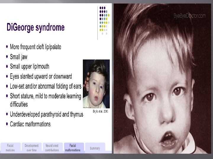
T CELL DEFICIENCY - Di. George's syndrome (Congenital Thymic Aplasia, Immune Deficiency with Hypoparathyroidism). - Purine Nucleoside Phosphorylase (PNP).
Di. George's syndrome - During 6 to 8 weeks of intrauterine life, the thymus and parathyroid glands develop from epithelial evaginations. Di. George syndrome is the result of interference with normal embryonic development at approximately 12 weeks of gestation. - The most frequent presenting sign in patients with this syndrome occurs in the first 24 hours of life with hypocalcemia that is resistant to therapy. Neonatal tetany and various types of congenital syndromes may also be present.
Di. George's syndrome - Most patients develop recurrent and chronic infections with viral, bacterial, fungal or protozoal organisms. Failure to thrive may be present. - Circulating T cells are reduced in numbers. - Delayed hypersensitivity and graft rejection are depressed. - Treatment is by transplantation of fetal thymus tissue.
Refrences - Immunology A Short Course (Seventh Edition 2015). Richard Coico&Geoffrey Sunshine. - Textbook of Immunology(Second Edition-2014). Sunil Kumar Mohanty& K Sai Leela
THANK YOU & GOOD LUCK