Human skin The human skin is the outer


- Slides: 15
Human skin The human skin is the outer covering of the body. In humans, it is the largest organ of the integumentary system. Pigments There at least five different pigments that determine the colour of the skin. 1 -Melanin: It is brown in colour. 2 -Melanoid: It resembles melanin but is present diffusely throughout the epidermis. 3 -Keratin: This pigment is yellow to orange in colour. 4 -Hemoglobin: It is found in blood and is not a pigment of the skin but develops a purple color. 5 -Oxyhemoglobin: It is also found in blood and is not a pigment of the skin. It develops a red color.
Melanin is a natural pigment found in most organisms. In animals melanin pigments are derivatives of the amino acid tyrosine. Melanin is brown, non-refractile, and finely granular with individual granules having a diameter of less than 800 nanometers. The most common biological melanin is eumelanin: This is a brownblack polymer of dihydroxyindole carboxylic acids and their reduced forms. Another common form of melanin is pheomelanin: a cysteine-containing red -brown polymer of benzothiazine units largely responsible for red hair and freckl.
Melanin synthesis Special cells known as melanocytes produce melanin in the outer layer of the skin. The greater expression of the gene creates an increase in the synthesis of melanin. Precursor L-tyrosine is a precursor of melanin. This means that certain biochemical pathways convert L-tyrosine into melanin through the use of numerous "intermediate molecules" that are systematically modified into the end product. Upon exposure to UV radiation, DNA damage triggers cytokines, growth factors and other inflammatory factors to stimulate melanin production. Melanocytes, by increasing the production of intracellular nitric oxide (NO), they trigger signal transduction cascades to initiate melanogenesis through a series of oxidative reactions involving the amino acid tyrosine in the presence of the enzyme tyrosinase.
The process of melanin synthesis
1. Synthesis of melanin. i. Melanin pigment gives the black colour to the skin and hair Tyrosinase and tyrosine hydroxylase: Both these enzymes will add hydroxyl group to tyrosine to produce dihydroxy phenylalanine (DOPA). Tyrosinase is present in melanoblasts. The enzyme produces DOPA, which is used for melanin synthesis. Tyrosine hydroxylase is present in adrenal medulla and the DOPA thus generated is used for epinephrine synthesis. Thus even in tyrosinasedeficient person (albinism), epinephrine synthesis is normal Melanocytes in the deeper layers of epidermis synthesise melanin in granular form in melano-somes. The extracellular granules are later dispersed under the influence of melanocyte stimulating hormone (MSH). Alpha MSH has 13 amino acids, which constitute the amino terminal part of ACTH. Therefore MSH has weak ACTH activity and ACTH has weak MSH activity.
Clinical applications of Melanin: i. Copper deficiency: Since tyrosinase is a copper containing enzyme, there may be disturbances in pigmentation during copper deficiency. Hair synthesised at the time of deficiency may be depigmented. If copper deficiency is intermittent, alternate black and white regions may be seen in the hair. ii. Malignant Melanoma: Melanoblasts, especially in junctional naevi, may multiply to give rise to malignant melanoma. Melanogen may be excreted through urine in such conditions. Such urine if kept in a test tube, the upper part of the tube becomes black due to oxidation to melanin. iii. Leukoderma: When tyrosinase or melanin forming cells or both are absent from epidermis, leukoderma (white patches) results. Graying of hair is also due to the disappearance of melanocytes from the hair root. iv. Albinism: Albinism and leukoderma are different. In albinism, tyrosinase is absent in melanocytes all over the body.
Photoprotection "The term Photoprotection designates the mechanisms that nature has developed to minimize the damages that the human body suffers when exposed to UV-irradiation. Photoprotection of the human skin is achieved by extremely efficient internal conversion of DNA, proteins and melanin. Internal conversion is a photochemical process that converts the energy of the UV-photon into small amounts of heat. This small amount of heat is harmless. If the energy of the UV-photon were not transformed into heat, then it would lead to the generation of free radicals or other harmful reactive chemical species (e. g. singlet oxygen, or hydroxyl radical). “
VITAMIN D AND MELANIN There is a relationship between the amount of melanin in your skin and vitamin D levels. Your body can synthesize vitamin D when your skin is exposed to direct sunlight. Because melanin blocks the effects of sunlight, increased levels of melanin can impair your ability to make new vitamin D. As a result, people with dark skin often have lower levels of vitamin D than lighter-skinned people.
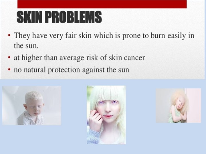
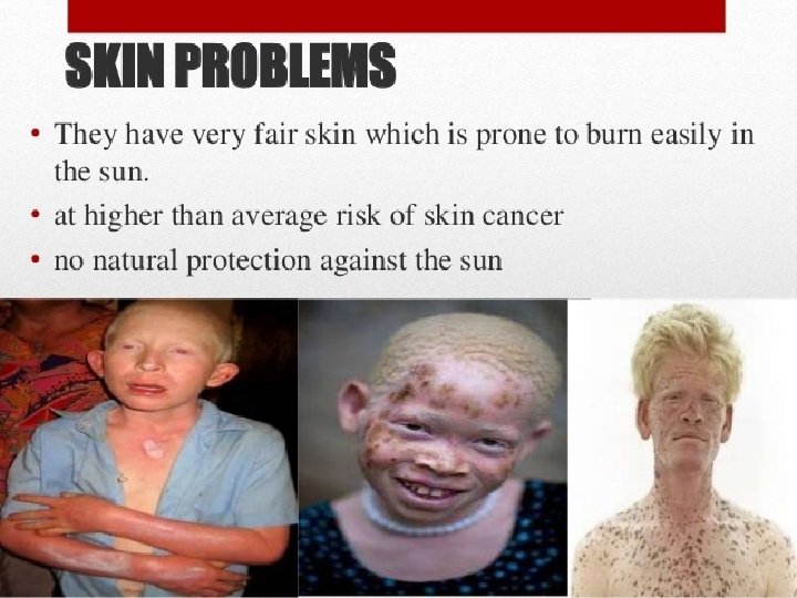
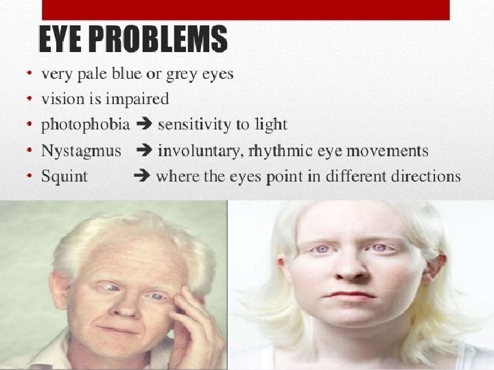
Albinism is a group of genetic conditions that causes a lack of pigment. It can affect only the eyes or both the eyes and skin. Most types of albinism are inherited when an individual receives the albinism gene from both parents. Description • Due to the defect in tyrosine metabolism it results in a deficiency of melanin production and partial or full absence of pigment from the skin, hair, and eyes • It may be inherited by one of several modes: autosomal recessive, autosomal dominant. • Affected people may appear to have white hair, skin & iris color. They may have vision defects and photophobia. • Oculocutaneous albinism is most severe form resulting from a deficiency of tyrosinase activity, causing a total absence of pigment from the hair, eyes & skin
Causes • Albinism is caused by an alteration of the gene that regulates the melanin pigment synthesis. Symptoms • Absence of pigment from the hair, skin, or iris of eyes • Lighter than normal skin and hair or complete albinism • Most forms of complete albinism have some of the following possible symptoms: – Rapid eye movements – Strabismus (eyes not tracking properly) – Photophobia (avoidance of light because of discomfort) – Decreased visual acuity – Functional blindness Complications • Skin cancer • Decreased vision, blindness
hyperpigmentation is the darkening of an area of skin or nails caused by increased melanin. Hyperpigmentation is a common, usually harmless condition in which patches of skin become darker in color than the normal surrounding skin. Causes Hyperpigmentation may be caused by sun damage, inflammation, or other skin injuries, including those related to acne vulgaris. Many forms of hyperpigmentation are caused by an excess production of melanin. As the body ages, melanocyte distribution becomes less diffuse and its regulation less controlled by the body. UV light stimulates melanocyte activity, and where concentrations of the cells are denser than surrounding areas, hyperpigmentation is effected.